




Abstract

T
Working in the field of cell–cell transfer of HIV-1 and protein–protein interactions at the virological synapse, we demonstrated by three-dimensional (3D) confocal imaging that the tetraspanin CD63 is predominantly present at the virological synapse (Fig. 1). Jurkat cells were nucleofected with the vector pHIV-1JR-FL Gag-iGFP expressing the Gag-internal green fluorescent protein (iGFP) and uninfected Jurkat cells were added. Endogenous expressed CD63 was immunostained and localized by confocal laser microscopy in order to obtain a 3D image from cells in contact. To note, we recently demonstrated that CD63 interacts with the transmembrane envelope protein gp41 of HIV-1. 4 We conclude that HIV virions are routed through the virological synapse utilizing host proteins such as CD63.
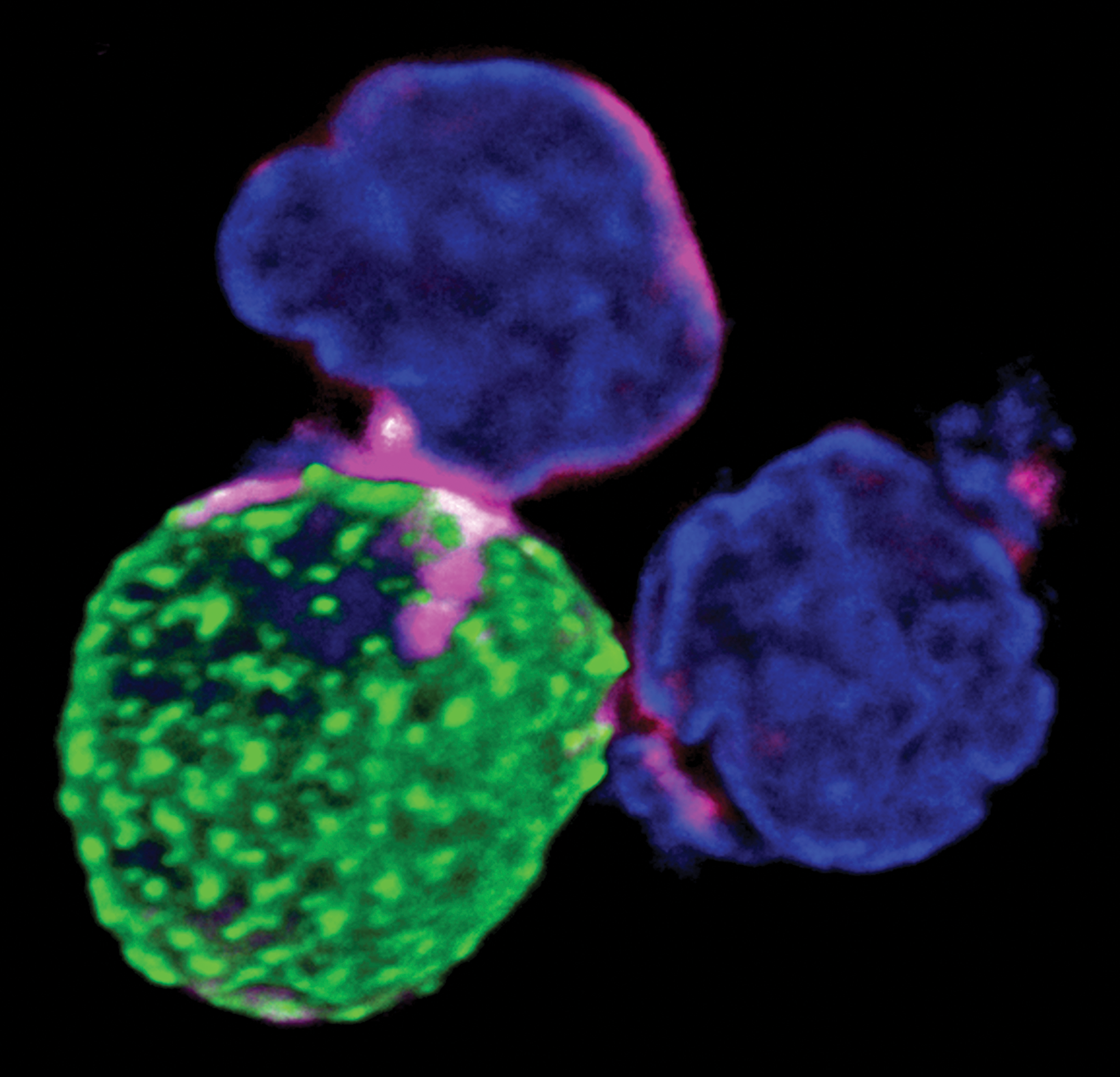
Three-dimensional (3D) imaging of CD63 and HIV-1 shows the formation of a virological synapse between cells. Jurkat cells were nucleofected with the molecular clone HIV-1JR-FL Gag-iGFP and produce viral particles (green). The endogenous CD63 was stained with anti-CD63 antibodies (magenta; the brightness level represents signal intensity) and nuclei were stained with DAPI (blue). HIV-1 cell–cell transfer occurs at the virological synapse, which is formed between HIV-1 producer and acceptor cells where CD63 accumulates. The image was obtained with an inverted confocal laser microscope (Zeiss cLSM 780) by taking a stack of optical sections through the sample (z-stack) and 3D projection using Zeiss ZEN software. We used a Gaussian filter as the unsharp mask (Adobe Photoshop) to produce a sharper image.
Footnotes
Acknowledgments
The work was supported by the Peter und Traudl Engelhorn Foundation, Germany with a grant to Daniel Ivanusic and Jung-Stiftung für Wissenschaft und Forschung, Hamburg/Germany.
Author Disclosure Statement
No competing financial interests exist.