
Abstract

H
Recent reports have shown that cell-to-cell spread of HIV-1 between T cells is more efficient than the equivalent cell-free infection. 1,2 Normally, T cells are not inherently polarized and do not display front–rear polarity in the absence of stimulation. However, T cells can adapt and show front–rear polarity during stimulation, including cell–cell contact with antigen-presenting cells at the immunological synapse (IS), T cell migration, and chemokine or cytokine stimulation. IS formation triggers cytoplasmic and membrane remodeling within the T cell and recruits organelles, such as the secretory apparatus, signaling machinery, and mitochondria, to the contact region.
Mitochondria has been reported to play an important role in the formation of IS by promoting sustained calcium influx to support synaptic signaling. 3 VS formation shares many similarities with the IS. 2 Mitochondria have long been known to be essential organelles that provide metabolic energy. They are involved in many physical functions and regulations including cellular signaling pathways, apoptosis, autophagy, and innate immunity. 3 There is limited information regarding the role of mitochondria in VS.
To address this question, we used transmission electron microscopy (TEM) to observe the location of mitochondria during HIV-1 VS formation using HIV-1-infected Jurkat T cells cocultured with uninfected Jurkat T cells. Our results illustrated in Fig. 1 showed that mitochondria were polarized and were engaged primarily in the contact regions of the HIV-1-infected T cells (effectors) during cell-to-cell contact. Recently, Groppelli et al. also indicated that polarization of mitochondria supports HIV-1 VS formation. 2 Lee-Huang and Huang used live-cell real-time imaging to observe mitochondria in HIV-1-infected cells. 4
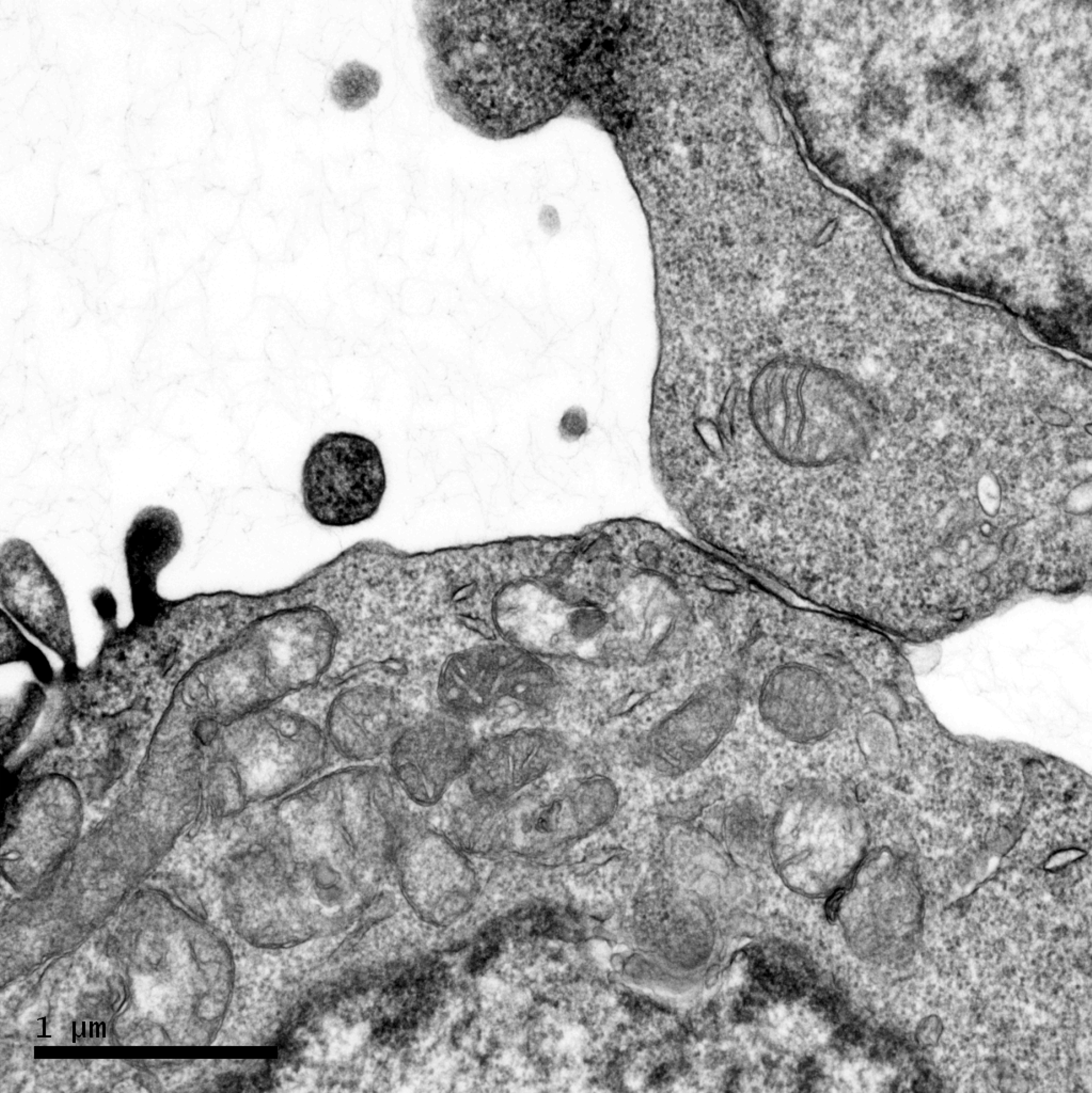
Cell-to-cell contact between HIV-1-infected (the lower cell) and HIV-uninfected (the upper cell) Jurkat T cells under transmission electron microscopy (TEM).
We therefore suggest that mitochondria are important for VS formation and supply energy for cell-to-cell transmission of HIV-1. At present, the detailed mechanism of VS formation and cell-to-cell transmission is still not fully understood. These findings provide further insight into intercellular interactions and organelle localization during HIV-1 VS formation.
Footnotes
Acknowledgments
The authors wish to thank the staff and their colleagues at the Institute of Biomedical Sciences, Academia Sinica, Taiwan for their technical assistance with TEM. This work was supported by grants from the Center for Infectious Disease and Cancer Research, Kaohsiung Medical University, grants KMUTP103E03 and KMU-TP103E20. This study was also supported by a grant from the Kaohsiung Medical University Research Foundation (KMU-Q104001).
Author Disclosure Statement
No competing financial interests exist.