
Abstract

K
Case Description
The patient, a 43-year-old man, had HIV infection and hepatitis C acquired through intravenous drug use. He was receiving cART with emtricitabine, tenofovir, atazanavir, and ritonavir. He had obtained undetectable HIV viral load and a partial immune recovery (CD4+ count between 400 and 500 cells/mm3).
The patient had been staying at our correctional facility for 2 years. He was sent for dermatological examination for the persistence since about 3 years of asymptomatic lesions located frontally and at the inside face of the left thigh. Such lesions were papular, infiltrated of purplish brown color. He had been previously prescribed topical treatments, mainly antibiotic and steroid, but no change of the lesions had occurred.
The lesions showed a slowly progressive character with the confluence of some elements in plaques. In addition, thigh appeared of in toto increased consistency (almost woody) on manual examination; the thigh was so stiffened to determine some difficulties in the movement.
After clinical evaluation, the dermatologist performed a skin biopsy (Fig. 1).
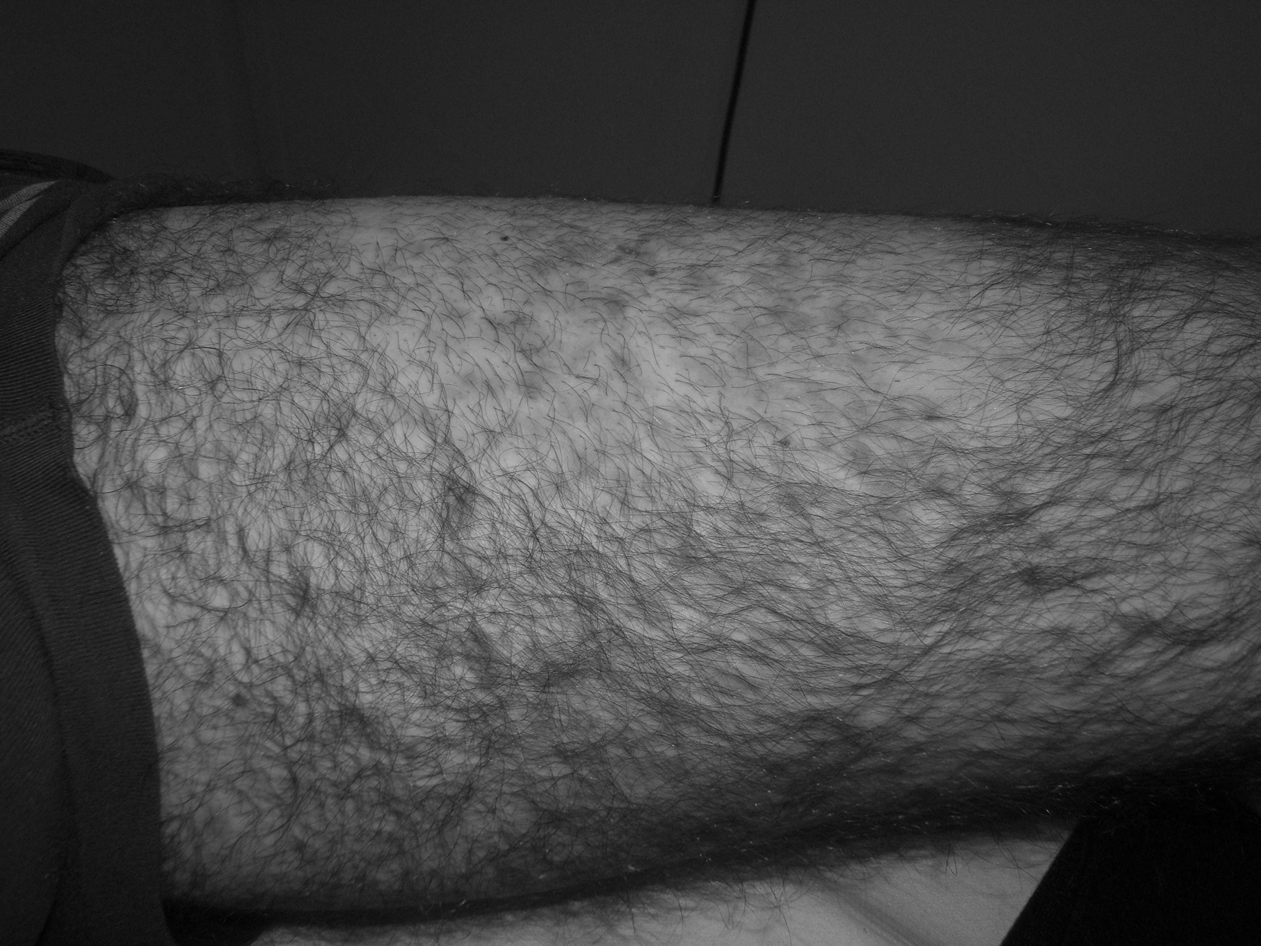
Left leg: papular lesions with confluence of some elements.
Histological examination showed epidermal acanthosis and perivascular and periannexial lymphoplasmocitary infiltrate in the dermis with presence of some spindle cells (hemosiderinic pigment Perls+). Immunohistochemistry examination showed human herpesvirus 8 (HHV8) nuclear expression and positive staining for CD34, CD31, and F VIII in vascular elements. Histopathologic findings were diagnostic of KS. 3
The patient was then released from prison for treatment at an Infectious Diseases Department. He received chemotherapy with vinblastine and bleomycin followed by local radiotherapy. He had an excellent clinical response with disappearance of the skin lesions and 4 years of relapse-free (and prison-free) follow-up. The patient did not present with any other HIV-related opportunistic manifestation or any other non-HIV-related cancer.
The patient had a presentation (type and site of lesions) of KS infrequent among immune-suppressed subjects, making difficult to suspect KS. 1 Thus, the occurrence of slowly progressive skin lesions of uncertain diagnosis, even if not immediately suggestive of KS, confirms the need for accurate skin checks in HIV-infected individuals, even if not severely immune suppressed and in any setting (including prison). The skin biopsy proved once again the cornerstone for the diagnosis of KS. 1,3
Footnotes
Author Disclosure Statement
No competing financial interests exist.