
Abstract

Protease is one of the three enzymes encoded within HIV's pol gene. Protease is a retroviral aspartyl-protease (retropepsin), which, unlike other aspartyl-proteases, must associate into a 22 kDa homodimer to exert its activity.
1
As all aspartic acid proteases, the catalytic residues (D25, T26, and G27) are highly conserved. Protease is responsible for the cleavage of viral Gag-Pol polypeptide into mature viral proteins and a target of current antiretroviral therapy.
2
HIV protease diversity has been widely studied in European, North American, and African countries due to the impact of resistance mutations against protease inhibitors. Although saquinavir was the first FDA-approved protease inhibitor, the current arsenal includes atazanavir, darunavir, fosamprenavir, ritonavir, and tipranavir (
This image maps Shannon entropy levels of variable sites as derived from the study of 777 Mexican protease nucleotide sequences. Shannon entropy was calculated employing amino acid equivalents representing increases in entropy with regard to HXB2 reference. 3 Entropy levels were arbitrarily classified as high (>0.6), mid (between 0.6 and 0.2), and low (<0.2). Protease sites having high (5 sites) and mid (10 sites) entropy levels were mapped into an X-ray diffraction crystal of HIV-1 protease dimer coupled to the antiretroviral drug saquinavir (PDB ID: 3OXC) employing The PyMOL Molecular Graphics System (Version 2.0 Schrödinger, LLC.).
In Figure 1, the protease dimer is represented as a blue cartoon with surface mesh, and saquinavir is shown in white. The flap domain regions (elbow and tip of the flap) are shown in orange, the core domain regions (C-terminal triad, catalytic site, 10s and 60s loop) are shown in red, and the dimerization domain regions (amino and carboxy terminus) are shown in green. The amino acid sites exhibiting high and mid entropy levels are indicated in red and orange residue numbers, respectively. All sites exhibiting high and mid entropy affected the surface of the protein and were confined to an area located between the elbow and the 60s loop. High levels of entropy were observed in residues surrounding the 10s loop, the elbow region, the 60s loop, and in the first half of the C-terminal domain.
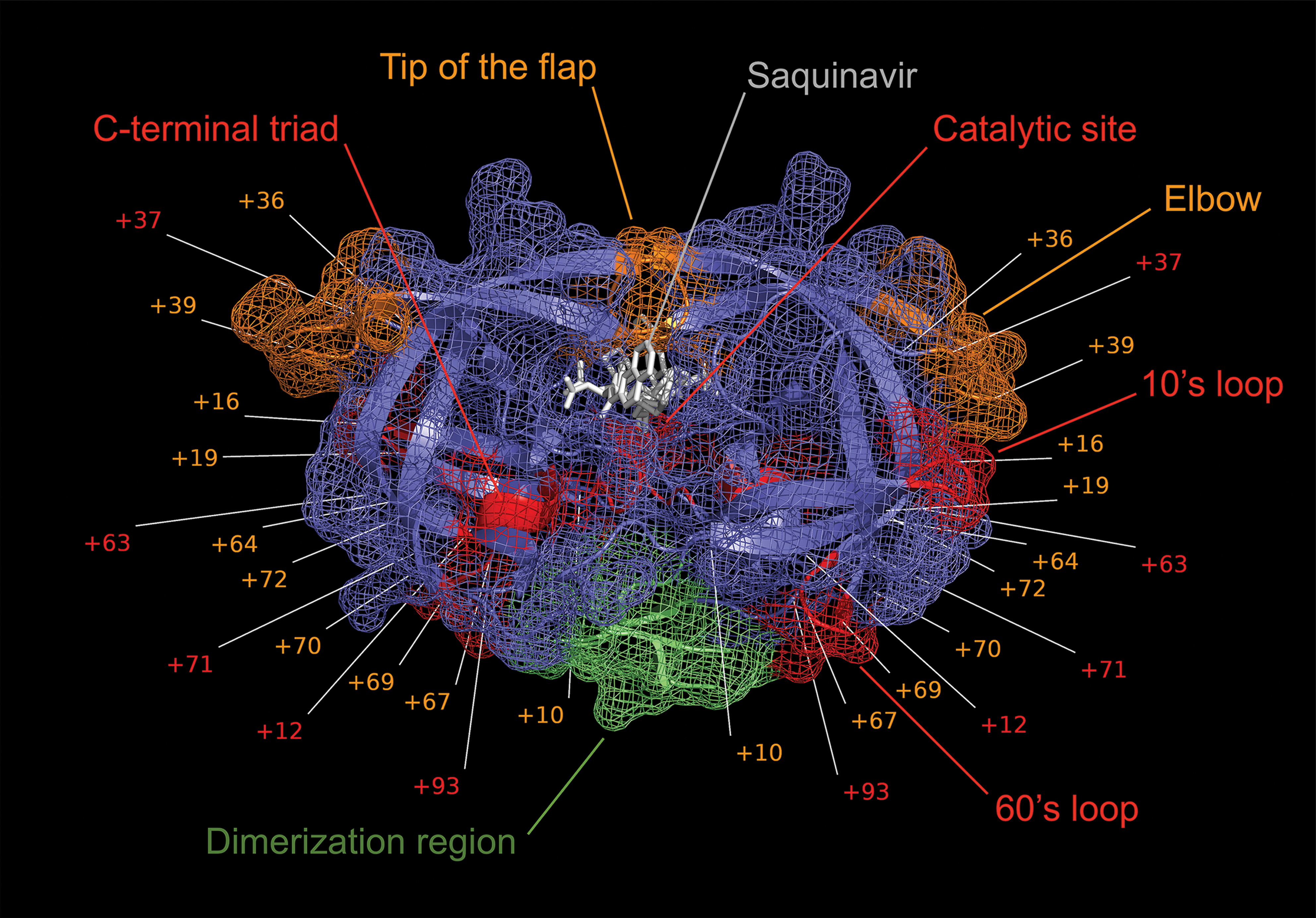
Protease amino acid sites exhibiting high and mid entropy levels are indicated in red and orange residue numbers, respectively. The protease dimer is represented as a blue cartoon with surface mesh, and saquinavir is shown in white. The flap domain regions (elbow and tip of the flap) are shown in orange, the core domain regions (C-terminal triad, catalytic site, 10s and 60s loop) are shown in red, and the dimerization domain regions (amino and carboxy terminus) are shown in green. Color images are available online.
Despite this, only five mutations affected functionally important regions, including the high-entropy Ser 37 and mid-entropy Pro-39, both located in the elbow region of the flap domain. The most conserved regions were the catalytic residues of the active site, the flap domain, and a region stretching from the 60s loop to the C-terminal triad. In addition, viral aspartic acid proteases have been shown to be functionally tolerant of mutations outside of the catalytic residues. 4 Given the comprehensive number of sequences analyzed, these findings further our understanding of HIV molecular diversity in Mexico. Understanding the molecular distribution of polymorphism can help develop better models of viral evolution, correlate disease progression with viral diversity, predict drug resistance, and guide vaccine development as well as provide insights into viral ancestry.
Footnotes
Author Disclosure Statement
No competing financial interests exist.