




Abstract
Abstract
Determination of acute inhalation toxicity is requested for hazard assessment of substances. In the regulatory-required test, otherwise toxicologically inert solid (dust) aerosols have to be tested up to a very high concentration limit of 5 mg/L of air. By testing a series of organic pigments at this concentration, we found that some pigments were well tolerated (hence, resulting in an LC50 >5 mg/L). For other pigments, we identified obstruction of the airways with subsequent suffocation as the cause of death at this concentration. In these cases, Organization for Economic Cooperation and Development TG 436 requires that additional animals are tested at 1 mg/L air. However, the mortality of animals at the high concentration could have been avoided if the suffocation by obstruction of the airways was predictable. Hence, we investigated the correlation of several physicochemical characteristics with the observed mortality. Test substances with the highest contact angle, a measure of hydrophobicity, produced mortality at high concentrations, whereas the more hydrophilic compounds did not. Therefore, the contact angle of test substances may serve as a predictive parameter for suffocation potential. We propose conducting this characterization before in vivo testing to reduce the number and suffering of animals until further in vitro and in silico approaches are developed to completely replace animal testing.
Introduction
D
Deposition and distribution of inhaled aerosols are dependent on chemical and physical properties, such as reactivity, solubility, lipophilicity/hydrophobicity, and surface structure. Since these properties are also important for the observed toxicity, they should be considered in the interpretation of the results of inhalation toxicity tests. Particularly at the concentration around 5 mg/L air, insoluble solid aerosols can lead to an obstruction of the airways and subsequently to suffocation. 9 This limit dose is very high and not considered meaningful to assess actual acute inhalation hazards 10 ; for example, it is 100-fold higher than the default concentration of 0.05 mg/L air assigned to charging or discharging of powders at nondedicated facilities in the ECETOC TRA tool (ECETOC TRAv3 tool: www.ecetoc.org/tra).
We are regularly performing acute inhalation toxicity studies in rats according to the Organization for Economic Cooperation and Development (OECD) guideline 4035 for regulatory purposes. Rats survived exposure to most of the organic pigments at ∼5 mg/L. In some of the experiments with organic pigments, however, we observed unexpected mortality in a concentration range around 5 mg/L. These compounds were nontoxic with regard to other toxicological endpoints. Histopathological examinations were carried out in three of these studies to establish the cause of death. Based on these results, it is evident that obstruction of the airways and consequently suffocation was in fact the cause of death after exposure to the test substance at 5 mg/L.
Considering animal welfare aspects, it seems unnecessary to expose animals to the limit concentration of 5 mg/L air of otherwise inert materials, only to detect suffocation by clumps of particles which obstruct the airways. It should be possible to identify this property by other means. Therefore, the aim of the present study was to find out a predictive physicochemical parameter for these compound characteristics.
Methods
General
The inhalation studies were conducted according to the OECD Principles of Good Laboratory Practice, 11 which principally meet the United States Environmental Protection Agency Good Laboratory Practice Standards [40 CFR Part 160 (FIFRA) and Part 792 (TSCA)]. The studies were conducted according to OECD guideline 403. 5 All animal studies were performed to meet requirements of national chemical legislations.
Test materials and characterization
Test materials
Pyrimidine azo yellow pigment (lot KL950, purity 95.6%), pyrimidinone black pigment (lot 2005-01, purity 99.9%), benzodifuran black pigment (lot 0008219266, purity 99.0%), benzimidazolone black pigment (lot Partie 053001, purity >99%), azomethine yellow pigment (lot 0005629903, purity 99.9%), diketopyrrolopyrrole red pigment (DPP red pigment, lot Op.1 + 03/06 R4), and diketopyrrolopyrrole orange pigment (DPP orange pigment, lot 0008679022) were manufactured by BASF Colors and Effects. The tested materials comprise all organic pigments, which were tested by BASF at ca 5 mg/L according to OECD guideline 403 and for which the original test material was available in the GLP archive. Pigment materials for which a stable test atmosphere of ca 5 mg/L could not be achieved, were considered to be not of interest to this investigation. Of note, no cases of mortality were observed in these cases. The relative density and the molecular weight of the test materials ranged between 1.51 and 1.92 and 324 g/mol and 636 g/mol, respectively. Their water solubility is below the limit of detection of generally <0.1 mg/L with the exception of the pyrimidine azo yellow pigment, which has a water solubility of 0.8 mg/L.
Characterization
Hydrophobicity
The contact angle of the test substances was used as a measure for their hydrophobicity. For this, the test substance powders were prepared onto adhesive, transparent tapes, which were analyzed in a Drop Shape Analyzer (DSA 100; Krüss GmbH, Hamburg, Germany) by using the static droplet method and water as wetting agent (Fig. 1). The preparation of nanomaterial powder surfaces and their assessment by the sessile drop technique was previously validated.12,13 A minimum of three droplets were measured for each sample. Conventionally, the contact angle of 90° separates hydrophilic from hydrophobic materials.
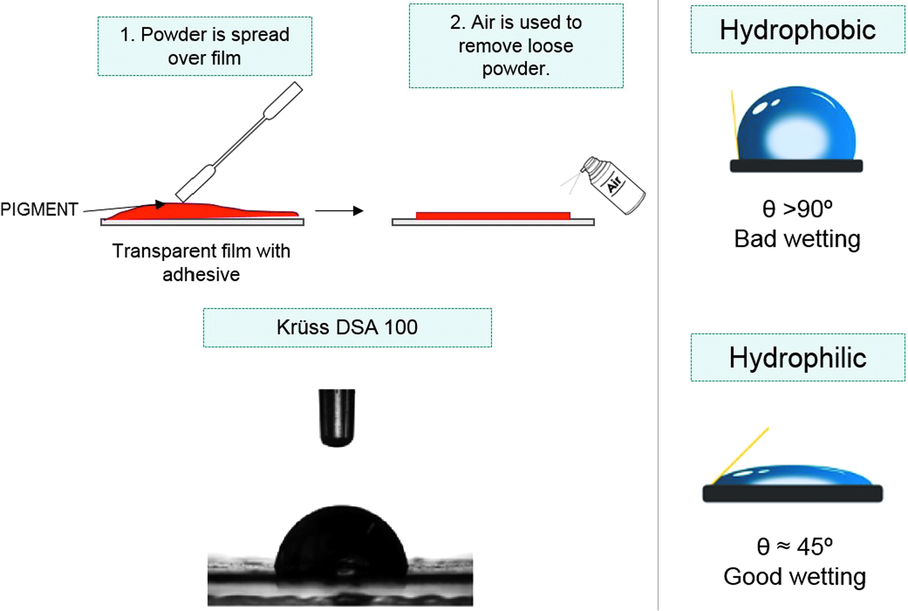
Preparation of surfaces and measurement of contact angle. DSA, Drop Shape Analyzer.
Surface charge
Zeta potential at pH 7 was determined by Laser Doppler Electrophoresis using Zetasizer Nano (Malvern) and a folded capillary cell (DTS 1060; Malvern). The samples were dispersed at room temperature in the background electrolyte solution (10 mmol/L KCl) and adjusted to the corresponding pH by 0.1 M NaOH or HCl. The concentration of the samples was adjusted to the signal intensity in the range of 3000–7000 kcps. The results are the average of two measurements. The instrument was calibrated with Malvern Standard DTS 1235. We recorded the electrophoretic mobility across the titration range of pH 3–10 to identify the isoelectric point where the electrophoretic mobility crosses zero. Using the dispersed size determined by analytical ultracentrifugation (AUC), mobility, and pH 7, we extracted the zeta-potential reported in Table 1.
Charge of DPP orange pigment is not detectable by the used measurement technique.
AUC, analytical ultracentrifugation; BET, Brunauer–Emmett–Teller; DPP, diketopyrrolopyrrole; N/A, not applicable; TEM, transmission electron microscope.
Bridging across lipid vesicles
The behavior of the pigments in contact with lipids was measured in D2O-based lipid-containing media (1.8 g pigments/L D2O, DPPC:DOPC:DPPG = 40:40:20, 1 mmol HEPES). For this, an AUC machine (Beckman Ultracentrifuge type XLI with integrated interference optics) was used, as described in detail earlier.14,15 The test substances were dispersed to the lipid-containing media in a concentration of 0.5 mg/mL by sonication in a cup horn sonifier (Branson Digital Sonifier, 17 minutes, 50% amplitude, 3141 MJ/m3). Afterward, the dispersion was stirred at 700 rpm for 1 hour at room temperature to allow for interaction to occur. During AUC measurements at 20,000 rpm, the speed of flotation of the lipid vesicles is monitored by interference optics, and is evaluated quantitatively by the freeware program Sedfit, with fitting model ls g(s*). Significant integration of the relatively low-density organic pigment particles with vesicles leads to a bridging and thus faster flotation of vesicles. The results in Table 1 are given in relation to a control sample of lipid-containing media without pigments. Thus, faster flotation due to bridging leads to a result >1 as the control sample results in 1.
Specific surface area
Specific surface area was determined by the Brunauer–Emmett–Teller method on 50–300 mg samples. First, samples were decontaminated under vacuum at 100°C. Nitrogen adsorption/desorption isotherms at 77 K were recorded at five pressures between 0 and 0.2 P/P0. The measurements were distributed between different instruments—Autosorb 6b (Quantachrome) or TriStar or ASAP 2420 (both Micromeritics)—all adhering to the standard DIN 66131.
Size (distribution), shape, representative image: dry
The primary size and shape were assessed using a transmission electron microscope (TEM) from FEI, Type Strata 400 DB, equipped with a field emission cathode. For TEM analysis, samples were wetted in ethanol, then gently spread on a sample holder, and transferred into vacuum for TEM imaging. Analysis of the resulting images was done visibly.
Animals
Permission for animal studies was obtained from the local regulatory agencies, and all protocols were in compliance with the federal guidelines. The laboratories of BASF's Experimental Toxicology and Ecology, where all the studies were performed (except DPP orange pigment, which was performed at Harlan Laboratories, Basel, Switzerland), are AAALAC certified. All procedures for animal care and exposure were conducted under the rule of the German Animal Welfare Act (1998). Male and female Wistar rats were obtained from Charles River Laboratories, Sandhofer Weg, Sulzfeld, Germany [7 weeks of age, strain Crl:WI (Han)], in case of benzimidazolone black pigment and pyrimidinone black pigment from RCC, Ltd. Laboratory Animal Services (Füllinsdorf, Switzerland) [9 to 10 weeks of age, strain HanRcc:WIST(SPF)], and in case of DPP orange pigment from Harlan Laboratories B.V. (Horst, Netherlands) [8 to 9 weeks of age, strain RccHan™:WIST(SPF)], and were allowed free access to mouse/rat laboratory diet (Provimi Kliba SA, Kaiseraugst, Switzerland) and water. The animals were housed in cages in accommodation maintained at 20°C–24°C, with a relative humidity of 30%–70%, a light/dark cycle of 06.00–18.00 hours light and 18.00–06.00 hours dark and were allowed to acclimatize to these conditions for ∼1 week before commencement of the study.
Exposure regimen/test groups
Groups of five male and female Wistar rats were head–nose exposed to respirable dusts for 4 hours. Fourteen days postexposure, the surviving animals were necropsied. In case of azomethine yellow pigment groups of one male and one female animal were exposed to 1.0 and 4.1 mg/L air, respectively, and the respiratory tract examined histopathologically.
Generation of the test atmospheres
Brush dust generators (dosing-wheel dust generator developed by Gericke in cooperation with BASF, Germany) served for generation of test atmospheres. With the exception of Dikeotpyrrolopyrrole Orange Pigment, between 1% and 2% of a fused silica (Aersosil 200 and/or Aerosil R972) was added as vehicle to improve flowability.
A prechamber and a cyclone preseparator were used to prevent entrance of larger aerosol particles into the inhalation chamber (except studies with benzimidazolone black pigment and pyrimidinone black pigment, where the dust was entered directly in the inhalation chamber).
Generated dusts were mixed with compressed air and passed through a cyclone into the inhalation system. The cylindrical stainless steel inhalation chamber was fed through a cone-shaped inlet at the top and exhausted at the opposite end. The desired inhalation chamber concentrations were achieved by withdrawing/exhausting and replacing a portion of the dust aerosol air with conditioned supply air immediately before entering the chamber.
In case of DPP orange pigment, the dust was generated through a CR3020 rotating brush aerosol generator connected to an AirVac TD110M. The aerosol generated was then discharged into a flow-past exposure chamber through a 63Ni charge neutralizer.
A schematic diagram of the inhalation system is shown in Figure 2.

Schematic picture of the inhalation exposure system.
Monitoring and characterization of the test atmosphere
Compressed and conditioned supply air and exhaust air flow rates, chamber temperature and humidity were measured automatically with appropriate sensors/orifice plates; data were saved every 10 seconds and retained for analysis. To quantify the atmospheric dust concentration, gravimetric measurements of air samples taken adjacent to the animals' breathing zone were performed four times per study (probe internal diameter 7 mm). A defined volume of sample air was drawn by vacuum pump across a binder-free glass-fiber filter paper (Macherey-Nagel MN 85/90 BF, diameter 4.7 cm).
Aerosol dust concentration was calculated as the increase in weight of the filter after sampling, divided by sample volume at test conditions (22°C, atmospheric pressure, 50% relative humidity). To determine the MMAD (the calculated aerodynamic diameter which divides the size distribution in half when measured by mass), cascade impactor measurements were performed with an Anderson stack sampler Mark III. The effective aerodynamic cutoff diameters were 29.5, 18.2, 8.5, 5.5, 2.8, and 1.2 μm. To capture the particles <1.2 μm, the impactor was equipped with a backup filter. The deposition on each impactor stage as well as on the backup filter was determined gravimetrically. Particle size distributions were calculated according to DIN 66141 and DIN 66161, that is, linear regression of cumulative percent (probit values) versus logarithms of effective cutoff diameters. Particle size distributions measured by cascade impactor were expressed as MMAD and GSD.
In case of DPP orange pigment, gravimetric determination of aerosol concentration was performed twice (5 mg/L air) or six times (1.0 and 0.5 mg/L air) during exposure. The samples were collected on a Millipore® Durapore filter, Type HVLP loaded in a 47-mm in-line stainless steel filter sampling device. The filters were weighed before and immediately after sampling using a calibrated balance. The test aerosol concentration was calculated from the amount of test item present on the filter and the sample volume. The particle size distribution of the test aerosol was determined twice (5.0 mg/L) or three times (0.5 and 1.0 mg/L) during exposure using a Mercer 7-Stage Cascade Impactor (Model 02-130, In-Tox. Products, Inc., Albuquerque, NM). The particle size distribution was measured by gravimetrically analyzing the test item deposited on each stage of the cascade impactor. Effective cutoff diameters were 4.6, 3.0, 2.13, 1.6, 1.06, 0.715, and 0.325 μm.
Animal exposure
During exposure, rats were restrained in glass tubes fixed to the inhalation chamber walls with their snouts projecting into the inhalation chamber (head/nose exposure). Overpressure was maintained inside the inhalation chamber to ensure that the aerosol in the animals' breathing zone was not diluted by laboratory air. The exposure systems were kept under exhaust hoods in an air-conditioned room.
Observations and postmortem examinations
Health status and cage-side clinical signs were checked at least once daily. Body weights were measured on study days 0, 1, 3, 7, and 14. Animals were euthanized by exsanguination under Narcoren® anesthesia. Gross necropsy was carried out.
Lungs and trachea were instilled with 4% buffered formaldehyde and fixated. In case of azomethin yellow pigment and DPP red pigment nasal cavity, trachea, larynx, pharynx, and lungs were paraffin embedded, sectioned, and stained with Hematoxylin–Eosin and examined microscopically. One animal which died after exposure to benzodifuran black pigment was examined likewise.
Statistical analysis
Due to the study design (acute toxicity) no statistical evaluations were necessary.
Results
Characterization of test materials
A comprehensive list of physicochemical characteristics is given in Table 1 (Fig. 3).
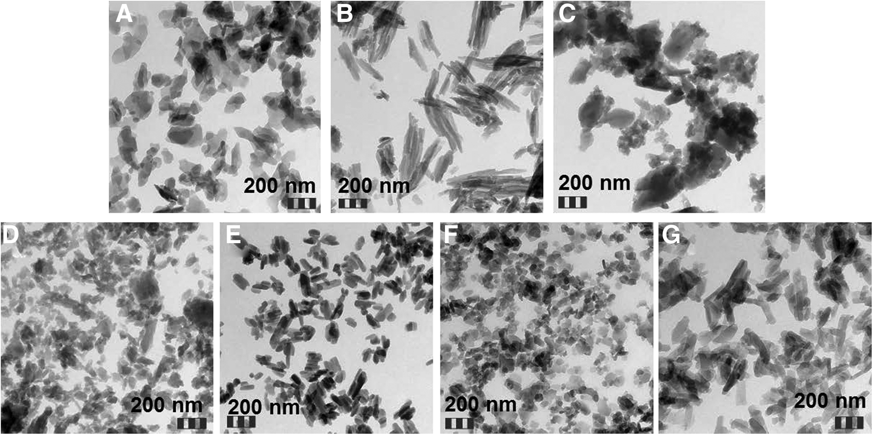
Scanning electron microscopy images of the test materials.
Characterization of the test atmosphere
Results of gravimetric determination of aerosol concentrations and particle size distribution are summarized in Table 2.
GSD, geometric standard deviation; MMAD, mass median aerodynamic diameter; SD, standard deviation.
The MMADs of the particles in the aerosols were small enough to reach the upper and lower respiratory tract of rats (the particles were respirable for rats).
Mortality, clinical observations, and body weight
Mortality data are summarized in Table 3.
f, female; m, male.
At concentrations of ∼1 mg/L air and below clinical signs were restricted to piloerection, decreased activity, and changes of respiration (increased respiration rate, labored and intermittent breathing, respiration sounds, and abdominal respiration).
At concentrations in the range of 5 mg/L air, piloerection, and intermittent and labored respiration were observed after exposure to pyrimidine azo yellow pigment. Additional signs were observed after exposure to benzimidazolone black pigment (squatting posture), pyrimidinone black pigment (attempts to escape, squatting posture), benzodifuran black pigment (red encrusted nose), and DPP red pigment (gasping).
Body weights of surviving animals decreased during the first day, but increased from study days 3 or 4 onward.
Macroscopic and microscopic examination
No macroscopically visible changes were observed after exposure to pyrimidine azo yellow pigment, and only contaminated fur was present in case of pyrimidinone black pigment and benzimidazolone black pigment.
Benzodifuran black pigment
Red-discolored lung and black-discolored skin in the nose region occurred in the animal that died. Black-discolored lungs, and enlarged, gray-discolored mediastinal lymph nodes were observed in the animals killed at the end of the 14-day observation period. Microscopic examination of the animal which died revealed severe amounts of the test compound in the larynx/pharynx, small amounts of pigment in the nose and lung, as well as moderate amounts of the test substance in alveolar macrophages; the lung was congested.
Azomethine yellow pigment
The animals which died showed red- to brown-colored foci in the lungs, incompletely collapsed lungs, and greenish-discolored pharynx and trachea. At 4.7 mg/L, microscopic examination revealed severe test substance deposition in the nasal cavity, moderate test substance deposition in trachea and larynx/pharynx. Severe test substance deposition with obstruction and diffuse emphysema was observed in the lungs. Slight test substance deposition in the nasal cavity and moderate-to-severe deposition in the lungs, with associated purulent inflammation and multifocal necrosis in the lungs were observed in the animals exposed to 1.0 mg/L air. No macroscopically visible changes were observed in the animals after the 14-day postexposure period.
DPP red pigment
Animals that died or were killed in moribund condition had red foci in the lungs, which were partly transparent in two animals. Histopathological examinations showed a moderate-to-severe multifocal red pigment deposition (presumably substance) in bronchi, bronchioli, and alveoli. The larynx showed a massive red pigment deposition (presumably substance) within airways with a total obstruction (Fig. 4). The nasal cavity (all levels) showed a slight-to-moderate multifocal red pigment deposition on the mucosa (presumably substance). The trachea was without findings. No macroscopically visible changes were observed in the animals after the 14-day postexposure period.
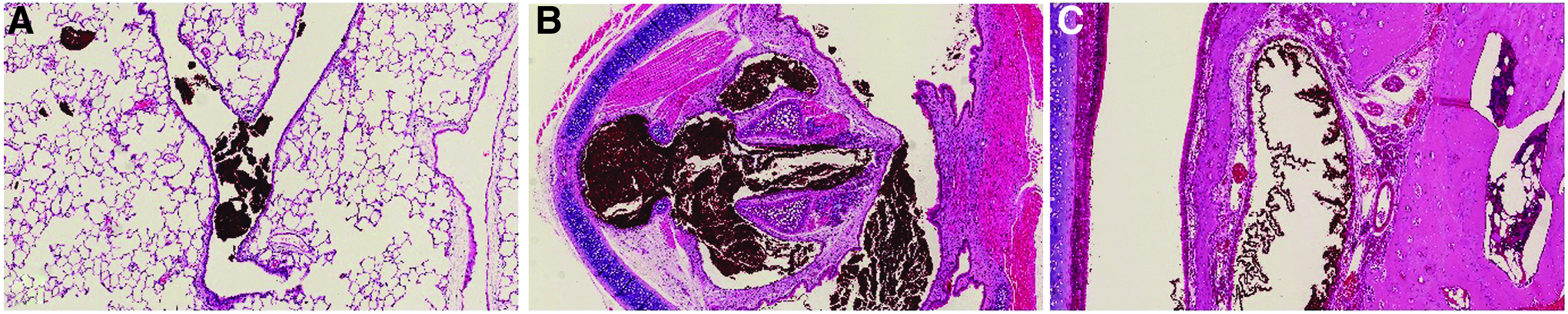
Microscopic findings after exposure to 5.2 mg/L air DPP red pigment.
DPP orange pigment
All animals exposed to 5 mg/L showed reddish discolored and incompletely collapsed lungs at necropsy. No macroscopic findings were present in animals exposed to 0.5 or 1.0 mg/L at scheduled necropsy.
Discussion
During the exposure period, the target concentrations were maintained constant and stable. Particle size distribution measurement demonstrated very high fraction of respirable particles.
In case of DPP orange pigment, all rats died within 2 hours of exposure at 5.0 mg/L air, whereas at 1.0 mg/L air. In case of classical toxicological concentration response curves, one would have expected more effects at 1.0 mg/L air. Therefore, a different mode of action is possibly present at 5 mg/L. Also, in the other experiments mortality happened early, that is, during exposure phase or shortly thereafter. Histopathological examinations of the animals which died revealed obstruction of the airways by large amounts of the inhaled pigment. Therefore, it is evident that suffocation was the cause of death in these cases. Although histopathological examinations were not performed in case of DPP orange pigment, suffocation is also considered as a cause of death in this study. The azomethine yellow pigment was the only test substance for which local inflammatory reactions were observed. These are likely to be related to the copper, which is contained in the pigment in a complexed form.
When screening REACH registration dossiers in general for acute inhalation studies of pigments, seven other organic pigments were identified for which stable concentrations in the relevant particle size could only be achieved in concentration ranges between 1 and 4 mg/L. In none of these studies, were incidences of mortality observed.
With regard to the observed LC50 values, there was no correlation with the particle size distribution of the aerosolized test materials and most of the physicochemical parameters. However, the contact angle, a measure of hydrophilicity/hydrophobicity, did correlate well with the obtained LC50 values. As shown in Table 4, DPP orange pigment was the most toxic compound, and had the highest measured hydrophobicity. On the other hand, pyrimidine azo yellow pigment, which was quite well tolerated at the concentration near the limit concentration of 5 mg/L air, showed a highly hydrophilic character. Its contact angle was much lower than the contact angle of the other test substances (Table 4) which is consistent with its significantly higher water solubility. For inorganic pigments, all acute inhalation studies known to the authors resulted in LC50 values of >5 mg/L. Although contact angles and observed toxicity did not match perfectly, it is noteworthy that the three compounds which produced 100% mortality around the limit concentration of 5 mg/L air had the highest contact angles, and that the only hydrophilic test substance had the lowest toxicity. Thus, the hydrophobicity as determined by the contact angle of a sessile water drop seems to be a suitable parameter to predict the acute inhalation toxicity of pigments at concentrations of 5 mg/L air. Of note, the surface charge as determined by zeta potential helps to understand the phospholipid vesicle interactions by high hydrophobicity and low net charge, but neither zeta potential assay nor the phospholipid vesicle assay can predict toxicity at a limited dose. This finding does not downplay the importance of interactions mediated by charge and lipids, but with the current experimental approaches to quantify interactions, the water contact angle correlates best to the in vivo findings. Further research is needed to understand the role of additional physicochemical factors in acute inhalation toxicity of otherwise toxicologically “inert” pigments.
Although the present database is limited to seven pigments, a contact angle above a certain threshold (e.g., 130°) could predict the probability of suffocation, whereas an angle below a certain threshold (e.g., 100°) could predict low or no toxicity. In the former case, testing could be performed at a lower limit concentration of 1 mg/L as alternative to the current limit concentration of 5 mg/L. In the latter case testing for acute inhalation, toxicity could be waived provided the pigment meets the criteria of poorly soluble, low-toxicity particles.13,14 In cases, where the contact angle is between these borders testing could be performed at the current limit concentration of 5 mg/L air.
Conclusion
In the present study, inhalation of pigments in acute toxicity tests at high concentrations around the limit concentration of 5 mg/L air caused mortality due to obstruction of the airways and subsequent suffocation. In the present study, this effect appears to be dependent on the hydrophobicity of the test substance, which can be measured by the contact angle. Thus, determination of the contact angle may serve as prediction of mortality due to suffocation at the limit concentration. Future refinements might include additional physical–chemical parameters, such as charge, but are currently not warranted by the experimental evidence.
The scientific value of physical obstruction of the airways with subsequent suffocation at these high concentrations is of limited value and does not represent a real hazard information of the test material. Therefore, the necessity of testing of hydrophobic pigments at these high concentrations should be discussed in general, also in view of animal welfare. After proof of high hydrophobicity, acute inhalation testing at a lower maximum concentration of 1 mg/L air is considered as useable 3R strategy to minimize animal testing and yet obtain sufficient hazard information.
Footnotes
Acknowledgments
This work was sponsored by BASF SE, BASF Colors and Effects, and BASF Switzerland.
Authors' Contributions
L.M. carried out the inhalation experiments. S.G. carried out the histopathological evaluation. J.C.A., N.N., and W.W. performed the physicochemical characterization of the materials. W.T. identified pigments and compiled existing data. N.E. and U.V. provided the test substances. T.H. is the author of the article. R.L. initiated and led the project and contributed to the interpretation of the results. B.R. contributed to the interpretation of the results. All authors read and approved the final article. All authors contributed to the text of this article and reviewed the final text.
Author Disclosure Statement
The authors are or were employees of BASF SE, BASF Schweiz AG, or BASF Colors and Effects Switzerland AG. BASF Colors and Effects produces and markets pigments.