




Abstract
Introduction:
Kombucha is not really a fungus but a community of several yeasts and bacteria. It has a wide range of vitamins and amino acids.
Materials and Methods:
The antioxidant activity of kombucha on rat bone marrow cells and human blood lymphocytes, anticancer effect on A549 and HepG2 cells, cytotoxicity effect on HGF cells, and growth and formation of biofilm on Pseudomonas aeruginosa and Staphylococcus aureus were evaluated.
Results:
Kombucha extract has inhibited cytotoxicity, genotoxicity, and oxidative stress induced by cisplatin and improved antioxidant factors. Different concentrations of the extract showed that concentrations of 50 μg onward induce a better antioxidant effect. Using kombucha as monotherapy in two categories of cancer cells showed that it can inhibit the growth of cancer cells. Increasing the concentration increases the anticancer capability, so we see the best effect at a concentration of 100 μg. On the other hand, exposure to kombucha alone in HGF showed that this extract causes little toxicity. The minimum inhibitory concentration (MIC) of P. aeruginosa and S. aureus was at concentrations of 250 and 500 μg/mL. The minimum bactericidal concentration test showed Kombucha for ATCC25923 and P. aeruginosa isolates at 4 MIC concentration with bactericidal ability. P. aeruginosa, S. aureus, and ATCC: 25923 expressed strong biofilm and also in ATCC27853 moderate biofilm. A concentration of 8 MIC in ATCC27853 and 2 MIC in ATCC25923 was able to inhibit biofilm.
Conclusion:
Kombucha extract has anticancer and antioxidant properties due to its wide range of vitamins and amino acids, especially glucuronic acid and polyphenols such as catechin.
Introduction
Kombucha is a combination of two Japanese words, “kombu” meaning seaweed, and “cha” meaning tea. 1 From the fermentation of black tea and sugar and a Symbiotic Culture of Bacteria and Yeast (SCOBY), a drink called Kombucha is generally obtained for 7–21 days.1,2 SCOBY contains different types of bacteria such as acetic acid bacteria and yeasts and several lactic acid bacteria.
The ingredients of Kombucha after fermentation are: sugars; organic food acids; tea polyphenols; ethanol; fiber; carbon dioxide; antibiotic substances; hydrolytic enzymes; amino acids, including lysine; essential elements such as Cu, Fe, Mn, Ni, and Zn; and water-soluble vitamins such as vitamin C, and several B vitamins. 2
The major metabolites identified in fermented beverages include lactic, acetic, ethanol, glycerol gluconic, and glucuronic acids. Concentration and metabolic composition depend on the source of the tea fungus, the sugar concentration, and the fermentation period. The fermentation process induces the synthesis of a complex of B vitamins. During the fermentation process, kombucha's pH decreases due to an increase in its organic acid content. Green and black tea are the best substrates for the production of acetic acid and gluconic acid.
Tea polyphenols (epigallocatechin gallate [EGCG], epigallocatechin [ECG], epicatechin [EC], and theophylline [TF]) and organic acids are the active ingredients in kombucha tea that have a broad variety of advantageous effects. 3
This fermented drink has many properties in human health, from antioxidant and anti-inflammatory properties, lowering cholesterol and regulating blood pressure to anticancer properties and improving kidney function, the gastrointestinal tract, and strengthening the immune system.1,4 In vitro and animal studies have shown that the effects of kombucha on improving human health are due to its compounds, which include acetic acid, glucuronic acid, phenols, polyphenols, and B-complex vitamins, including folic acid. 5
During normal cellular activity, reactive species produce oxygen and nitrogen. Reactive oxygen species (ROS), which is a general term, expresses O2-derived free radicals such as anion superoxide (O2•−) and hydroxyl radicals (HO•), and non-radical O2-derived species such as hydrogen peroxide (O2). They are constantly produced in organisms. 6
Cellular antioxidants are involved in detoxifying these species, but oxidative stress conditions occur when the balance is lost. If oxidative stress persists, oxidative damage is done to vital biomolecules (such as the genome) and the accumulation of this damage leads to some biological effects such as changes in message transmission, changes in gene expression, mitogen, mutation, mutation, and cell death. 7
One of the studies examined the effects of Kombucha tea on oxidative stress-induced alterations in rats exposed to chromate treatment. Kombucha tea consumption alone did not affect malondialdehyde (MDA) and reduced glutathione (GSH) levels, but it significantly enhanced humoral response and Delayed-type hypersensitivity (DTH) response as compared with the control group.
Chromate exposure resulted in increased MDA levels in plasma and tissues, decreased DTH response, and elevated activities of glutathione peroxidase (GPx) and catalase, whereas GSH, superoxide dismutase (SOD), and antibody titers remained unchanged. However, feeding with Kombucha tea entirely reversed the chromate-induced effects. These findings suggest that Kombucha tea possesses robust antioxidant and immunopotentiation properties. 8
A biofilm is a bacterial community composed of bacteria that adhere to a living or nonliving surface and are surrounded by an extracellular layer.8,9 Biofilms can be produced by gram-positive and gram-negative bacteria. Among the most important bacteria that can form biofilms are gram-positive bacteria, Staphylococcus epidermidis Staphylococcus aureus, Streptococcus viridans, Enterococcus faecalis, and gram-negative bacteria named Klebsiella pneumoniae, Escherichia coli, and Pseudomonas aeruginosa. 10
The Pseudomonas family consists of a large group of gram-negative bacilli or non-fermented aerobic bacilli that are oxidase positive and motile and can grow in very simple environments. Among the isolated bacilli in clinical specimens, 12 to 16% belong to this family.11–13 P. aeruginosa are gram-negative environmental bacteria that have minimal nutritional requirements and can adapt to a wide range of environmental conditions. The pathogen infects various organs such as the skin, ears, eyes, heart, soft tissues, bones, and joints, and the gastrointestinal, urinary, respiratory, and central nervous systems. However, direct contact between epithelial barriers of the skin, eyes, and respiratory tract and the external environment increases the risk of infection.14,15
Treatment of P. aeruginosa infection with antibiotics was often difficult due to the high potential of this pathogen to develop resistance. Multidrug-resistant strains have been classified by several health care organizations as a serious threat to public health. 16
S. aureus is a gram-positive opportunistic pathogen that threatens humans and animals frequently and without specific symptoms. This disease can cause various types of infections and is considered an important nosocomial pathogen. S. aureus is an important cause of nosocomial infections that has shown resistance to antibiotic treatment in recent years.
One reason for this is the ability of these bacteria to produce biofilms. The high susceptibility of S. aureus strains in obtaining antibiotic resistance genes from other bacteria and thus increasing antibiotic resistance along with high pathogenicity and biofilm formation has become a major concern in patients' health. 17
This study is divided into several parts:
Antioxidant effect of Kombucha mushroom extract on rat bone marrow cells and human blood lymphocytes (by micronucleus method). Anticancer effect of Kombucha mushroom extract on A549 and HepG2 cells. Toxicity of Kombucha mushroom extract on HGF cells. Evaluation of growth and formation of biofilm on gram-positive bacteria (Pseudomonas aerogenes) and gram-negative bacteria (S. aureus).
Materials and Methods
Materials
All chemical reagents were purchased from Sigma-Aldrich.
Extract of kombucha
The fungus was purchased from reputable centers and after washing, it was dried in the laboratory and away from direct sunlight. Then, all the mushroom parts were crushed by the mill. The methanolic extract of the fungus was obtained by soaking. First, the various components of the Soxhlet device are connected. After that, about 1.3 volumes of the cartridge are filled separately from the desired sample and placed in its special place, and an extraction test is performed with different solvents for 6 hours.
As the first solvent, 500 mL of distilled water is poured into the Soxhlet balloon. The dried sample of powder is poured into the grain finger and closed in it. Finally, the Soxhlet device is closed and the faucet is returned. After 25 minutes, the water is boiled by an electric heater and the extraction operation continues for 6 hours. The siphon operation is performed and the aqueous extract is separated, then it is placed in a glass jar in the refrigerator.
Extraction and culture of stem cells from bone marrow
In this experimental study, male Wistar rats aged 50 days and weighing 200 ± 20 g were used. The animal used in this study was purchased from Mazandaran University of Medical Sciences at a temperature of 27°C and with good access to food and water. Following the ethical principles of working with laboratory animals, the mice were anesthetized with diethylene ether, their femurs and calves were removed, and then the connective tissues around the bones were completely removed.
The bones were placed in a Dulbecco's modified Eagle medium (DMEM) containing 15% fetal bovine serum (FBS) and 1% penicillin-streptomycin at 4°C and transferred to a laminar hood. The two ends of the bone were cut with sterile scissors, and the bone marrow was removed by flushing and guided into a falcon tube containing a complete culture medium and then centrifuged at 2500 rpm for 5 minutes.
The supernatant was removed, and the cell sediment was suspended in 5 mL of fresh medium, cultured in a T25 flask, and incubated in a CO2 incubator (37°C, 5% CO2). After 24 hours, the supernatant containing non-adherent cells was removed and then the cell environment was changed every 3 days for 14 days. When the bottom of the flask reached a high density of cells, the cells were separated from the bottom of the flask with the help of (Trypsin/EDTA) and transferred to new flasks. Three passage steps were repeated to obtain high purity from these cells.
MTT assay
Ninety microliters (5 × 10⁴ cells per well) of bone marrow cells were seeded in a 96-microwell plate and incubated for 24 hours. After 24 hours of incubation, the non-toxicity of kombucha extract was evaluated at 12, 24, and 36 hours on rat bone marrow cells at concentrations of 25, 50, and 100 μg/mL.
After incubation, 10 μL of MTT solution was added to each well, followed by a 4-hour incubation. After removing and washing the contents of each well with phosphate-buffer sulfide (PBS), 30 μL of dimethyl sulfoxide (DMSO) was added to dissolve the formazan dye, and the Biotek ELx800 Microplate Reader was used to measure absorption at max = 490 and 630 nm. 18
Cell lines and cell culture
A549, HepG2, and HGF cell lines were purchased from Pasteur Institute of Iran in Tehran. Cell suspensions containing DMEM, 1% PenStrep, and 10% FBS were placed in a 75 cm3 cell culture flask and incubated at 37°C in a humid atmosphere of 5% CO2 to reach their logarithmic growth stage. When primary cultures become almost confluent, 1 cc of trypsin-EDTA was added to the flask and incubated for 5 minutes to completely separate the cells from the flask. Then, to evaluate the oxidative stress and cell survival tests, the amount of 100 μL of cell suspension in each well was added to 96-well plates, and the plates were incubated for 24 hours. 19
MTT assay
Ninety microliters (5 × 10⁴ cells in per well) of cells were seeded in a 96-microwell plate and incubated for 24 hours. After 24 hours of incubation, the cell line was exposed to a single dose of cisplatin (IC50 = 2 μg) as positive control and Kombucha was then added to the cells in different doses (25, 50, 100 μmol/mL) and then incubated for 48 hours.
Then, 10 μL of MTT solution was added to each well, and it was incubated for 4 hours. After that, all the contents of each well were removed and washed with PBS; then, 30 μL of DMSO was added to dissolve the formazan dye; and absorption was determined at max = 490 and 630 nm by the Biotek ELx800 Microplate Reader. 20
Reactive oxygen species
Intracellular oxygen free radicals were measured by the DCFH-DA reagent. When DCFH-DA enters living cells, it is converted to DCFH by intracellular esterases. And when it reacts with oxygen radicals, it converts to DCF, which can be detected and read by a spectrophotometer. Twenty microliters of DCFH solution were added to each well and incubated in the dark for 15 minutes. The absorbance was then read as 485 and 530 nm, using a fluorescence microplate reader.21,22
Measurement of lipid peroxidation
Lipid peroxidation was evaluated by determining the production rate of thiobarbituric acid reactive substances (TBARS) and was expressed as MDA equivalents. For measuring lipid peroxidation, 100 μL of cell suspension, 100 μL of TBA reagent, and 100 μL of phosphoric acid were mixed well and incubated in a warm water bath for 30 minutes after cooling. Then, 0.2 mL of n-butanol was added, shaken well, and then centrifuged at 3500 rpm for 10 minutes. The n-butanol layer was separated for measurement at 532 nm by the Biotek ELx800 Microplate Reader.23,24
Measurement of intracellular glutathione (GSH)
In the falcons' tube containing the studied cells, which were pre-treated with different concentrations of substances, 1.5 mL of TCA and EDTA (10%) were added to precipitate the proteins in the next step. The samples were centrifuged at 3500 g for 15 minutes. Then, 1 mL of supernatant was removed, 2.5 mL of Tris buffer was added with pH = 8.9, and then 0.5 mL of DTNB (40%) was added and incubated for 15 minutes. For finishing the reaction, the tube was shaken well to obtain a uniform yellow color. Finally, the absorbance was measured by spectrophotometry at 412 nm.25,26
Micronucleus assay
Blood samples were taken from 4 healthy men without underlying disease and smoking and alcohol consumption. All samples were placed in a 37°C hot water bath. Then, 0.5 mL of blood was added to each well along with 4.5 mL of DMEM culture medium. To accelerate blood cells' growth, 2% of the total volume, PHA was added and incubated for 24 hours. Kombucha was then added to the cells in different doses (25, 50, 100 μM) with a single dose of cisplatin (0.1 μg/mL) 27 and incubated for 48 hours.
Forty-eight hours after adding PHA, 3.6 μL of Cytochalasin B (Cyt-B) was added to each well to inhibit cellular cytokines. At the end of the incubation time, each well's contents were transferred to a centrifuge tube and centrifuged for 6 minutes, after which the lymphocytes were centrifuged with KCL solution and methanol-acetic acid fixation solution, respectively.
About 2–3 drops of the remaining suspension were taken and poured on the slides, and after drying, they were placed in Giemsa paint solution for 20 minutes. Light microscopes were used to examine the number of cells with two nuclei and micronuclei with a magnification of × 40 and × 100.24,28
Statistical population and number of samples
In this study, two clinical isolates (one P. aeruginosa and S. aureus) from the wound and P. aeruginosa ATCC: 27853 and S. aureus ATCC: 25923 prepared from the microbial bank of Pasteur Institute of Iran were used as the quality control. Laboratory identification of P. aeruginosa isolates was performed by standard microbiological and biochemical methods, including oxidase and catalase tests, reactions in triple sugar iron (TSI) agar, SIM (sulfide, indole, motility), and oxidative-fermentative (OF) media (Merck, Darmstadt, Germany), and for S. aureus isolate catalase, oxidase, DNase, and coagulase test were used. 29
Investigation of inhibitory effect by minimum inhibitory concentration and minimum bactericidal concentration method
To determine the minimum inhibitory concentration (MIC) of Kombucha mushroom extract against S. aureus and P. aeruginosa isolates, the Microdilution method was used. First, 100 μL from the Müller-Hinton Broth environment were added to 96-well microplate wells, respectively. Consecutive concentrations of 62.5, 125, 250, 500 μg/mL, and 62.5, 125, 250, 500, and 1000 μg/mL diluted mushroom extract for S. aureus and P. aeruginosa isolates were then added to Müller-Hinton broth culture medium.
Then, 1.5 × 10⁵ colony-forming unit (cfu)/mL isolates were inoculated separately in the medium and after incubation for 24–18 hours at 37° C bacterial growth in the medium was examined and the minimum concentration that inhibited growth was recorded. 30
After the MIC determination, to determine the minimum bactericidal concentration (MBC) of the kombucha extract, 50 μL of concentrations of MIC, 1/2 MIC, 1/4 MIC, 2 MIC, and 4 MIC were cultured on Müller Hinton agar, and the plate was placed in an incubator at 37°C for 24 hours. Finally, the growth and non-growth of bacteria indicate the lowest lethal concentration. 30
Investigation of biofilm formation detection by micro-titer plate microtiter method
Micro-Titer Plate (MTP) biofilm assay was used to identify isolate biofilms. Isolates of S. aureus and P. aeruginosa culture were incubated overnight to TSB containing 1% glucose and then incubated at 37°C to reach half McFarland turbidity. Then, 200 μL of suspension bacteria was added to the 96-well microtiter plates wells as duplicate and the plate was incubated at 37° for 20 hours.
Then, the supernatant of each well was poured out and the wells were washed with PBS. The plate was dried completely. In the next step, staining was done with violet crystal and then it was washed with physiological serum. Then, 100 μL of 70% ethanol with 10% isopropyl alcohol was added to the wells. In the final stage, the absorption optical density (OD) at 570–630 nm was read by the enzyme-linked immunosorbent assay (ELISA) reader. Samples with OD ≤0.1 have no biofilm, 0.1 < OD < 0.2 weak biofilm, 0.2 < OD < 0.3 moderate biofilm, and OD ≥0.3 strong biofilm.31,32
Evaluation of minimum biofilm removal concentration by minimum biofilm eradication concentration method
S. aureus and P. aeruginosa culture overnight were incubated to TSB containing 1% glucose and then incubated at 37°C to reach half McFarland turbidity. Then, 200 μL of suspension bacteria was added to the 96-well microtiter plates wells as duplicate and the plate was incubated at 37° for 20 hours. In the next step, the top phase was removed from each well and the wells were washed with physiological saline. We left the plate to dry completely.
Then, concentrations of MIC, 2 MIC,4 MIC, and 8 MIC of Kombucha extract were added to each well and incubated at 37°C for 24 hours. Then, the kombucha extract was emptied and washed with saline. The culture medium was then added to it and incubated at 37°C for 24 hours. In the final stage of absorption, (OD) at 570–630 nm was read by an ELISA reader. 33
Statistical analysis
Statistical analysis was performed by Prism ver.8 Software, data were compared with one-way analysis of variance and related post-test (Tukey-Kramer multiple comprehension tests), and the same graphic program was used to make the graphs.
Results
Effect of kombucha mushroom extract on A549 and HepG2 cells
Cisplatin at IC50 concentrations in A549 and HepG2 cells inhibited the growth of cancer cells by 44.66% and 44.91%, respectively. Exposure of lung cancer cells to kombucha extract at concentrations of 25 μg 4.21%, 50 μg 8.2%, and 100 μg 14.79% inhibited the growth of cancer cells and it is clear that with increasing concentration the inhibition of growth has increased (Table 1).
Mean and Standard Deviation of A549 Cell in Different Tests
GSH, reduced glutathione; ROS, reactive oxygen species; SD, standard deviation.
There were statistically significant differences between 25 and 50 μg (p < 0.001) and 100 μg (p < 0.0001) in comparison with the cisplatin group (Fig. 1A). Similarly, the exposure of liver cancer cells to different concentrations of kombucha extract inhibited the growth of cancer cells by 6.5%, 15.63%, and 22.59% growth inhibition (Table 2). In the comparison of HepG2 treatment groups with the cisplatin group, the concentration of 25 (p < 0.0001) and 50 μg (p < 0.001) and the concentration of 100 μg (p < 0.01) were significantly different (Fig. 2A).

Anticancer effect of kombucha extract on inhibiting the growth of cancer cells
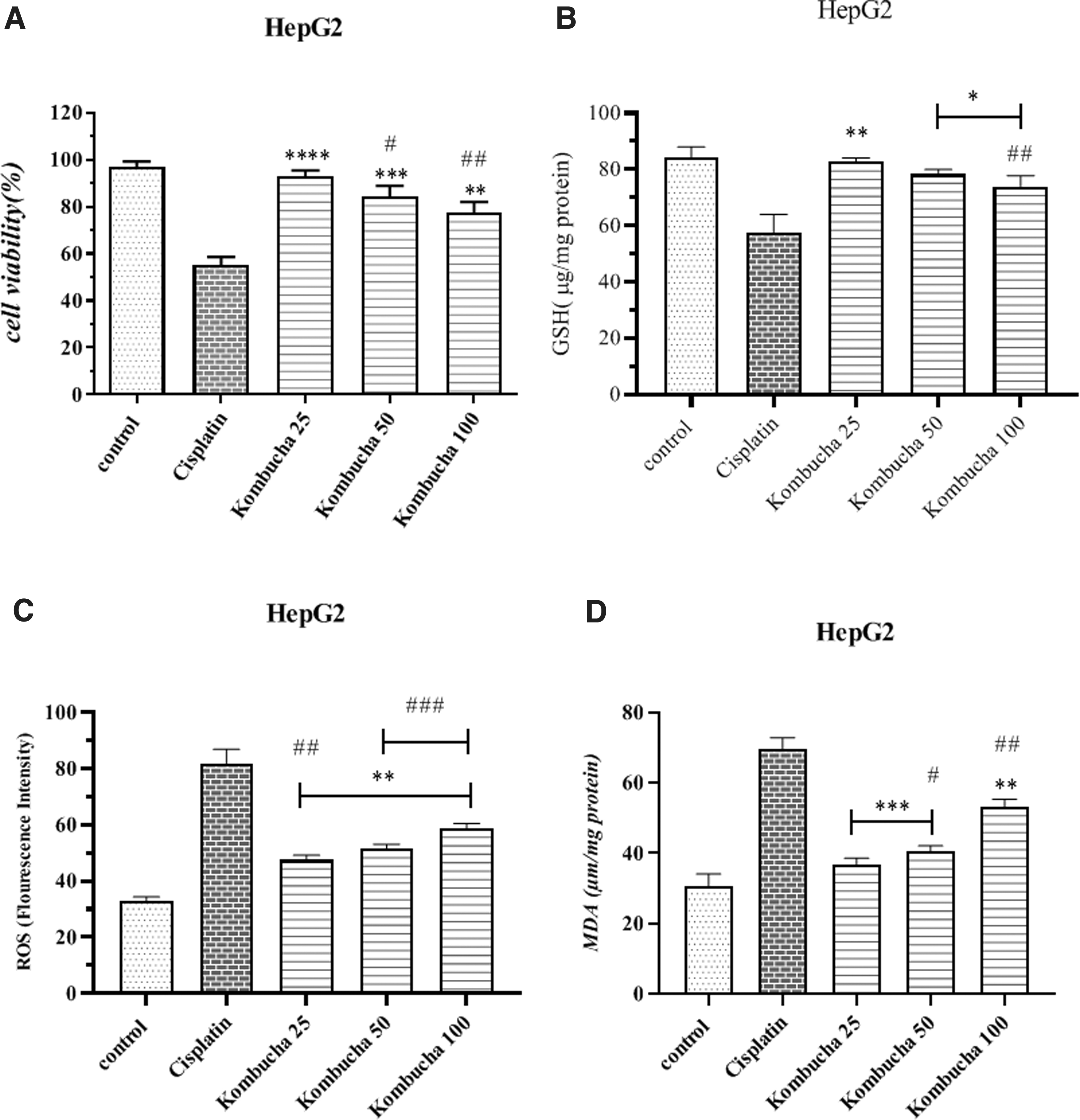
Anticancer effect of kombucha extract on inhibiting the growth of cancer cells
Mean and Standard Deviation of HepG2 Cell in Different Tests
Cisplatin reduced the glutathione of lung cancer cells by 34.62%. Exposure of lung cancer cells to different concentrations of kombucha extract GSH stores by 12.1%, 16.29%, and 20.75%, respectively (Table 1). Statistically, the concentration was 25 μg (p < 0.01), and concentrations of 50 μg and 100 μg (p < 0.05) were significantly different from the cisplatin group.
Compared with the negative control group, the concentration of 50 μg (p < 0.05) and the concentration of 100 μg (p < 0.01) had a significant difference (Fig. 1B). The level of glutathione of liver cancer cells in the cisplatin group decreased by 15.64%. In the groups exposed to different concentrations of kombucha, the amount of glutathione decreased 17.33% at the lowest concentration and 26.07% at the highest concentration, respectively (Table 2).
The statistical comparison showed that the group receiving 25 μg of kombucha had a significant difference from the cisplatin group (p < 0.01). Also, the two groups receiving concentrations of 50 and 100 μg had significant differences with the cisplatin group (p < 0.05) (Fig. 2B).
The amount of ROS produced in A549 cells exposed to cisplatin is 77.41%, which has increased from 43.30% at 50 μg to 55.86% at 100 μg in cells exposed to the extract (Table 1). Statistically, in comparison with the cisplatin group, the concentration was 25 and 50 μg (p < 0.001) and the concentration of 100 μg (p < 0.05) was significantly different from the cisplatin group (Fig. 1C).
Reactive oxygen radicals formed in liver cancer cells exposed to cisplatin are 32.78%. The ROS production is equal to 47.57%, 51.58%, and 58.78% in the treatment groups exposed to different concentrations of kombucha extract in concentrations of 25, 50, and 100 μg, respectively (Table 2). Statistically, concentrations of 25, 50, and 100 μg (p < 0. 01) had a significant difference with the cisplatin group (Fig. 2C).
Then, 64.73% of MDA was produced in cisplatin-exposed cells and in the groups treated with kombucha extract, the amount of lipid damage was from the lowest concentration to the highest concentration, respectively: 29.50%, 36.74%, and 41.46% (Table 1). Statistically, in comparison with the cisplatin group, the concentration was 25 and 50 μg (p < 0. 01) and a concentration of 100 μg (p < 0.001) was significantly different from the cisplatin group (Fig. 1D).
The rate of lipid damage in HepG2 cells is 69.56. Cancer cell exposure to kombucha extract at a concentration of 25 μg 36.62% at a concentration of 50 μg 40.54% and at a concentration of 100 μg 53.26% increased the MDA of liver cancer cells Is. It is known that with increasing concentration, more lipid peroxidation occurred (Table 2). There were statistically significant differences between 25 and 50 μg (p < 0.001) and 100 μg (p < 0.01) in comparison with the cisplatin group (Fig. 2D).
Cytotoxicity of kombucha mushroom extract on HGF cells line
To evaluate the cytotoxicity of the extract, we exposed different concentrations to HGF cells. The results showed that the highest extract concentration of only 8.57 inhibited cell growth (Table 3). Compared with the cisplatin group, all groups had significant differences (p < 0.0001) (Fig. 3A).
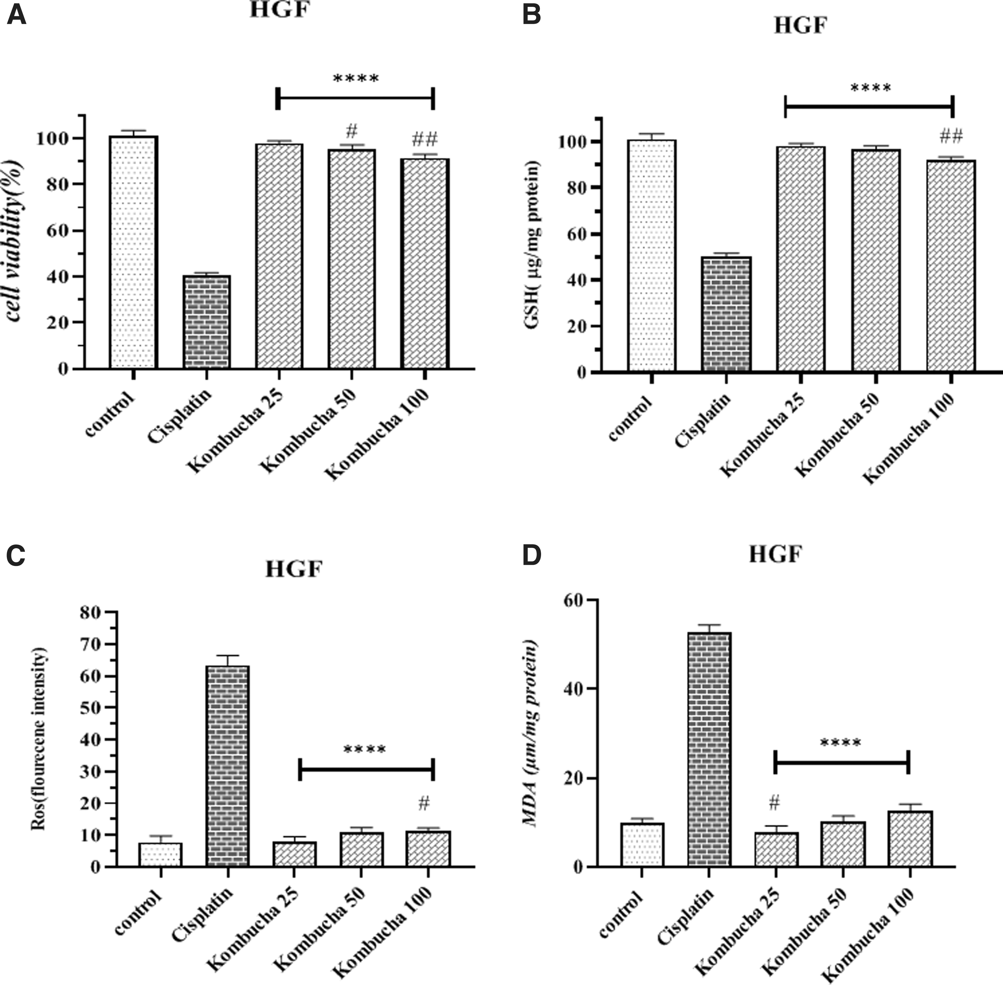
Cytotoxicity of Kombucha extract on cell growth rate
Mean and Standard Deviation of HGF Cell in Different Tests
The glutathione reserves of normal gingival cells decreased in the face of different concentrations of kombucha extract at the lowest concentration of 25 μg, 1.75%, and at the highest concentration of 100 μg, 7.79% (Table 3). Compared with the cisplatin group, all treatment groups had a significant difference (Fig. 3B).
The amount of ROS produced in cells exposed to different concentrations of kombucha extract is as follows: 25 μg concentration 8.17%, 50 μg concentration 10.98%, and 100 μg concentration 11.23% (Table 3). Compared with the cisplatin group, all treatment groups had a significant difference (Fig. 3C).
The amount of MDA in normal gingival cells exposed to cisplatin is 52.73%. In the groups treated with kombucha extract, the amount of MDA produced in the lowest concentration is 7.82%, and in the highest concentration is 12.66% (Table 3). All treatment groups had a significant difference from the cisplatin group (Fig. 3D).
Antioxidant activity of kombucha mushroom extract on bone marrow cells
Exposure of rat bone marrow cells to cisplatin and treatment with kombucha extract inhibited cisplatin-induced cytotoxicity. In other words, cell survival after exposure to cisplatin is 32.95%. After treatment with kombucha extract, cell life increased from 38.41% to 65.93% (Table 4). All treatment groups showed a significant difference compared with the cisplatin group. 25 μg (p < 0.05), 50 μg (p < 0.01) and 100 μg (p < 0.001) (Fig. 4A).

Antioxidant effect of kombucha extract on growth rate of rat bone marrow cells exposed to cisplatin
Mean and Standard Deviation of Stem Cell in Different Tests
Intracellular glutathione stores are reduced when exposed to cisplatin. Receiving kombucha extract inhibits toxicity and improves glutathione stores. The amount of glutathione in the lowest concentration of kombucha increased by 5% compared with the cisplatin group, from 63.14% to 68.72%. With increasing concentration, the toxicity of cisplatin decreased and the amount of glutathione reached 88.28% (Table 4). Compared with the cisplatin group, concentrations of 50 and 100 μg were significantly different (p < 0.001 and p < 0.0001, respectively) (Fig. 4B).
The amount of free radicals produced by cisplatin in mouse bone marrow cells is 86.72%. Different concentrations of kombucha extract were given to the cells to inhibit cisplatin-induced ROS production. As a result, ROS decreased to 54.62% at a concentration of 100 μg (Table 4). Statistically, the groups of 50 μg (p < 0.01) and 100 μg (p < 0.001) were significantly different from the cisplatin group (Fig. 4C).
The rate of cisplatin-induced lipid damage in cells receiving kombucha extract has improved. This rate is 72.39% in the cisplatin group and after receiving kombucha extract, this amount has increased from 69.42% to 50.26% in the highest concentration of the extract (Table 4). Compared with the cisplatin group, 50 μg and 100 μg groups had a significant difference (p < 0.01) and (p < 0.0001) (Fig. 4D).
The antigenotoxic activity of kombucha mushroom extract on human blood lymphocytes
In the micronucleus test, cisplatin was used as a clastogenic agent. According to Table 5, the amount of micronucleus produced by cisplatin is 21 ± 1 When lymphocytes were exposed to different concentrations of kombucha, the results showed that kombucha was able to reduce the amount of cisplatin-induced genetic toxicity due to its antioxidant properties.
Mean and Standard Deviation of Micronucleus Test
At a concentration of 25 μg, the number of micronuclei decreased to 19.33 ± 1.15, and with increasing the concentration to 100 μg, it decreased to 14.33 ± 0.57. The kombucha group of 100 μg also indicates that this extract did not cause genetic toxicity at this concentration (Table 5). Statistically, compared with the cisplatin group, only 100 μg of kombucha showed a significant difference (p < 0.05) (Fig. 5).

Antioxidant effect of Kombucha extract on cisplatin-induced genetic toxicity in human blood lymphocytes. Kombucha extract has antioxidant properties and inhibits the genetic toxicity caused by cisplatin in human blood lymphocytes. *p < 0.05, compared with the cisplatin group. ##p < 0.01, ####p < 0.0001 compared with the control group. $p < 0.05, $$p < 0.01, and $$$$p < 0.0001 compared with the Kombucha 100 group.
Antimicrobial activity of kombucha extract on gram-positive and -negative bacteria
MIC and MBC
The results showed that the MIC of the ATCC27853, P. aeruginosa, and S. aureus was 250 μg/mL and also the MIC for the ATCC25923 was 500 μg/mL. The MBC test showed Kombucha extract for ATCC25923 and P. aeruginosa isolates at 4 MIC concentration with the bactericidal ability (Table 6).
Antimicrobial Activity of Kombucha Extract on Gram-Positive and -Negative Bacteria
MBC, minimum bactericidal concentration; MBEC, minimum biofilm eradication concentration; MIC, minimum inhibitory concentration; MTP, Micro-Titer Plate.
Micro-titer plate
The results of a quantitative study of biofilm formation were obtained in P. aeruginosa, S. aureus, and ATCC: 25923 strong biofilm and also in ATCC27853 moderate biofilm (Table 6).
MBEC
Kombucha extract at a concentration of 8 MIC in ATCC27853 and 2 MIC in ATCC25923 was able to inhibit biofilm but in P. aeruginosa, S. aureus it was ineffective on biofilm removal (Table 6).
Discussion
Kombucha is a traditional fermented beverage that is very popular and full of properties. 34
Kombucha is a non-dairy fermented beverage, the raw material of which is usually sweetened tea sweetened with sugar. Kombucha is produced by the coexistence of yeast and bacteria during the fermentation process of 7–10 days from black sweet tea. Yeast can use the source of nitrogen in brewed tea, and the sucrose in tea is hydrolyzed to glucose and fructose by yeast, which converts glucose to ethanol and carbon dioxide by yeast.
In the next step, ethanol is converted to acetic acid by acetobacter. During fermentation and oxidation, yeasts and bacteria produce valuable substances such as lactic acid, glucuronic acid, ethanol and vitamins, amino acids, and other metabolites. Ethanol concentrations in Cambodia rarely reach more than one percent. If the fermentation time is long, the amount of acetic acid can increase to 3%. 35
Various in vitro and in vitro studies have identified different functions and properties of kombucha. Kombucha is effective in cardiovascular disease, diabetes, and neurological diseases, and it has also improved liver and gastrointestinal function. It has also improved diseases by improving and strengthening the immune system. This fermented extract also has antimicrobial, anticancer, antioxidant, and detoxifying properties. 5
Antioxidant activity
Kombucha, like black tea, contains polyphenols and other antioxidant compounds, but fermented kombucha tea is far more beneficial than regular black tea. Experts have found that the antioxidant activity of kombucha is 100 times higher than that of vitamin C and 25 times higher than that of vitamin E. Therefore, drinking it helps to treat chronic diseases caused by oxidative stress. 36
In a study, the hypocholesterolemia effects and antioxidant effects of Kombucha tea in hypercholesterolemic mice fed the results showed that Kombucha tea was able to reduce serum MDA levels and increase serum antioxidant capacity and gluten peroxidase. 37 In a study, the beneficial effects of Kombucha tea on oxidative stress caused by lead acetate were investigated.
Oral administration of kombucha tea to lead-exposed rats reduced lipid peroxidation and DNA damage by simultaneously increasing glutathione levels and GPx activity. The results of this study showed that the treatment of rats with Cambodian tea reduced serum MDA and decreased DNA fragmentation, as well as increased serum glutathione levels and increased SOD activity. 38
The hepatoprotective effects of Cambodia against carbon tetrachloride were evaluated. In this study, kombucha tea with black tea and tea processed with ET enzyme (tea fungus enzymes [enzyme-processed tea, ET]), in terms of hepatic and therapeutic properties against toxicity induced by CCl4, was studied in male albino mice as an experimental model. The results of this study showed that treatment with Cambodian tea reduces the level of enzymes such as ALP, AST, and ALT and also reduces the level of serum MDA. 39
Investigating the molecular mechanisms involved in the liver protection effect of kombucha showed that: Tertiary butyl hydroperoxide introduced apoptosis as the primary phenomenon of cell death. In addition, ROS production, changes in mitochondrial membrane potential, cytochrome c release, activation of caspases (3 and 9) and Apaf-1 were detected, confirming the involvement of the mitochondrial pathway in this pathophysiology.
Co-treatment of kombucha with tert-butyl hydroperoxide, on the other hand, protects cells from oxidative damage and maintains their normal physiology. In conclusion, kombucha, a fermented tea, was found to modulate oxidative stress-induced apoptosis in rat hepatocytes, possibly due to its antioxidant activity and its action through mitochondria-dependent pathways, and could be beneficial against liver diseases, where oxidative stress plays an important role. 40
The study investigated the impact of oral administration of various doses of Kombucha-tea on albino rats. The results suggest that Kombucha-tea does not pose significant toxicity, as evidenced by several biochemical and histopathological parameters. Moreover, it was observed that Kombucha-tea can effectively prevent lipid peroxidation and a decline in GSH levels induced by cold and hypoxia in simulated chamber conditions.
Further, Kombucha-tea was found to reduce Wrap-restraint fecal pellet output in rats, indicating its anti-stress properties. In addition, it was observed that Kombucha-tea significantly decreases paracetamol-induced hepatotoxicity, suggesting its hepato-protective activities. In summary, this study provides evidence for the antistress and hepato-protective effects of Kombucha-tea. 41
The aim of another study was to investigate the potential of Kombucha-tea in preventing lead-induced oxidative stress. The experiment involved administering 1 mL of a 3.8% lead acetate solution to Sprague Dawley rats daily, either alone or in combination with oral Kombucha-tea for a period of 45 days. The researchers then evaluated the levels of lipid peroxidation and antioxidant enzymes, as well as humoral immunity and DNA fragmentation in the liver.
Results showed that administering lead acetate to rats increased lipid peroxidation and the release of creatine phosphokinase while reducing GSH levels and antioxidant enzyme activity, specifically SOD and GPx. There was no effect on humoral immunity, but DTH response was inhibited compared with the control.
Moreover, lead administration increased DNA fragmentation in the liver. On the other hand, oral supplementation of Kombucha-tea to lead-exposed rats resulted in decreased lipid peroxidation, DNA damage, and increased levels of GSH and GPx activity. Kombucha-tea also relieved the immunosuppressive effects of lead exposure to a significant extent. These findings indicate that Kombucha-tea has promising antioxidant and immunomodulatory properties against lead-induced oxidative stress. 38
As the results of this study showed, Kombucha extract, due to its amino acids, minerals, and various vitamins, has been able to inhibit cytotoxicity, genotoxicity, and oxidative stress induced by cisplatin and improve antioxidant factors. The various studies mentioned also confirmed that kombucha extract has strong antioxidant properties that are consistent with the results of this study. The use of different concentrations of the extract showed that concentrations of 50 μg onward induce a better antioxidant effect.
Antimicrobial activity
Tea brewing xanthan and caffeine increase cellulose synthesis by stimulating bacteria. Kombucha drink is an acid-containing probiotic culture that is used to help maintain a balanced metabolism and strengthen the immune system. This pragmatic drink is the only known food source containing glucuronic acid, which plays an important role in detoxifying the body by the liver. 42
One study found that different bacteria tested were sensitive to different Kombucha analogues. The antimicrobial activity of Kombucha against S. aureus, E. coli, Helicobacter pylori, and Agrobacterium tumefactions is related to the acetic acid content. 43 The inhibitory effects of Kombucha tea against bacteria are related to polyphenols, especially catechins. Green tea has a higher catechin content than black tea. As a result, green tea may have more antimicrobial activity than black tea. 44
According to studies and experimental results, Cambodia, with its polyphenolic compounds and rich chemical content including organic acids, minerals, and vitamins, which in many experiments has shown its desired effect, however, has not been able to inhibit biofilm growth in the clinical specimen of P. aeruginosa, but was reported in the standard MIC 8 specimen.
In this study, the lowest inhibitory concentration (MIC) and the lowest lethal concentration (MBC) in the clinical sample of P. aeruginosa and the ATCC sample of P. aeruginosa were investigated. Both clinical and standard isolated MICs were reported to be 250 μg/mL. Also, the lethal concentration (MBC) of the fungal extract for ATCC isolation could not kill bacteria, but at 4 MIC, it reduced the number of bacterial colonies compared with other concentrations and was reported in the 4 MIC clinical sample.
Then, biofilm production and the effect of the lowest lethal concentration of antibiotics on biofilm (MBEC) were investigated. The results of the MBEC test were applied to ATCC clinical sample strains of 8000 μg/mL kombucha extract. The results showed no effect of the studied extract on the prevention of biofilm formation in the clinical sample and in the standard sample in MIC 8 in P. aeruginosa. The MTP test showed strong biofilm in clinical strains and moderate biofilm in ATCC.
The result of this study showed that the MIC of the ATCC27853, P. aeruginosa, and S. aureus was 250 μg/mL and also the MIC for the ATCC25923 was 500 μg/mL. The MBC test showed Kombucha extract for ATCC25923 and P. aeruginosa isolates at 4 MIC concentration with bactericidal ability. Also, the results of a quantitative study of biofilm formation were obtained in P. aeruginosa, S. aureus, and ATCC: 25923 strong biofilm and also in ATCC27853 moderate biofilm. Kombucha extract at a concentration of 8 MIC in ATCC27853 and 2 MIC in ATCC25923 was able to inhibit biofilm but in P. aeruginosa, S. aureus it was ineffective on biofilm removal.
Anticancer activity
The Scientific Center for Cancer Research in Moscow has found that daily consumption of kombucha is associated with extremely high resistance to cancer. It has been observed that patients suffering from cancer do not have L-lactic acid in their connective tissues and have a blood pH higher than 6 or 7.5. Kombucha can regulate blood pH and lactic acid concentration. In summary, the beneficial effects of kombucha are related to the presence of tea polyphenols, gluconic acid, glucuronic acid, lactic acid, vitamins, amino acids, antibiotics, and a range of micronutrients produced during fermentation.45,46
In a study that examined the expression of the surviving gene in the DU145 cell line of prostate cancer treated with kombucha, the results showed that Kombucha extract affected the cell line. In the MTT study of cells treated with this extract, a significant decrease in the number of cells was observed after 48 hours and also had a decreasing effect on the expression of the Surviving gene, which increased with increasing concentration and time.
The highest decrease in expression was observed in 75% of the extract at 72 hours after treatment. The results showed that kombucha extract reduces the rate of cell division by affecting the main genes of the apoptotic pathway and this reduction prevents the development of cancerous tissue. 47
Kombucha tea is a rich source of glucuronic acid and its consumption helps to heal the body. According to research, the Kombucha mushroom, which is involved in the production of Cambodian tea as a yeast, prevents the growth or development of cancer and is known to be useful in preventing heart attacks. All these effects result from the presence of abundant glucuronic acid in this fungus. Some studies show that there is a strong antibiotic in Kombucha that strengthens the immune system. 48
The use of kombucha extract as monotherapy and its exposure to two categories of cancer cells (A549, Hepg2) showed that it can inhibit the growth of cancer cells. By increasing the concentration from 25 to 100 μg, the anticancer capability increases, so we see the best effect at a concentration of 100 μg. On the other hand, exposure to kombucha extract alone in normal gingival cells (HGF) showed that this extract causes little toxicity in normal cells.
As previous studies have shown, kombucha extract has anticancer properties due to its wide range of vitamins and amino acids, especially glucuronic acid and polyphenols such as catechin, and the results of this study confirm that kombucha extract has anticancer properties.
Conclusion
The use of natural compounds as a complementary therapy in the treatment of cancer is a new approach and requires further research and studies to accurately understand the mechanisms involved and also to identify the optimal conditions for their effectiveness. The findings of this study indicate the positive effects of treatment with kombucha extract at a specific time and preventing the growth of cancer cells and inhibiting cytotoxicity and genetic toxicity.
According to the above collection, it seems that kombucha extract can cause these positive effects due to glucuronic acid, acetic acid, vitamin C, and also active antioxidant compounds. Due to the anticancer effects of this extract, it is suggested that in future studies, the combined treatment of this extract with anticancer drugs and apoptotic factors be investigated.
Footnotes
Authors' Contributions
M.S.: contributed to conception and study design and management. F.M.: contributed to data collection and analysis data, writing the article, and drafting the manuscript. R.S., M.S.: contributed to cellular part, oxidative stress tests. H.M.-B.: contributed to microbial test. P.M.: contributed to editing and reviewing the manuscript. All authors read and approved the final manuscript.
Author Disclosure Statement
All authors declare that they have no conflict of interest.
Funding Information
This study was supported by a grant from the Research Council of Mazandaran University of Medical Sciences (IR.MAZUMS.RIB.REC.1400.034).