




Abstract
Introduction:
Sulforaphane inhibits tumor cell growth in vitro. A recently patented broccoli sprouts extract from Clojjic, LLC, contains sulforaphane stabilized by lactic acid fermentation driven by 12 specifically targeted gut microbiota, which also produce bacteriocins with cytotoxic effects. Here, we examined the cytotoxic effect of Clojjic extract on cultured human colorectal tumor cells.
Materials and Methods:
HCT-116 human colorectal tumor cell monolayers were treated with serial dilutions of Clojjic extract preparations for up to 48 hours. Cytotoxicity, viability, and apoptosis were measured with bioluminescence- and fluorescence-based assays. The effect of Clojjic extract on histone deacetylase (HDAC) enzyme was measured in HeLa cell nuclear extracts.
Results:
Clojjic extract was cytotoxic to HT-116 cells at high concentrations (1:4 dilution of the extract containing up to 130 μg/g1 of sulforaphane), as evidenced by increased CellTox Green fluorescence; however, the culture medium pH was substantially reduced and may have contributed to the observed cytotoxic effects. Exposure to Clojjic extract, but not purified sulforaphane, showed a modest dose-dependent cytotoxic effect on HT-116 cells at 24 and 48 hours, with reduced viability per CellTiter-Fluor assay and increased markers of apoptosis. The Clojjic extract also demonstrated HDAC inhibitory activity in HeLa cell extracts, suggesting that it contains an HDAC inhibitor(s), possibly a sulforaphane metabolite or probiotic byproduct.
Conclusion:
Clojjic extracts have a powerful cytotoxic effect via dual bioactive components: lactobacilli metabolites (bacteriocins) and stabilized sulforaphane from broccoli sprouts. Further in vitro and in vivo studies are warranted to investigate the utility of the Clojjic extract as a potential cancer therapeutic.
Introduction
Sulforaphane, a natural isothiocyanate derived by the action of myrosinase on glucosinolate glucoraphanin via a Lossen-like rearrangement, is found in broccoli sprouts and as a metabolite in gut microbiota. Sulforaphane has been reported to have certain human health benefits,1,2 as well as an inhibitory effect in vitro in tumor cells.3–6 Sulforaphane induces cell death through G2/M phase arrest and triggers apoptosis in HCT-116 human colon cancer cells. 7 It is a known histone deacetylase (HDAC) inhibitor, which may contribute to its mode of action in vivo. HDAC inhibition by sulforaphane likely requires prior biotransformation to a cysteine or N-acetylcysteine conjugate. 8 The cytoprotective role of dietary phytochemicals such as sulforaphane against cancer development via induction of phase II and antioxidant enzymes has also been well documented.4,7,9,10
Broccoli sprouts are an exceptionally rich source of various enzyme inducers that protect against chemical carcinogens, including glucosinolates. 9 The amount of glucosinolates in broccoli sprouts appears to far exceed that in broccoli florets. 11 As the shelf-life of broccoli sprouts has proven challenging for the food industry,12–14 various groups have prepared extracts of broccoli sprouts as sulforaphane supplements. However, processing of broccoli sprouts has a negative impact on the bioavailability of glucosinolates and their breakdown products, including sulforaphane.14–18 Therefore, research has focused on stabilizing sulforaphane in broccoli sprout extracts to preserve its toxicity against cancer cells. 9
Clojjic, LLC (Clarkston, MI), recently developed and patented a broccoli sprouts extract containing stabilized sulforaphane end product. 19 Sulforaphane is stabilized by lactic acid fermentation using 12 specifically targeted gut microbiota. 19 The 12 selected gut microbiota also produce bacteriocins with cytotoxic effects,4,20 which may enhance the toxicity of the Clojjic extract. In this study, we aimed to understand the potential dual effect of the Clojjic extract as a stabilized formulation of sulforaphane with cytotoxic bacteriocin on cell proliferation, cell death, and apoptosis.
Materials and Methods
Test substance
Clojjic, LLC, provided a fermented broccoli sprout probiotic preparation (US Patent 10,195,171) 19 containing up to 130 μg/g1 of sulforaphane for testing (Fig. 1). The soluble fraction of the probiotic preparation was collected by centrifugation at 400 × g for 10 minutes and sterile filtered using a 0.8/0.2-μM filter to remove the lactobacilli component. The pH was recorded, and the supernatant was divided into two aliquots: a test preparation was refrigerated for 6 hours at 4°C and an excipient control was created by heating to 90°C for 6 hours per Wu et al. 21 Note that, due to sample loss during sedimentation, insufficient test substance was available to prepare heat-treated excipient control samples for this study.
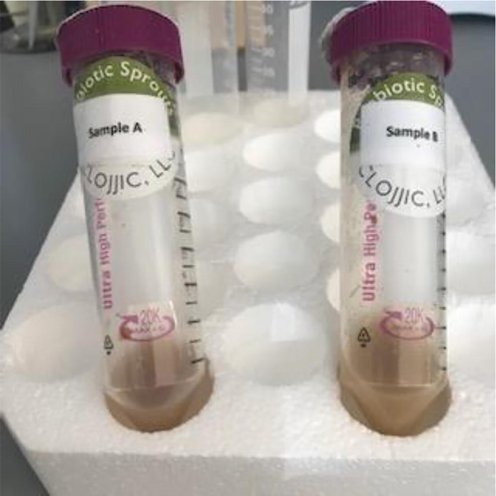
Test samples before centrifugation and filtration.
Serial dilutions of the test preparation were created in DMEM cell culture medium (Gibco #11995-065, Grand Island, NY) supplemented with 10% FBS (Avantor Seradigm #89510-194, Radnor, PA) to evaluate pH before cell treatment, using phenol red pH indicator. Medium alone served as an untreated control. Purified sulforaphane (Sigma Chemical, St. Louis, MO) was used as a positive control and as a calibrator to estimate sulforaphane concentration in the test preparation. Bortezomib (SelleckChem, Houston, TX) was used as an apoptosis-inducing positive control.
Cell culture
The Clojjic extract test preparations and controls were used to treat the cultured human colorectal tumor cell line HCT-116 (ATCC, Manassas, VA) and measure biomarkers of viability, cell death, and apoptosis. HCT-116 cells were chosen because of their known sensitivity to sulforaphane and its metabolites.8,21,22 HCT-116 cells were cultured as monolayers in DMEM cell culture medium (Gibco) supplemented with 10% FBS (Avantor Seradigm) in a humidified incubator at 37°C under 5% CO2. The cells were plated at 5000 cells per well, which provided a subconfluent starting density for accommodating growth during the 48 hours experimental conditions.
Cytotoxicity assay
Cytotoxicity, viability, and apoptosis assays were performed as a multiplex assay suite, starting with the CellTox™ Green Cytotoxicity Assay. Monolayers of HCT-116 cells were cultured at a subconfluent density in the presence of a CellTox™ Green Cytotoxicity Assay probe (Promega, Madison, WI), a fluorescent dye that selectively labels dead cells. Serial dilutions of test preparation, negative control media, and positive control (pH adjusted to test pH) were applied to the monolayers. CellTox™ Green fluorescence intensity was measured (485–500nmEx/520–530nmEm) immediately after treatment to ascertain the extent of any fluorescence quenching by the test preparation. The treated cells were returned to the incubator for 48 hours. CellTox Green fluorescence intensity was then measured at 24 and 48 hours to quantify cell death using a Glomax® Discover Microplate reader (Promega).
Cell viability
After the 48-hour treatment period, a 5x solution of CellTiter-Fluor™ cell viability assay reagent (Promega) was added to each well and fluorescence intensity was measured as a marker of live cells (380–400nmEx/505Em). The cells were placed at 37°C for 30 minutes, and a final measure of fluorescence intensity was collected using the Glomax Microplate Reader (Promega) to assess cell viability.
Apoptosis
Caspase-Glo®3/7 Assay System Reagent (Promega) was added to all wells and mixed to ensure homogeneity. Luminescence intensity as a marker of apoptosis induction was read as unfiltered light on a Glomax Microplate Reader (Promega) after a 30-minute incubation at RT.
HDAC activity assay
HeLa cell nuclear extract as a source of HDAC enzyme activity was used to test HDAC inhibition by Clojjic extract. HeLa cell nuclear extract (Enzo Life Sciences, East Farmingdale, NY) was prepared by dilution in DMEM cell culture medium. Serial dilutions of test preparation were created in cell culture medium. Medium alone served as an untreated control, and purified sulforaphane was a positive control and calibrator. The known HDAC inhibitor suberoylanilide hydroxamic acid (SAHA) (SelleckChem, Houston, TX) was also used as a positive control.
Serial dilutions of test preparation and positive control (pH adjusted to test pH) were applied to the HeLa nuclear extract and incubated at room temperature for 30 minutes. HDAC-Glo™ I/II luminescent histone deacetylase assay reagent (Promega) was added, and the plates were incubated at room temperature for 30 minutes. Luminescence was measured on the Glomax Microplate Reader (Promega) to quantify histone deacetylase inhibition.
Data analysis
The potency of the Clojjic extract effect on HCT-116 cell death, viability, proliferation, apoptosis induction, and HDAC inhibition was calculated with GraphPad Prism software (Boston, MA) using curve fit analysis.
Results
Assessment of Clojjic extract pH
The test preparation of the Clojjic extract had a pH of 4.0, consistent with a fermented product. This level of acidity exceeded the buffering capacity of the DMEM cell culture medium at lower dilution factors, as shown qualitatively by a phenol red color change to yellow at low dilution factors of a twofold dilution series (Fig. 2, columns 1–6). When diluted test preparations were added to an equal volume of HCT-116 cells in DMEM, only the 1:4 and 1:8 dilutions demonstrated acidic color changes with the phenol red pH indicator (not shown). DMEM medium adjusted to pH 4.0 with 1 M HCl and diluted as described (far right column) was largely buffered when added to cells. Sulforaphane and SAHA did not change the pH of the medium when added to cells.

Visual pH assessment of diluted Clojjic test preparations and controls in DMEM media before addition to cells. Columns 1–3: dilution series of Clojjic test preparation A in triplicate; Columns 4–6: dilution series of Clojjic test preparation B in triplicate; Columns 7–9: dilution series of purified sulforaphane in triplicate (row A = 150 µM); Columns 10–11: dilution series of the HDAC inhibitor SAHA in duplicate (row A = 1 µM); and Column 12: dilution series of DMEM medium adjusted to pH 4.0 with 1M HCl (pH control). Dilutions start at twofold in row A and increase by factors of 2 through row H. HDAC, histone deacetylase; SAHA, suberoylanilide hydroxamic acid.
Dose-dependent initial cytotoxicity of Clojjic extract in colorectal cancer cells
CellTox Green signal and round cell morphology indicate the cytotoxic effect of the Clojjic extract on HT-116 cells (Fig. 3). The cytotoxic effect was seen at a high extract concentration (1:4 dilution; Fig. 3D) where the culture medium pH is substantially reduced (see Fig. 2). Therefore, it was not possible to separate the potential cytotoxic effect of low pH from that of the bioactive extract components. No indication of lactobacilli was observed in the 1:4 dilution test preparation. The cytotoxic effect of the test preparation at 1:8 dilution was significantly decreased compared to the 1:4 dilution (Fig. 4E).
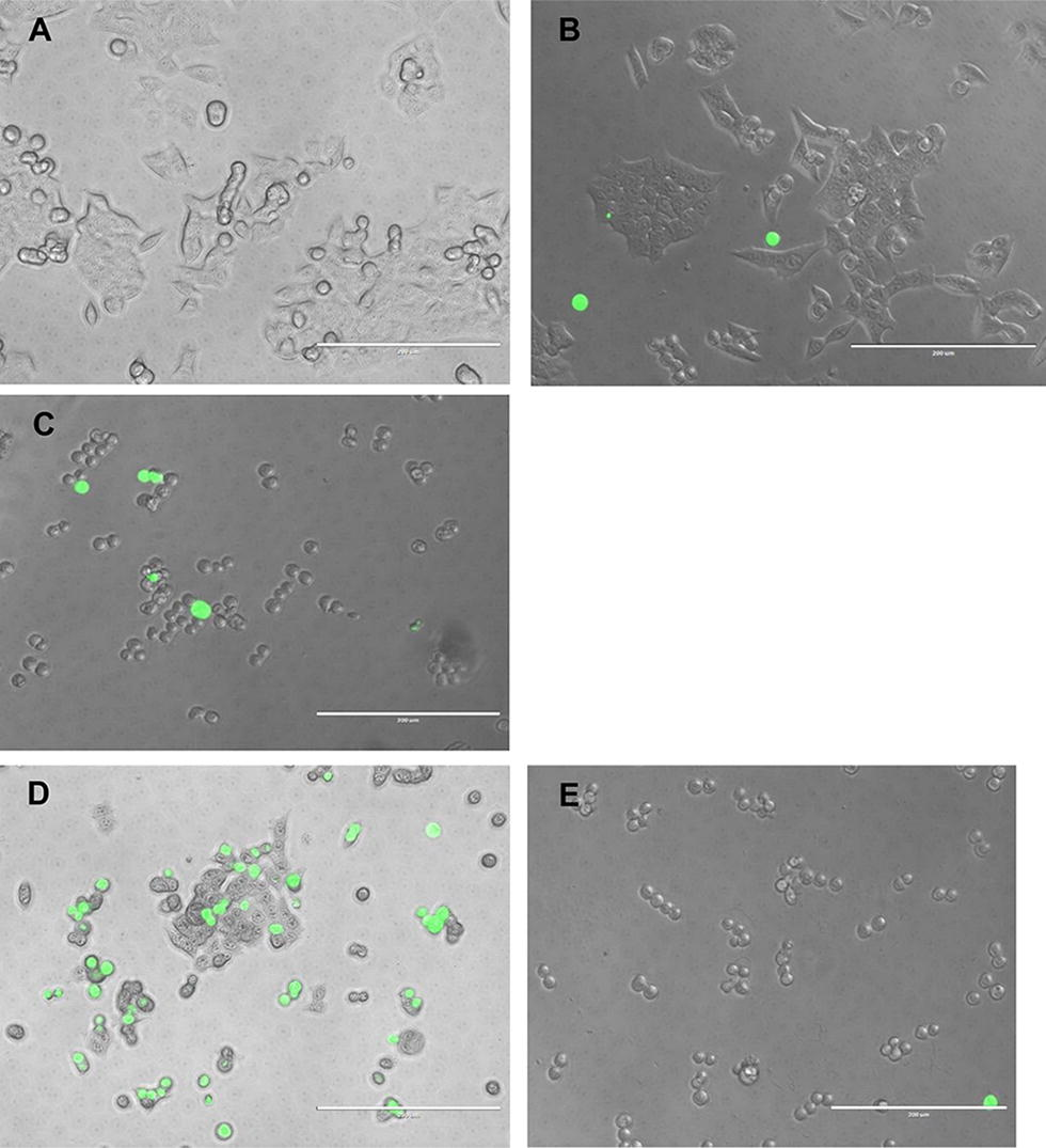
Cytotoxicity of Clojjic extract on HT-116 cells after 24 hours.

Effect of the Clojjic extract on HDAC inhibition.
Clojjic extract effects on HDAC inhibition
Two Clojjic extract test preparations inhibited HDAC activity in a dose-dependent manner (Fig. 4), suggesting that the extract contains an HDAC inhibitor(s), possibly a sulforaphane metabolite(s). 8 However, due to the mismatch of pH between the pH control dilution series and the Clojjic sample dilution series shown in Figure 2, we cannot rule out the contribution of reduced pH to the observed inhibitory effect on HDAC activity at the higher end of the curve where the dilution factors are low. Purified sulforaphane had no inhibitory effect on HDAC, consistent with the need for biotransformation. 8
Duration of cytotoxic effect of Clojjic extracts on colorectal cancer cells
Exposure to Clojjic extracts, but not sulforaphane, for 24 hours had a modest dose-dependent cytotoxic effect on HT116 cells as evidenced by the increase in CellTox Green fluorescence (Fig. 5). After a 48-hour incubation with two Clojjic extract test preparations, a dose-dependent increase in CellTox Green cytotoxicity signal and a reciprocal decrease in CellTiter Fluor viability signal was noted, consistent with a cytotoxic effect (Fig. 6A). For both the 24- and 48-hour timepoints, the effects of the Clojjic extract at the highest concentration should be interpreted with caution due to the potential impact of reduced pH. Also, at 48 hours, a dose-dependent increase in apoptosis was observed (Fig. 6B), implying that apoptosis contributes at least in part to the cytotoxic effect of the Clojjic extracts on cultured colorectal cancer cells. Note that the peak apoptosis marker signal may have occurred earlier than 48 hours when a more pronounced apoptosis induction might be observed. Treatment of HT-116 cells with bortezomib, a known cytotoxin that acts via apoptosis, had clear dose-dependent cytotoxic effect, reducing cell viability (Fig. 6C) and inducing apoptosis (Fig. 6D). In contrast, purified sulforaphane had little to no effect on cell viability and did not induce apoptosis (Fig. 6C, D).

Cytotoxicity of Clojjic extract preparations after 24 hours.

Cytotoxicity of Clojjic extracts and cell viability after 48 hours.
Discussion
In this study, we found that the Clojjic broccoli sprout extract containing stabilized sulforaphane and gut lactobacilli-produced bacteriocins had a dose-dependent cytotoxic effect on cultured HT-116 human colorectal cancer cells, reducing viability and promoting apoptosis 24 and 48 hours after treatment. The Clojjic extracts also had a dose-dependent inhibitory effect on HDAC, which may underlie its cytotoxic effects. The low pH of higher concentration Clojjic extracts may contribute to the observed cytotoxicity.
The test preparations in this study contained only the soluble portion of the Clojjic extracts. Adding insoluble particulates would have interfered with cell-based assay readouts and interpretations. Nevertheless, the insoluble portion may contain substantial sulforaphane or other unidentified or synergistic phytochemical components that become bioavailable after ingestion in vivo, such as niacin, a powerful antioxidant involved in cell signaling. 23 Indeed, in vitro approaches do not recapitulate the in vivo biotransformation of phytochemicals such as sulforaphane or their effects on a multicellular tumor environment, including bystander effects on cells providing immunity function.
Other studies have demonstrated the limitation of monolayer cultures for in vitro toxicology assessments, such as the HT-116 cells used here. Three-dimensional (3D) multicellular tumor spheroids (MCTs) are emerging as an essential tool in cancer research that more accurately recapitulates the complex interactions of in vivo solid tumors. 24 It is possible that Clojjic extract cytotoxicity may be different in 3D MCTs versus 2D monolayer cultures. Furthermore, while tandem filtration with a 0.8/0.2-μM filter was employed to eliminate/reduce the lactobacilli in the test preparations, the preparations likely contained active lactobacilli, which are incompatible with standard mammalian cell culture models. Ultimately, in vivo studies will be needed to verify our in vitro findings.
The test preparation was a fermented product of low pH. A highly buffered cell culture medium was employed to reduce fluorescent and luminescent assay interference and effects on cell health caused by deviation from a standard pH 7.2 environment. Future studies will need to uncouple the cytotoxic effects of pH from those of the bioactive components of the extract. In addition, significant sample loss occurred during sedimentation and filtration due to membrane-wetting void volumes and poor filtration efficiency. More starting material will be needed to create excipient control preparations, such as heat-inactivated control samples, for future studies.
The Clojjic extract 19 contains a combination of 12 lactobacilli strains that produce metabolites such as bacteriocins that may enhance the cytotoxicity of the preparation. It will be important to identify the nature of sulforaphane metabolites and bacteriocins present in the Clojjic extract to understand their contribution to the cytotoxic effect in HT-116 colorectal cancer cells as well as other cancer cell types. Previous studies have described the effect of bacteriocins on melanoma cell death 25 and have shown that bacteriocins from lactic acid bacteria are toxic to certain pathogens26,27 and cancer cell types.10,22,28 Studies on lactic acid bacteria have suggested their use as delivery vehicles for a range of molecules and applications, including for antiinfectives, 29 therapies for allergic diseases, 30 and therapies for gastrointestinal diseases. 31 Interleukins have been coexpressed with antigens in lactic acid bacteria to enhance the immune response raised against the antigen. 25 Future work will need to characterize the antipathogenic and antioncogenic substances produced by the selected human gut microflora in the Clojjic extract.4,20
Conclusions
Clojjic extract consists of a combination of broccoli sprouts-derived sulforaphane and active Bifidobacterium–Lactobacillus–Streptococcus strains. Despite the technical challenges posed by the insoluble materials in the Clojjic extracts, this study demonstrated that it is a powerful cytotoxic agent with dual bioactive components: lactobacilli metabolites (bacteriocins such as plantaricin) and sulforaphane from broccoli sprouts (Fig. 7). Together, these components promoted HT-116 colorectal cancer cell death and induced apoptosis, partially through inhibition of HDAC. Further studies are warranted to investigate the Clojjic extract as a potential cancer therapeutic, including identifying specific bacteriocins, testing other cancer cell lines or 3D models, assessing synergistic effects of components, and elucidating molecular mechanisms.
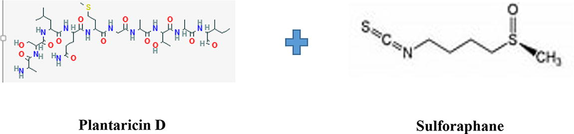
Sulforaphane and plantaricin D as the potential bioactive ingredients in Clojjic broccoli sprouts extract.
Footnotes
Authors’ Contributions
A.-M.K.-K. conceptualized the study, analyzed the data, and wrote the article; P.M. conducted the study and reviewed the article; J.J.C. collected and analyzed data and reviewed the article. All authors approved the final version of the article for submission.
Author Disclosure Statement
P.M. and J.J.C. are the employees of Promega Corporation. A.-M.K.-K. has no conflicts of interest to declare.
Funding Information
This study used Promega’s facility, technical expertise, and equipment infrastructure. The authors did not receive any monetary support from Promega to conduct the study.