




Abstract
Background:
The growing global production and application of nanomaterials have brought nanotoxicology into the spotlight, particularly regarding their potential health risks. Despite these advancements, the potential adverse effects of these nanoparticles on human health remain largely underexplored.
Materials and Methods:
This study evaluated the toxicity of Silver nanoparticles (AgNPs) and silica nanoparticles (SiO2-NPs) on blood cells using two.
Methods:
Assessing cell viability in the Jurkat blood cell line via the MTT assay at nanoparticle concentrations ranging from 1 to 1000 μg/mL over 24, 48, and 72 hours, and examining hemolysis in peripheral blood using the Acid Citrate Dextrose method. IC50 values were determined through web-based programs and results were analyzed statistically.
Results:
The MTT assay revealed that the cytotoxic effects of AgNPs and SiO2-NPs varied based on their physicochemical properties, concentration, and duration of exposure. Hemolysis tests indicated that SiO2-NPs resulted in an increased percentage of hemolysis with rising concentrations. At lower concentrations, AgNPs induced a higher level of hemolysis compared with SiO2-NPs; however, at concentrations of 500 and 1000 μg/mL, SiO2-NPs exhibited significantly greater hemolytic activity.
Conclusion:
This study highlights important implications for biomedical applications, particularly in drug delivery and cancer therapy, by comparing the toxicity of AgNPs and SiO2-NPs. It emphasizes the necessity for a deeper understanding of toxicity mechanisms to effectively translate in vitro findings into clinical practice. Future research should focus on bridging the gap between in vitro and in vivo studies to ensure the safe application of these nanoparticles.
Introduction
In nanotechnology, particle size plays a critical role, as the physicochemical properties of nanomaterials are significantly influenced by their dimensions. Smaller particle sizes enhance the biological activity of nanomaterials, thereby increasing their potential for absorption and interaction with biological tissues.1,2 Nanoparticles represent a novel category of biomedical products, offering promising applications in medical devices and drug delivery systems for the treatment of complex conditions such as cancer, as well as inflammatory and neurological diseases.3,4 Given their expanding biomedical applications, it is essential to establish a clear understanding of how nanomaterials interact with biological systems to ensure their efficacy and safety. Currently, nanomaterials are extensively utilized across various fields, including medicine (e.g., reducing microbial skin infections, treating burn wounds, and preventing bacterial accumulation on medical devices such as prostheses). Furthermore, they play a significant role in advancements in pharmaceuticals, cosmetics, transportation, energy, and agriculture, becoming a crucial component of societal and industrial development.1,2 Despite their numerous advantages in applications such as biomedical equipment and electronic devices, nanoparticles can pose significant risks to human health. 5 Given their expanding applications, it is crucial to understand the properties of nanoparticles and their effects on human cells prior to their widespread use. Investigating the toxicity of nanomaterials is essential, not only to elucidate their functional mechanisms but also to facilitate their safe translation into clinical applications.
The extensive use of nanomaterials, without sufficient consideration of their consequences, has unfortunately resulted in significant environmental risks that directly affect plants, animals, and humans. Nanomaterials can penetrate human cells through inhalation, ingestion, skin contact, or injection. Once inside the body, they readily enter the bloodstream and distribute to sensitive organs. Research has highlighted the adverse effects of nanomaterials on blood cells,6,7 with their systemic distribution potentially triggering acute cardiovascular risk factors such as hemodynamic changes (e.g., alterations in blood pressure and heart rate), disruption of homeostasis, or inflammation. Consequently, nanoparticles intended for biomedical applications must undergo rigorous biocompatibility testing before being approved for patient use. While ongoing efforts aim to establish critical parameters for the clinical evaluation of engineered nanoparticles, harmonized protocols for biocompatibility testing remain limited. 8 Addressing this gap is necessary to develop standardized methodologies that ensure the safety and efficacy of nanomaterials in medical applications.
Silver nanoparticles (AgNPs) are widely recognized for their antimicrobial properties and are commonly utilized in consumer and healthcare products. Despite their extensive application, including therapeutic uses that promote cellular entry, there is a significant lack of comprehensive information regarding their molecular and cellular toxicity in humans. Composed of 20–15,000 silver atoms and typically measuring less than 100 nm in diameter, AgNPs demonstrate considerable antimicrobial activity even at low concentrations due to their high surface-to-volume ratio. 9 Among metal pollutants, silver ions are particularly toxic, ranking highest in toxicity classification. However, their effectiveness against a broad spectrum of microorganisms, coupled with relatively low toxicity to humans, has facilitated their development for treating burn wound infections.10,11 AgNPs, due to their nanoscale size, can more easily penetrate tissues, cells, and biological molecules in living organisms. 12 However, their widespread use presents potential health risks to both humans and animals, potentially resulting in tissue damage and functional disruption. Among the affected tissues, the liver—being the largest gland and playing a crucial role in metabolism, detoxification, and glycogen storage—is particularly susceptible. Repeated exposure to AgNPs can lead to liver damage, which may initially be reversible but could progress to acute liver failure (FHF) and even result in death.13,14 While several studies have investigated the in vitro toxicity of AgNPs, further research is needed to bridge the gap between laboratory findings and their clinical implications. In vitro studies are frequently the initial step in understanding the interactions of AgNPs due to their controlled environments. Research has examined the toxicity of AgNPs using various cell types, including mouse hepatocytes, 15 mouse progenitor stem cells, 16 human fibroblasts, 17 and mouse adrenal cells. 18 Recent studies provide growing evidence that AgNPs exhibit toxic effects on a variety of cultured cells.
Crystalline silica, the crystallized form of silica (SiO2) found in quartz, cristobalite, or tridymite, is one of the most abundant compounds in the Earth’s crust. 19 Recent epidemiological studies conducted by the International Agency for Research on Cancer have classified the quartz and cristobalite forms of crystalline silica as Group 1 carcinogens due to their association with lung cancer resulting from occupational exposure. 20 Despite these risks, silica nanoparticles (SiO2-NPs) are widely used in cancer diagnosis and treatment, the development of advanced nanomedicines as carriers for drug or gene delivery, and imaging applications.21,22 Potential exposure routes for SiO2-NPs include ingestion, dermal absorption, and injection. 5
Numerous studies have investigated the toxic effects of nanoparticles on animals; however, research regarding their impact on human cells and tissues remains limited. 23 Hussain et al. addressed this knowledge gap by evaluating the acute toxic effects of metal oxide nanoparticles using a liver-derived cell line from laboratory mice (BRL 3A). The study assessed nanoparticles of silver (Ag, 15 and 100 nm), molybdenum (MoO3, 30 and 103 nm), aluminum (30 and 103 nm), iron oxide (FeSO4, 30 and 47 nm), and titanium dioxide (TiO2, 40 nm). Among these, silver exhibited high toxicity, molybdenum demonstrated moderate toxicity, while aluminum and iron oxides showed minimal to no toxicity at the tested doses. 15 Yahyai et al. investigated the effects of magnetic iron nanoparticles on blood groups in four groups of six healthy male mice. Although the iron nanoparticles exhibited low toxicity under normal test conditions, the presence of an electromagnetic field resulted in liver coagulation at the tested doses, suggesting that electromagnetic exposure could exacerbate tissue changes. 24 Despite the widespread use of silica and AgNPs in various applications, their potential adverse effects on human health remain largely underexplored. This study aims to evaluate the effects of different concentrations of silver and SiO2-NPs on the viability of a blood cell line (Jurkat) and their influence on peripheral blood hemolysis. By integrating in vitro toxicity data with biomedical applications, this research seeks to contribute to a better understanding of the safety profile of these nanoparticles, ultimately facilitating their safe translation into clinical settings.
Materials and Methods
This study evaluated the toxicity of nanoparticles on blood cells using two approaches: first, by assessing the impact of varying nanoparticle concentrations on the survival of a Jurkat blood cell line over 24, 48, and 72 hours; and second, by examining their effects on peripheral blood hemolysis.
Jurkat blood cell lines were purchased from the Pasteur Institute of Iran. This work was approved by the Ethics Committee from the
Preparation of nanoparticles used in this study
After purchasing SiO2 and silver Ag nanoparticles, a stock solution of each of the nanoparticles was prepared with a concentration of 3000 μg/mL using deionized water. Then, using the stock solution, concentrations of 1, 10, 100, 500, and 1000 µ/mL of each of the nanoparticles were prepared.
Investigation of hemolysis of blood cells
The Acid Citrate Dextrose (ACD) method was employed to assess the hemolysis of blood cells. Peripheral blood samples (n = 3) were collected in heparin-containing tubes and 4.5 mL of ACD solution was added to 0.5 mL of each blood sample. The ACD solution was prepared by dissolving 0.544 g of anhydrous citric acid, 1.65 g of trisodium citrate dihydrate, and 1.84 g of dextrose monohydrate in 75 mL of distilled water. Nanoparticles at concentrations ranging from 1 to 1000 μg/mL were incubated with the blood samples for 1 hour at 37°C. A control sample was prepared without the addition of nanoparticles. After incubation, all samples were centrifuged at 800 rpm for 8 minutes. The supernatant was then collected, and the absorbance was measured at 545 nm. The degree of hemolysis was calculated relative to the control sample using the following formula:
Each experiment was conducted in triplicate, and the results are presented as the mean ± standard deviation.
Blood cell line culture
Cell culture experiments were conducted using a Jurkat blood cell line. The cell lines were delivered in 25 cm2 flasks, which were disinfected with 70% alcohol before being transferred to the laboratory. After verifying the cell density under a microscope, the flasks were incubated at 37°C in a humidified environment containing 5% CO2 and 95% air. Daily microscopic examinations were conducted to monitor cell growth, density, and morphology, as well as to ensure the absence of bacterial or fungal contamination. The culture medium was refreshed every 1–2 days under a laminar flow hood. During medium changes, the old medium and cells were transferred into sterile tubes, centrifuged at 1500 rpm for 5 minutes, and the supernatant was discarded. The sediment was then resuspended in fresh sterile RPMI-1640 culture medium containing 10% fetal bovine serum, bicarbonate buffer, amino acids, vitamins, and antibiotics (100 U/mL penicillin and 100 U/mL streptomycin). The refreshed medium was returned to the culture flasks to ensure optimal growth conditions.
Evaluation of cytotoxicity of nanoparticles by 2,5-diphenyl-2H-tetrazolium bromide (MTT) method
The Jurkat cell line (Clone E6-1; NCBI Code) is a suspension-type T lymphocyte characterized by lymphoblastic morphology. It was derived from the peripheral blood of a male patient diagnosed with acute T-cell leukemia. This cell line can be cryopreserved in liquid nitrogen. To evaluate cytotoxicity, approximately 5 × 10³ Jurkat cells were seeded into each well of 96-well plates. Following a 24-hour growth period, the cells were treated with varying concentrations (1–1000 µg/mL) of silver and SiO2-NPs, which were prepared in RPMI medium. Each concentration was tested in triplicate. Negative control wells contained only complete culture medium, while positive control wells included dimethyl sulfoxide (DMSO). After incubation for 24, 48, and 72 hours, 20 µL of 2,5-diphenyl-2H-tetrazolium bromide (MTT) dye was added to each well, and the plates were incubated at 37°C for 4 hours. Subsequently, DMSO was added to dissolve the resulting formazan crystals, and the optical absorbance of each well was measured at 490 nm using a multiplate reader (Biotech, Synergy, USA). The absorbance of all samples was normalized to the positive control and the percentage of viable cells was calculated using the following equation:
Finally, the concentration of nanoparticles required to inhibit the growth of 50% of the cells (IC50) was calculated using an online IC50 calculator available at [AAT Bioquest] (https://www.aatbio.com/tools/ic50-calculator). The results from both the hemolysis assay and the MTT cytotoxicity test were directly analyzed to interpret the effects of varying nanoparticle concentrations on the Jurkat cells. This approach provided a comprehensive understanding of the toxicological impact of silver and SiO2-NPs on blood cells under different experimental conditions.
Results
Results of hemolysis of blood cells
The results of the hemolysis assay for silver and SiO2-NPs at concentrations of 1, 10, 100, 500, and 1000 μg/mL are presented in Tables 1 and 2. The light absorption values for the positive control samples were 0.081, 0.078, and 0.067, yielding an average of 0.075333. These values were subtracted from all negative control results. The average absorbance for the negative control of the AgNP samples, after subtracting the positive control, was 0.908433, while for the SiO2-NP samples, it was 1.008433. For each concentration, the optical absorbance was measured for three samples, and the average absorbance value of the positive control was subtracted. The optical absorbance results of the treated (test) samples were then divided by the absorbance results of the control samples to calculate the percentage of hemolysis.
Results of Hemolysis of Blood Cells When Exposed to Silver Nanoparticles
Results of Hemolysis of Blood Cells When Exposed to Silica Nanoparticles
Figure 1 illustrates the hemolysis results of blood cells exposed to AgNPs. As shown, an increase in the concentration of AgNPs corresponds to a higher average percentage of hemolysis. The highest percentage of hemolysis was observed at a concentration of 1000 μg/mL, with an average of 1.275848 ± 5.283822. This trend indicates a concentration-dependent effect of AgNPs on blood cell hemolysis.
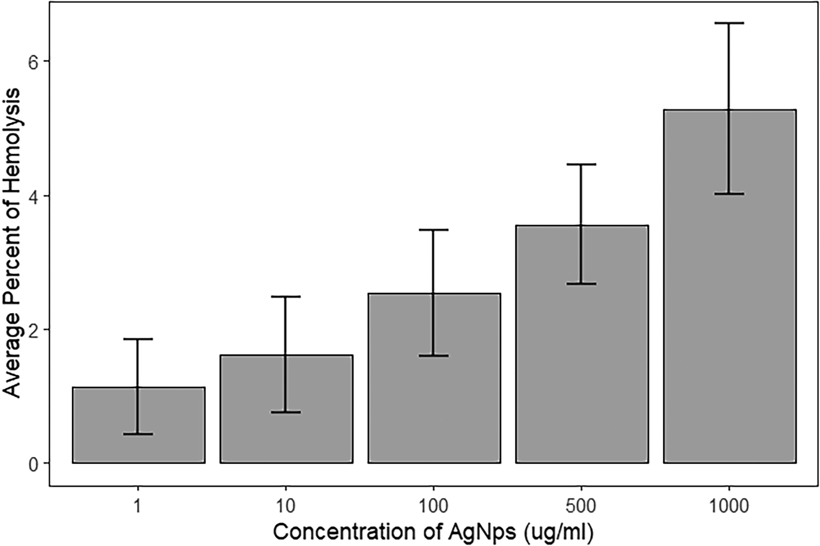
Concentration-dependent hemolysis induced by AgNPs. Dose-response plot showing the percentage of hemolysis caused by Ag-NPs across a range of concentrations (μg/mL). Error bars indicate standard deviation. AgNPs, silver nanoparticles.
Figure 2 presents the hemolysis results of blood cells exposed to SiO2-NPs. The data indicate that the average percentage of hemolysis increases as the concentration of SiO2-NPs rises. At the 1000 μg/mL concentration, the highest percentage of hemolysis was observed, with an average of 1.14933 ± 7.040624, demonstrating a concentration-dependent effect on blood cell hemolysis.

Concentration-dependent hemolysis induced by SiO2-NPs. Dose-response plot showing the percentage of hemolysis caused by SiO2 nanoparticles across a range of concentrations (μg/mL). Error bars indicate standard deviation. SiO2-NP, silica nanoparticle.
Figure 3 compares the hemolysis results of blood cells exposed to silver and SiO2-NPs. At lower concentrations, the average percentage of hemolysis induced by AgNPs is higher than that caused by SiO2-NPs. However, as the concentration increases, the average percentage of hemolysis from SiO2-NPs also rises. The most significant difference in the average percentage of hemolysis between silica and AgNPs was observed at concentrations of 500 and 1000 μg/mL.
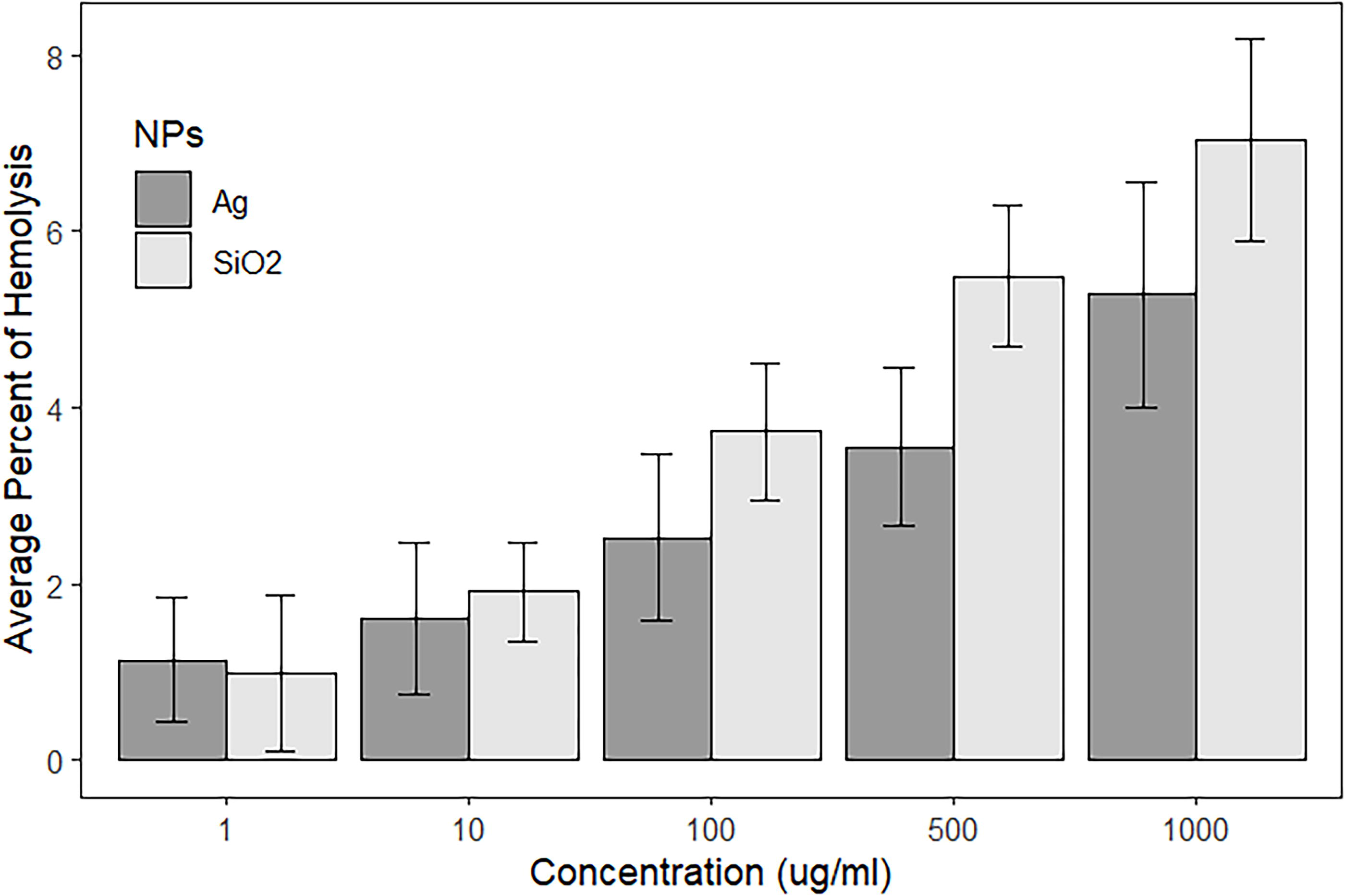
Effect of NPs on blood hemolysis. Bar graph comparing the effects of AgNPs and SiO2-NPs blood hemolysis at different concentrations (μg/mL). Data represent mean percent hemolysis ± SD. NP, nanoparticles, SD, standard deviation.
Cytotoxicity of nanoparticles
Tables 3 and 4 present the results of the cytotoxicity evaluation of silver and SiO2-NPs on the Jurkat cell line (Clone E6-1: NCBI Code) at concentrations ranging from 1 to 1000 μg/mL, observed over 24, 48, and 72 hours. The results were obtained by subtracting the absorption values of the positive control (0.036677, 0.1, and 0.106667 for 24, 48, and 72 hours, respectively) from the absorption values of the negative control and the samples. The average absorption values of the treated samples were then divided by the negative control values, and the resulting percentage of viable cells was calculated.
Cytotoxicity Results of Silver Nanoparticles on Jurkat Cell Line
SD, standard deviation.
Cytotoxicity Results of Silica Nanoparticles on Jurkat Cell Line
Dose-response studies utilizing the MTT assay indicate that treatment with nanoparticles for 24 hours does not result in significant toxicity at concentrations of 1 μg/mL or lower. However, as the concentration and treatment duration increase, notable toxicity is observed. When the treatment duration is extended to 72 hours, AgNPs exhibit approximately 87% toxicity, while SiO2-NPs demonstrate around 95% toxicity in the JURKAT cell line at a concentration of 1000 μg/mL.
Figures 4–6 illustrate the comparison of the cytotoxicity results of silver and SiO2-NPs on the Jurkat cell line (Clone E6-1: NCBI Code) at concentrations ranging from 1 to 1000 μg/mL, observed over 24, 48, and 72 hours.
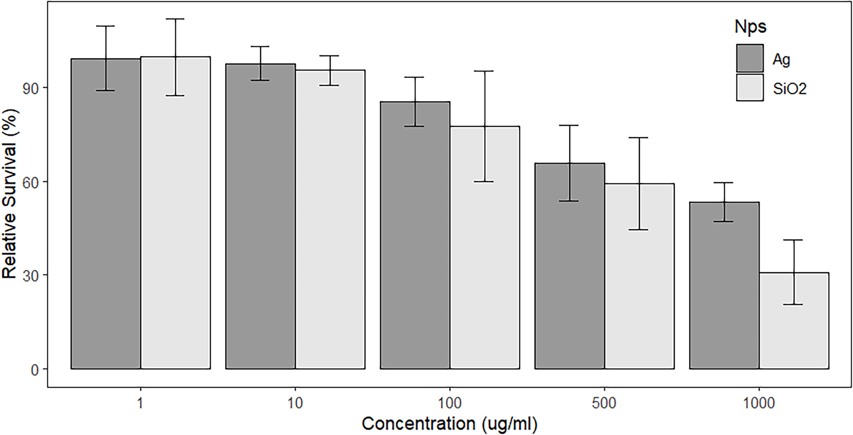
Cytotoxicity of NPs after 24-hour exposure. Dose-dependent effects of AgNPs and SiO2-NPs on cell viability after 24-hour treatment. Data show relative survival (%) compared with untreated controls (mean ± SD).
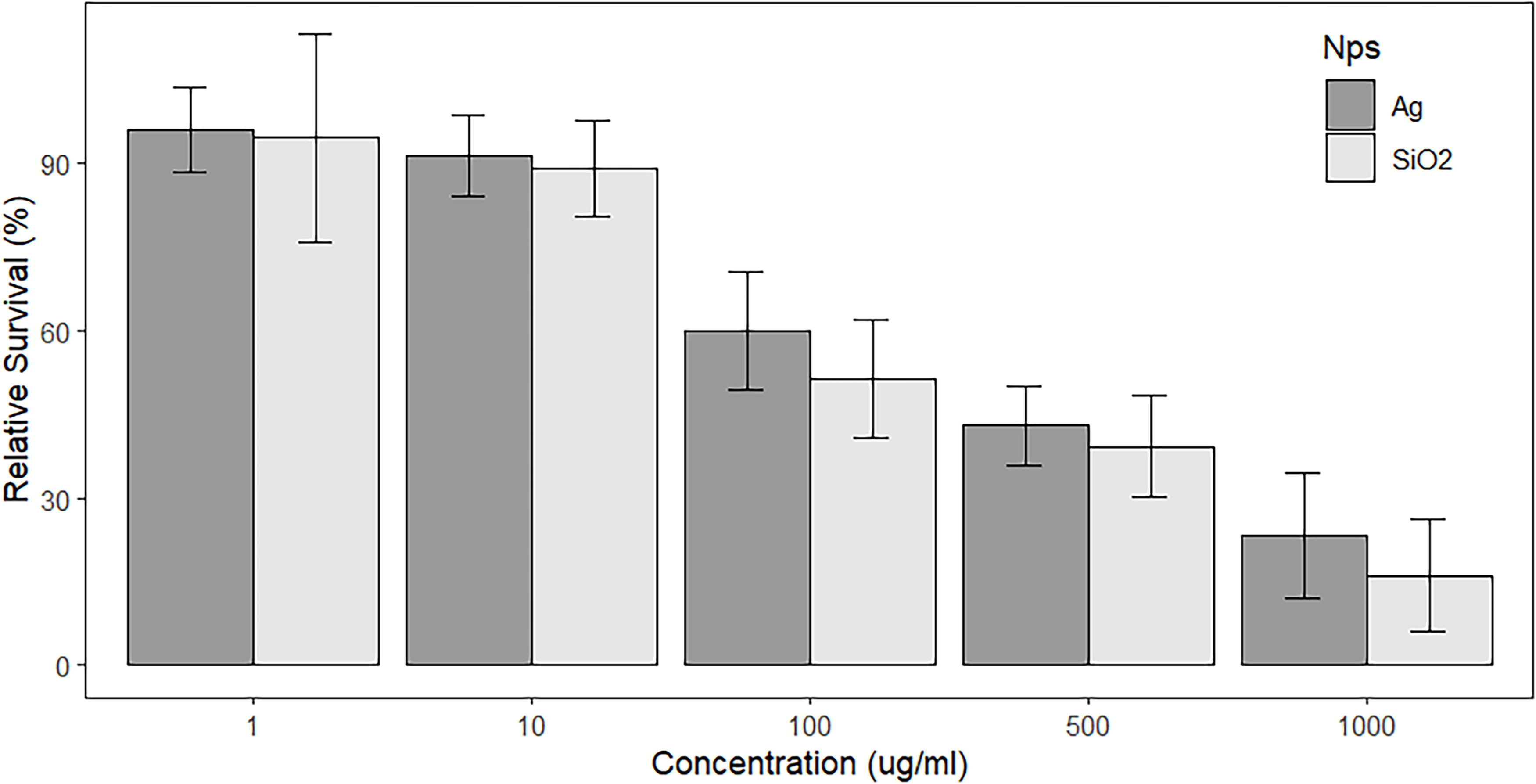
Cytotoxicity of NPs after 48-hour exposure. Dose-dependent effects of AgNPs and SiO2-NPs on cell viability after 48-hour treatment. Data show relative survival (%) compared with untreated controls (mean ± SD).
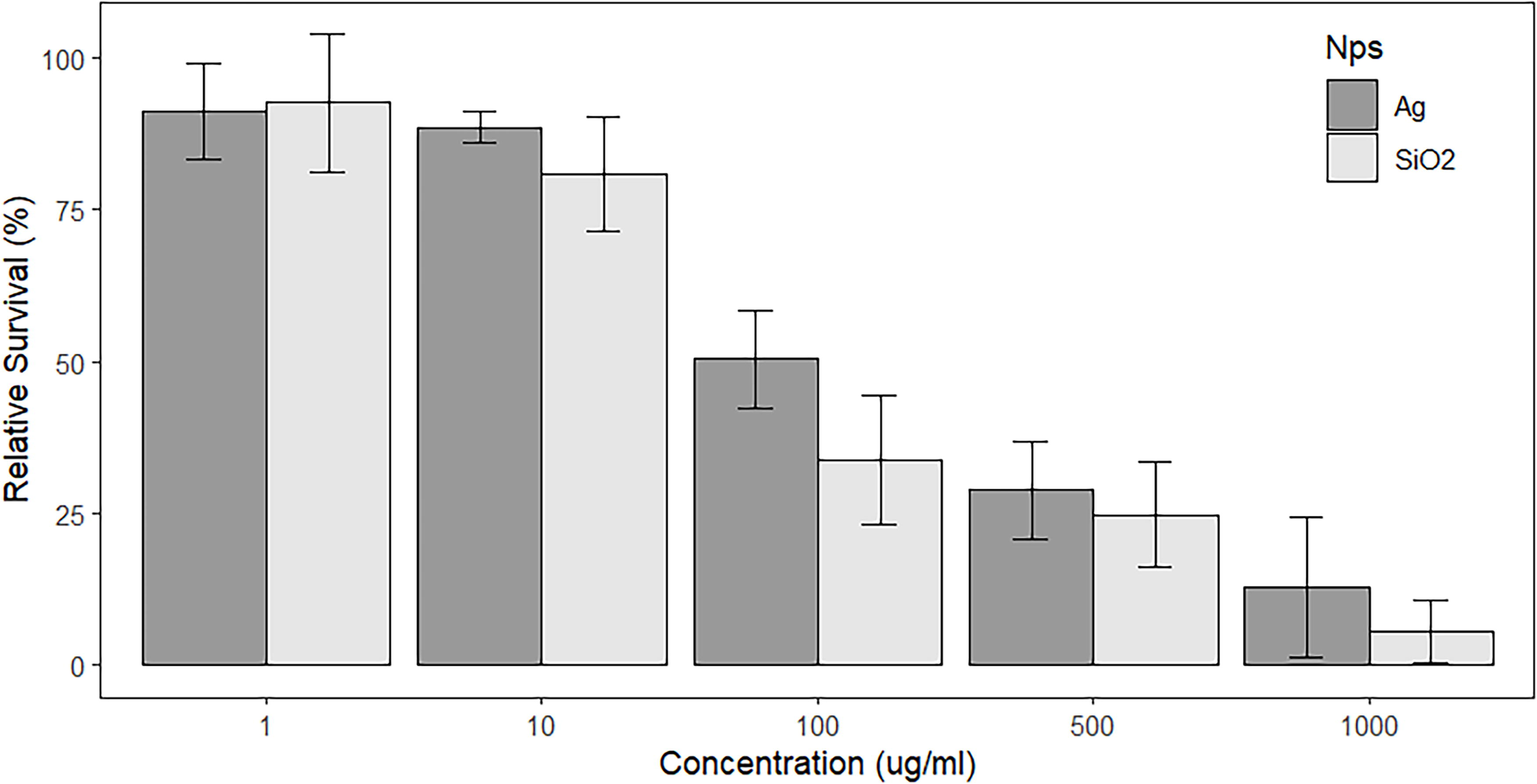
Cytotoxicity of NPs after 72-hour exposure. Dose-dependent effects of AgNPs and SiO2-NPs on cell viability after 72-hour treatment. Data show relative survival (%) compared with untreated controls (mean ± SD).
As indicated by the data, the percentage of viable cells decreases with increasing concentration and treatment duration. Furthermore, the results suggest that the impact of SiO2-NPs is more significant than that of AgNPs, as the percentage of viable cells is lower in samples treated with SiO2-NPs compared to those treated with AgNPs.
The concentration that inhibits the growth of 50% of cells (IC50) was determined using web-based tools (https://www.aatbio.com/tools/ic50-calculator). The results of the IC50 calculations are presented in Figures 7 and 8. The IC50 value after exposure to AgNPs was 58.0467 µg/mL, whereas the IC50 value for SiO2-NPs was lower, at 40.4074 µg/mL. This indicates that SiO2-NPs exert a stronger cytotoxic effect on the Jurkat cell line compared with AgNPs.

IC50 determination of AgNPs. Dose-response analysis of AgNPs cytotoxicity, yielding an IC50 value of 58.05 μg/mL. Curve fitted using nonlinear regression.

IC50 determination of SiO2-NPs. Dose-response analysis of SiO2-NPs cytotoxicity, yielding an IC50 value of 40.41 μg/mL. Curve fitted using nonlinear regression.
Discussion
The results of the MTT assay revealed that silver and SiO2-NPs exhibit varying levels of cytotoxicity depending on their physicochemical properties, concentration, and exposure duration. These findings align with previous toxicity studies.23,25 Given the significant potential of silver and SiO2-NPs in nanotechnology, our results highlight the urgent need for more comprehensive research, both in vitro and in vivo, to assess the potential health and environmental risks of these materials. When nanoparticles are absorbed through the skin, ingested, or inhaled, they can easily enter the bloodstream or interact with blood cells, including mononuclear cells. This exposure can lead to complications such as hemolysis, thrombosis, and possibly cell death. The observed cell death may be due to the high permeability of the cell membrane, which allows nanoparticles to bind to lipids and proteins, inhibit enzymes, and ultimately cause cellular damage. The increase in cytotoxicity over time could be linked to a greater binding of nanoparticles to surface or intracellular proteins, a time-dependent property. Previous studies have also shown that SiO2 nanoparticles can induce cell differentiation, inflammation, and changes in protein expression.26,27 These findings underscore the necessity of understanding the cellular mechanisms underlying nanoparticle toxicity to develop safer biomedical applications.
The study results indicate that the toxicity of SiO2-NPs for the Jurkat cell line and the blood hemolysis rate increase with higher concentrations and longer exposure times. At lower concentrations (1 μg/mL), the lack of a significant increase in toxicity may be due to the low nanoparticle concentration, which initially triggers a toxic shock response in the cells but does not sustain prolonged cytotoxic effects. Understanding these concentration-dependent effects is crucial for determining safe exposure levels in biomedical applications.
In a study by Barkhordari et al., titled “Assessing the cytotoxic effects of SiO2 nanoparticles on mononuclear cells of human blood,” a suspension of mononuclear cells from 10 healthy young men was exposed to various nanoparticle concentrations (1, 10, 100, 500, 1000, and 1500 µg/mL) for 6 and 24 hours. The study showed a significant difference in cell death rates between various concentrations of nanoparticles (p < 0.05). As the concentration of nanoparticles increased, the rate of cell death also increased, with a 6-fold rise in cell death at a concentration of 10 µg/mL after 24 hours compared with 1 µg/mL. This study was the first to determine the lethal effect of SiO2 nanoparticles at concentrations as low as 1 µg/mL for blood mononuclear cells, and it concluded that the cytotoxic effects are concentration- and time-dependent. 23 These data further support the idea that nanoparticle-induced toxicity is a complex process influenced by multiple factors, including dose and exposure duration.
Similarly, Gurr et al. investigated the damage caused to bronchial epithelial cells by titanium oxide nanoparticles using the MTT method and found that toxicity increased with prolonged exposure, particularly after three days. 28 In 2007, Dechsakulthom and colleagues used the MTS method to study the toxicity of skin fibroblast cells exposed to ZnO and TiO2 nanoparticles, observing a significant increase in toxicity after 24 hours compared with 4 hours of exposure. 29 These studies, along with our findings, emphasize that both the concentration and duration of nanoparticle exposure significantly affect their toxicity. Moreover, these results provide valuable insights for evaluating the risks of nanoparticle exposure in clinical and environmental settings.
However, the wide application of nanoparticles in the pharmaceutical, medical, and other sectors of agriculture and industry requires more attention to human safety and health and the investigation of the toxic effects of nanoparticles on cells, especially at the cellular-microscopic level. Future studies should aim to elucidate the precise molecular mechanisms by which silver and SiO2-NPs exert their toxic effects, particularly in different cell lines relevant to human health. Additionally, the exploration of nanoparticle surface modifications and targeted delivery systems may help mitigate toxicity while enhancing their biomedical efficacy. Understanding these mechanisms will be essential for the development of safer and more effective nanoparticle-based therapeutics.
Conclusion
In conclusion, this study demonstrates the significant cytotoxic and hemolytic effects of silver and SiO2-NPs on human blood cells and the Jurkat cell line. The results indicate that both types of nanoparticles enhance hemolysis in blood cells in a concentration-dependent manner, with AgNPs exhibiting a more pronounced hemolytic effect at lower concentrations. However, as the concentration increases, SiO2-NPs also display increased hemolytic activity, revealing a notable distinction between the two types of nanoparticles at higher concentrations. Furthermore, the cytotoxicity assessment of both silver and SiO2-NPs on the Jurkat cell line reveals a decrease in cell viability with increasing nanoparticle concentration and exposure time. Notably, SiO2-NPs exhibit a more potent cytotoxic effect than AgNPs, as evidenced by the lower IC50 value for SiO2-NPs. These findings have critical implications for the development of biomedical applications, particularly in drug delivery and cancer therapy. While silver and SiO2-NPs hold great promise for clinical use, their cytotoxic and hemolytic effects emphasize the necessity of carefully evaluating their biocompatibility and potential risks. A more in-depth understanding of the mechanisms underlying these toxic effects is essential for translating in vitro findings into safe and effective clinical applications. Moreover, this study contributes to the growing body of research on nanoparticle toxicity by offering a comparative perspective on silver and SiO2-NPs, distinguishing their effects from previous studies. Future investigations should focus on bridging the gap between in vitro and in vivo studies to ensure the safe integration of these nanomaterials into biomedical practice.
Footnotes
Acknowledgment
The authors acknowledge the support provided by
Authors’ Contributions
S.R.: Conceptualization, methodology, investigation, data curation, and data analysis. S.J.: Methodology, formal analysis, validation, investigation, and writing—review and editing. A.G.: Methodology, formal analysis, validation, and investigation. M.H.: Investigation and writing—review and editing. D.S.: Writing—review and editing and software. All authors read and approved the final article.
Author Disclosure Statement
The authors have no competing interests to declare that are relevant to the content of this article.
Funding Information
This project was approved and financially supported by Neyshabur University of Medical Sciences (Grant number: 155).