
Abstract
Nuclear factor E2–related factor 2 (Nrf2) is a transcription factor known to induce expression of a variety of cytoprotective and detoxification genes. Several of the genes commonly regulated by Nrf2 have been implicated in protection from neurodegenerative conditions. Work from several laboratories has uncovered the potential for Nrf2-mediated transcription to protect from neurodegeneration resulting from mechanisms involving oxidative stress. For this reason, Nrf2 may be considered a therapeutic target for conditions that are known to involve free radical damage. Because common mechanisms of neurodegeneration, such as mitochondrial dysfunction and build-up of reactive oxygen species, are currently being uncovered, targeting Nrf2 may be valuable in combating conditions with variable causes and etiologies. Most effectively to target this protein in neurodegenerative conditions, a description of the involvement of Nrf2 and potential for neuroprotection must come from laboratory models. Herein, we review the current literature that suggests that Nrf2 may be a valuable therapeutic target for neurodegenerative disease, as well as experiments that illustrate potential mechanisms of protection. Antioxid. Redox Signal. 11, 497–508.
Introduction
Neurodegeneration in humans is known to occur as a result of several primary causes, including expression of certain gene alleles, toxicant administration, and aging. As a consequence, researchers have used these primary causes to establish laboratory models for neurodegeneration in single cells as well as in organisms ranging from Caenorhabditis elegans and Drosophila melanogaster to nonhuman primates. In general, oxidative stress and mitochondrial dysfunction have been firmly established as elevated in these models of neurodegeneration. Several excellent reviews on the subject have been published recently (2, 14, 42). The causal nature of mitochondrial dysfunction and oxidative stress in neurodegeneration is yet to be proven indisputably; however, a long history of evidence suggests that free radicals are extremely important in causing neuronal death.
The earliest degenerative condition to be associated with oxidative stress was aging [recently reviewed by Muller et al. (45)]. The connection between free radicals, and oxidative stress by association, with aging was first presented >50 years ago (18), with mitochondrial involvement first hypothesized in 1972 (19). More recently, these potentially causal connections have been extended to many diseases for which age is a major risk factor, including Parkinson's disease, amyotrophic lateral sclerosis (ALS), and Alzheimer's disease, as well as nonneurodegenerative conditions such as cancer and heart disease (5, 61). If one is to grant the validity of the free radical theory of aging, as well as the involvement of free radicals in neurodegeneration, then it is possible to reduce neurodegenerative conditions to symptoms of aging in which the underlying cause (free radical buildup) and effect (cell and consequently organism death) is the same, but the manifestations are context dependent.
Targeting the prevention of oxidative stress and mitochondrial dysfunction may be best achieved by stimulation of endogenous cellular mechanisms known to serve this purpose. One such mechanism centers around transcription via the antioxidant response element (ARE). The ARE is a cisacting enhancer found in the 5′ flanking region of many phase II detoxification and cytoprotective genes. It was first identified by Rushmore et al. (54, 55) and has subsequently been found to be important in controlling myriad genes in most tissues (29). The putative transcription factor acting on the ARE is Nrf2 (20), a basic leucine zipper transcription factor that belongs to the cap'n’collar family. Cytoplasmic repression of Nrf2 activity is dependent on Kelch-like ECH-associated protein 1 (Keap1) (21), which sequesters Nrf2 in the cytoplasm and controls its ubiquitin-dependent degradation (30). On activation by small molecules, the interaction between Keap1 and Nrf2 is disrupted. Nrf2 protein turnover is thereby attenuated, and the transcription factor translocates to the nucleus, where it can bind small Maf proteins and modulate transcription through the ARE (20).
Based on previous and current data from this laboratory and others, targeting Nrf2 activity is becoming widely regarded as a strong candidate for neurodegenerative therapy. Several key points are presently being addressed such that an appropriate therapeutic strategy may be devised. First, a description of Nrf2 response to neurotoxic insult and Nrf2-dependent prevention of damage is being compiled. This serves as a proof of concept that Nrf2 activation protects in cell and animal models of neurodegeneration, while revealing appropriate strategies for induction of Nrf2 response. Second, targeting mechanisms are being addressed in laboratory models. Tissue-specific delivery of therapeutics and molecular targeting of the signal-transduction pathway are important considerations in this regard. Third, the point of intervention is being investigated for multiple disease states. It is becoming evident from published literature that Nrf2 is able to protect from oxidative toxicity when activated before or coincident with toxic exposure; however, current publications are beginning to address the potential benefits of Nrf2 activation after initiation of injury. Two studies using the Nrf2-activating chemical sulforaphane showed that administration after injury could attenuate damage in an Nrf2-dependent manner (70, 71). In one case, sulforaphane was administered 30 min after intracerebral hemorrhage, and in the other, 15 min after traumatic brain injury. These studies indicate that Nrf2 activation after insult may be beneficial therapeutically for acute toxicity.
It will be important to extend this concept to more-chronic neurotoxic events. Possibly the most conspicuous illustration of this would be Parkinson's disease, in which significant loss of dopaminergic neurons precedes any clinical appearance of the disease. Diseases like Parkinson's require therapies that not only can attenuate damage but also may aid in regeneration, and therefore, it will be interesting to delineate the potential role of Nrf2 in regeneration.
Theoretic Justification of Nrf2 as a Therapeutic Target
Current treatments for neurodegenerative conditions are lacking. In the case of Alzheimer's disease, only four drugs are currently approved by the U.S. Food and Drug Administration. Three are acetylcholine esterase inhibitors (donepezil, galantamine, and rivastigmine), and one is the NMDA-receptor antagonist memandine. For Parkinson's disease, therapeutic approaches are generally limited to either L-dopa or MAO-B inhibitors. Furthermore, ALS has only one approved therapeutic, the antiexcitotoxic Na+ channel blocker, riluzole. Huntington's disease has no approved therapeutics. In all cases, the approved therapeutics target neuronal activity, generally by affecting neurotransmitters or their receptors. The aim is usually to compensate for damage that has already occurred. Therefore, present therapeutic options do not address the underlying processes that contribute to cell death, such as oxidative stress and mitochondrial dysfunction. Clinical trials are being conducted to establish the potential of antioxidant therapeutics; however, to date, no major breakthroughs have been reported in this regard.
Oxidative damage is complex and may involve lipid peroxidation, protein modifications, and DNA adducts that lead to mutations. Thus, it is appropriate to address this multifaceted insult with a multifaceted response. Nrf2 is known to drive transcription of a variety of genes involved in combating not only oxygen radicals but products of oxidation as well (protein and DNA adducts from carbonyls, malondialdehyde, or hydroxyl radicals). The coordinate upregulation of defense genes has been described in detail by using several cell-culture and in vivo models [(34, 41, 52, 63) reviewed by Lee et al. (37)]. Profiles of Nrf2-dependent gene expression have been produced by using microarrays in neural systems, including neuroblastoma cells (40), human neural stem cells (39), primary rat glial cells (59), primary murine glial cells [summarized in Fig. 1 (35)], primary murine cortical neurons (38), primary murine cortical mixed cultures (31), and murine hippocampus (32).
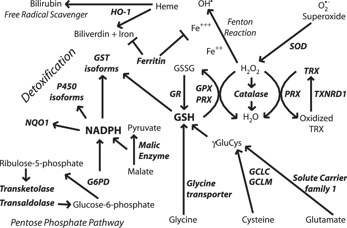
One clear example of the multifaceted nature of Nrf2-driven genes is in detoxifying and limiting production of reactive carbonyls. Reactive carbonyls are known to influence protein function by alkylation and crosslinking, contributing to mitochondrial dysfunction and disrupting the cytoskeleton. Furthermore, they have been implicated in neurodegenerative disease as intermediaries in toxicity [reviewed relating to Alzheimer's disease by Picklo et al. (51)]. Carbonyls are generated as a result of lipid peroxidation, which begins with a free radical attack on polyunsaturated lipids. It is apparent from the gene-expression profiling cited earlier that Nrf2 drives transcription of many genes important in free radical scavenging, especially those involving the critical tripeptide glutathione or production of the free radical scavenger bilirubin. Moreover, glutathione conjugation of 4-hydroxy-2-nonenol (HNE) is enhanced not only by the upregulation of glutathione synthesis and maintenance genes but also by upregulation of the putative HNE-metabolizing enzyme glutathione S-transferase A4 (GSTA4). HNE is one of the major reactive carbonyls produced as a result of lipid peroxidation. Additionally, Nrf2 regulates aldehyde dehydrogenase II (aldh1a1). This enzyme is also known to detoxify HNE as well as malondialdehyde, another highly reactive byproduct of lipid peroxidation.
Several other examples can be found in Fig. 1. In short, Nrf2-driven genes play a major role in homeostasis of glutathione, NADPH, iron, and reactive oxygen species (ROS). In all cases, driving Nrf2 transcription leads to an increase in the overall reducing potential of the cell. This may be at the expense of normal energy production in the case of upregulating the pentose phosphate pathway; however, it is clear that cells that transcriptionally activate Nrf2 will be resistant to oxidative insult by a variety of mechanisms. This multifaceted response makes Nrf2 an attractive target for prevention of neurodegeneration.
Nrf2 Protects from Neurodegenerative Conditions in Laboratory Models
Nrf2 provides antioxidant defense in cell culture and in vivo and is considered to be one of the major regulators of cytoprotective genes. Induction of Nrf2-mediated transcription has been shown to protect against neurotoxicity in a variety of models. In primary neural cultures, Nrf2 activation is known to be neuroprotective against oxidative stressors and mitochondrial toxins, including hydrogen peroxide, tert-butyl hydroperoxide, 6-hydroxydopamine, 3-nitropropionic acid (3-NP), 1-methyl-4-phenylpyridinium (MPP+), and rotenone (7, 23, 38, 58, 59). In all cases, the presence of induced Nrf2 was shown to contribute to neuroprotection, or conversely, Nrf2 deficiency led to increased sensitivity. These observations demonstrate the key point that Nrf2 response is a critical tool used by cells to combat toxic insults. Moreover, activation of Nrf2 before the insult or concurrent with the insult better equips cells to survive in an oxidative environment.
This paradigm follows in vivo as well. In general, acute neurotoxic events lead to Nrf2 activation, and Nrf2 activation has been shown to limit the extent of neurotoxicity resultant from the initial insult. Evidence is presented in the literature from several models of acute or subchronic neurotoxicity such as malonate, 3-nitropropionic acid, kainic acid, 6-hydroxydopamine, 1-methyl-2-phenyl-1,2,3,6-tetrahydropyridine (MPTP), and ischemia/reperfusion injury (6, 24, 32, 56 –58, 60).
Taking mitochondrial complex II inhibitors as an example, we can see that systematic Nrf2 deficiency sensitizes neurons in cell culture and in whole animals to 3-NP toxicity. 3-NP was applied to cortical neurons for 48 h, after which cell death (LDH release) was measured (Fig. 2A). We found that Nrf2 deficiency in primary neurons potentiates 3-NP toxicity. This occurs in whole animals as well. Nrf2-deficient mice are more vulnerable to striatal toxicity resulting from systemic 3-NP administration. Nrf2-deficient mice receiving 3-NP (50 mg/kg every 12 h for seven injections) performed poorly on the rotarod task (Fig. 2C) and lost an average of 12% of their starting weight over the course of 4 days (7). Furthermore, measurable striatal lesions were present in five of the eight Nrf2-deficient mice, whereas no wild-type mice and only one Nrf2 heterozygote exhibited lesions, as measured by Fluorojade-B staining (Fig. 2B and D). This increased sensitivity to complex II inhibition was validated by using intrastriatal malonate injections as well.

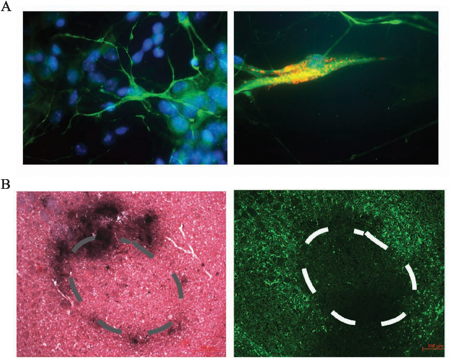
In light of the data supporting the crucial role of Nrf2 in prevention of toxicity, it is reasonable to expect either that Nrf2 is either an inducible response to toxic exposure or that basal Nrf2 activity is responsible for protection. Again we can use mitochondrial complex II inhibitors as an example to clarify this point. With primary cortical neuronal cultures from ARE-hPAP–containing reporter mice, we find that Nrf2-mediated transcription is increased after 3-NP toxicity. By using a histochemical fluorescent stain for hPAP, we can colocalize the reporter exclusively to GFAP-colabeled astrocytes (Fig. 3A), which are the only surviving cells in these cultures. Moreover, after intrastriatal administration of malonate, we can see that the ARE-hPAP reporter is expressed in the penumbra of the lesion. In serial sections, we see that this area contains a large number of GFAP-expressing astrocytes (Fig. 3B). Taken together, these data provide evidence that neuroprotection from basal Nrf2 activity is relatively minimal and that the induced response is likely the reason for protective capacity in this acute model of toxicity.
The mechanism by which Nrf2 protects neurons from toxicity is presently under examination. An early observation describing the Nrf2 protective response in neural conditions comes from cell-culture experiments. When Nrf2-activating chemicals were added to cultures of neurons and astrocytes, the vast majority of Nrf2 activation occurred in the astrocytes (26). The hypothesis that Nrf2 in astrocytes is sufficient to protect neurons from toxicity was subsequently tested directly in primary systems. Shih et al. (59) infected primary astrocyte cultures with Ad-GFP or Ad-Nrf2, after which they were lifted and seeded onto naïve neuronal cultures. Ad-Nrf2 infected astrocyte-protected neuronal cultures from oxidative glutamate toxicity, indicating that indeed Nrf2 activation in astrocytes is sufficient to protect neurons from cytotoxicity.
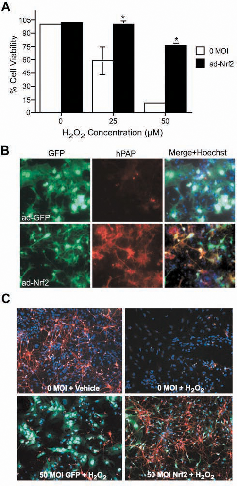
In another experiment, primary mixed neural cultures were prepared from ARE-hPAP reporter mice and infected with either Ad-GFP or Ad-Nrf2 at a multiplicity of infection that was determined preferentially to infect astrocytes. Only cells infected with Ad-Nrf2 were found to exhibit hPAP activity (Fig. 4B). Overexpression of Nrf2 in astrocytes was sufficient to protect neurons from H2O2 toxicity (Fig. 4A and C).
In vivo modulation of Nrf2 before the insult has proven to be an effective way to limit toxicity. One approach to modulating Nrf2 in vivo was to transplant Nrf2-overexpressing astrocytes into striatum and then follow with neurotoxic lesions of either the mitochondrial complex II inhibitor, malonate (7), or the mitochondrial complex I inhibitor, 6-hydroxydopamine (24).
In both cases, the Nrf2-overexpressing cells were able to protect from lesions compared with GFP-overexpressing transplants. Specifically, in the malonate experiments, cortical astrocytes were isolated from P1 pups and infected with either Ad-GFP or Ad-Nrf2. Astrocyte infection rates approached 100%, as visualized by GFP expression, and only those astrocytes infected with Ad-Nrf2 demonstrated hPAP activity (Fig. 5A). Cells were lifted by trypsinization and grafted into striatum at 100,000 cells per hemisphere. Five weeks later, the GFP-expressing cells were found mainly in the vicinity of the injection site. On subsequent malonate lesioning, the Ad-Nrf2–transduced astrocyte grafts provided significant protection to the surrounding neurons (Fig. 5B and C). These experiments verify the model that astrocytic Nrf2 activation is sufficient to protect neurons from cytotoxicity in vivo.

Two other approaches were pioneered by using 3-NP or cerebral ischemia to produce lesions (58, 60). The first was dietary supplementation of tert-butylhydroquinone (tBHQ) to protect from 3-NP lesions. In this study, 1% tBHQ was administered in the diet for 7 days before 3-NP administration. In Nrf2 heterozygous mice, tBHQ supplementation completely protected from lesioning; however, no effect was seen in Nrf2-deficient mice. Dietary supplementation of tBHQ was also found to reduce ischemic lesions in wild-type mice but not Nrf2-deficient mice. The second approach was direct infection of striatal tissue with Nrf2-overexpressing adenovirus. The major infection occurred in GFAP-expressing cells, with some infection of cells expressing the oligodendrocyte marker O4. In this study, Ad-GFP was delivered to one hemisphere, while Ad-Nrf2 was delivered to the contralateral hemisphere. After bilateral lesioning by systemic 3-NP, it was observed that Ad-Nrf2–receiving hemispheres were protected from toxin.
Others have used pharmacologic manipulation of Nrf2 to protect against neurotoxicity as well. Oral administration of 3H-1,2-dithiole-3-thione before toxicant administration partially protected from MPTP lesioning in wild-type mice. In Nrf2-deficient mice, this chemical provided no protection from MPTP (6). Additionally, several studies reported that known ARE activators can protect from neurodegenerative conditions. One recent publication showed that dietary sulforaphane protects from dopaminergic neuron degeneration in α-synuclein–expressing Drosophila (64). Additionally, specific prostaglandin (NEPP) compounds (57) and carnosic acid (56) have been used to protect from ischemia/reperfusion injury in vivo.
Mechanism of Protection
Mounting evidence suggests that neurodegeneration is not simply an event that occurs within neurons, distinct from influence by surrounding cells. This concept may be best illustrated in the context of the familial form of ALS, in which several groups have found major astrocyte and microglial involvement in disease progression. Researchers studying this disease have found that expression of the mutated allele in astrocytes alone is sufficient to cause motor neuron degeneration in vitro (46, 65). Furthermore, knocking down expression of the mutated allele in astrocytes leads to diminished microglial activation and overall disease progression (67). The mechanism by which astrocyte dysfunction leads to neural degeneration in ALS was explored by Barbieto (3). Several potential routes exist by which reactive astrocytes may potentiate neurodegeneration, including downregulation of glutamate transporters, release of reactive nitrogen species, or active release of proapoptotic proteins such as Fas-ligand or NGF. By using this developing model of neurodegenerative processes, we can easily see that targeting nonneuronal cells may be a valuable option for therapeutic intervention, not only to prevent toxicity mediated by glia, but also to stimulate glial protective responses.
Interestingly this hypothesis has been extended and tested in cell culture by using the ALS model with Nrf2 as a protective agent. The therapeutic potential of Nrf2 was explored in a model of ALS in which astrocytes from mice overexpressing the gene coding for superoxide dismutase-1 glycine to alanine at position 93 mutation (G93A-SOD) were isolated. Wild-type motor neurons were plated on top of the astrocytes. It was previously found that this coculture scheme leads to motor neuron apoptosis that is not observed when the astrocytes were derived from wild-type mice (65). The investigators coupled this knowledge to the observation that astrocyte-induced apoptosis in similar systems could be mediated by NGF and nitric oxide (49) and hypothesized that decreasing reactive nitrogen species could rescue the motor neurons. Follow-up experiments showed that activation of Nrf2 in the G93A-SOD–containing astrocytes was able to prevent apoptotic signaling. Moreover, glutathione-mediated nitric oxide detoxification was implicated as the likely mechanism of prevention (65).
It was later found that SOD-containing motor neurons are more sensitive to NGF toxicity via p75 receptor activation, likely because of a reduced Nrf2 expression and activity. In these cultures, Nrf2 activation can prevent NGF toxicity (50). Further linking Nrf2 in the G93A-SOD model, it was observed that in muscle and in spinal cord, the ARE-hPAP reporter is activated in concert with disease progression. This activation localizes to muscle, especially type I muscle fibers and spinal cord astrocytes and motor neurons (33). Currently, the potential for Nrf2 activation for prevention of G93A-SOD mediated motor neuron toxicity is being evaluated in transgenic mice.
It was previously suggested that this astrocyte-mediated protection is largely dependent on glutathione secretion from astrocytes (59, 65). It is becoming clear however, that this is not the only mechanism by which Nrf2 activation in astrocytes might influence neurons. It is likely that the relative importance of glutathione secretion in neuroprotection is context and model dependent.
Studies that probe this interaction have included microarray comparisons by Kraft et al. (31) by using the preferential infection of astrocytes by adenovirus. Primary cultures from ARE-hPAP mice were infected with either Ad-GFP or Ad-DN-Nrf2 virus followed by treatment with tBHQ or vehicle. Transduction was performed at an MOI that selectively infects astrocytes at nearly 100% while virtually no neurons are infected. Based on the astrocyte-specific expression of GFP, fluorescent-activated cell sorting (FACS) of mixed neural cultures was performed by using a FACS Vantage SE cell sorter. Postsort analysis of the isolated cell populations showed that both populations were >95% pure, based on GFP expression. The study was designed to test the differential genetic changes in astrocytes and neurons after tBHQ activation of Nrf2. It was previously known that tBHQ should specifically activate Nrf2 in astrocytes; thus, knockdown of Nrf2 signaling with Ad-DN-Nrf2 was performed in an astrocyte-specific manner as well.
Microarray analysis showed that 97 genes were increased and 37 decreased in the astrocyte (GFP-positive) pool by tBHQ treatment. In the presence of Ad-DN-Nrf2, nearly all of the gene changes were significantly attenuated. Only 17 of the increased genes and none of the decreased genes were not. Interestingly, the genetic changes identified in astrocytes had very little overlap with those identified in neurons. Only four of the increased genes and nine of the decreased genes changed in both cell types. Furthermore, selective Ad-DN-Nrf2 infection of astrocytes attenuated 100% of the mRNA increases in neurons and 79% of the decreases. These data show that after tBHQ treatment, the astrocytic and neuronal genetic response is extremely different. Moreover, the genetic response in neurons is dependent on Nrf2-ARE activation in the astrocyte.
Gene changes in the astrocyte (GFP-positive) pool were similar to those identified in primary astrocyte cultures. They include enzymes associated with glutathione synthesis and conjugation reactions (GSTs) as well as other detoxification genes (Figs. 1 and 6). Additionally, tBHQ induction of Nrf2 led to astrocyte-specific upregulation of genes associated with energy production, including the majority of the 10 genes catalyzing the glycolysis of glucose to pyruvate (Fig. 6).
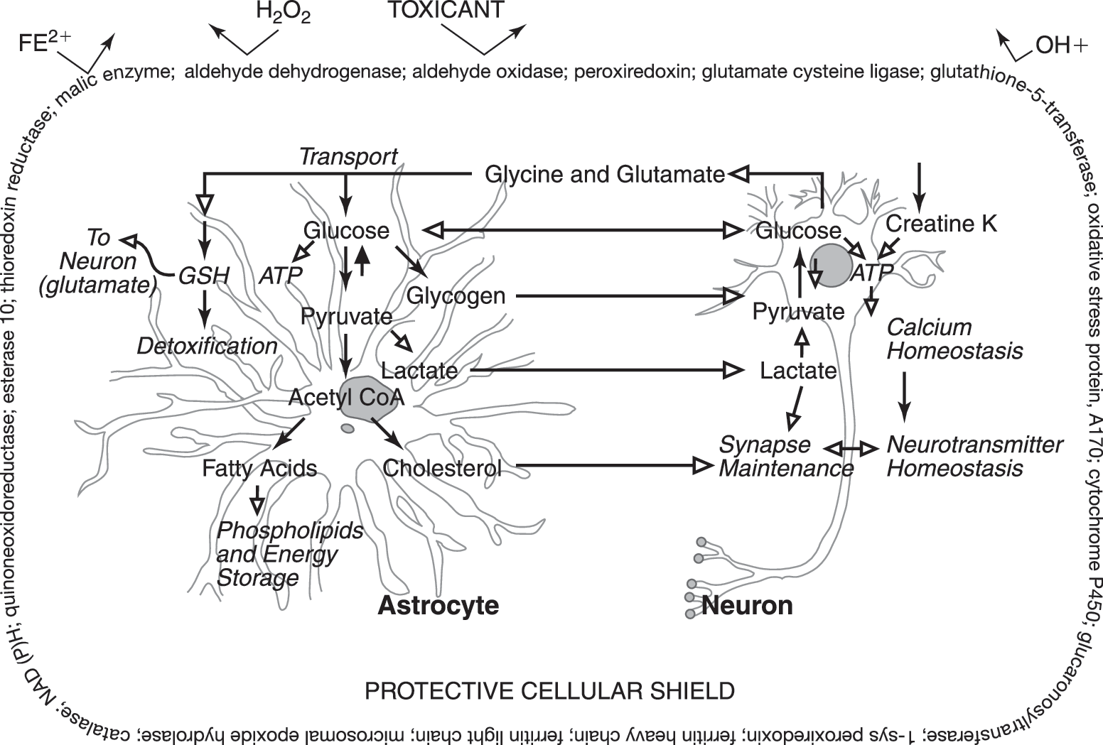
Potential mechanisms by which tBHQ-induced genetic changes in astrocytes and neurons lead to neuroprotection can be identified by using the microarray data. First, glucose conversion to pyruvate is extremely important in brain energetics. ATP resultant from glucose metabolism is essential for calcium homeostasis in the brain, and pyruvate can be further metabolized to lactate, which is the preferred energy substrate for neurons. Lactate also aids in the maintenance of synaptic transmission. Genetic increases leading to improved astrocytic glycogen synthesis (glucan branching enzyme), gluconeogenesis (GPI), and neuronal creatine production (Gatm and creatine kinase) may also indicate an enhanced maintenance of neuronal activity by tBHQ treatment. Furthermore, astrocytic metabolism of pyruvate to acetyl CoA can contribute to cholesterol synthesis (via squalene epoxidase and lanosterol synthase) and production of fatty acids for phospholipids and energy storage (via malic enzyme, fatty acid synthase, Scd1 and 2). It is hypothesized that neurons require glia-derived cholesterol to form efficient synaptic connections. Overall, we can see that induction of Nrf2 in astrocytes leads to genetic changes in astrocytes and neurons that are reflective of healthier, more functional neurons. The mechanism by which these changes occur is not completely understood; however, it is a point of active investigation.
Simply attributing these complex genetic changes to glutathione secretion may underestimate the potential value of Nrf2 as a therapeutic target. New evidence from our laboratory indicates that indeed, mechanisms besides glutathione secretion are involved in astrocyte mediated protection of neurons. In culture models using glutamate-cysteine ligase modifier subunit (GCLm) knockout mice, glutathione is decreased. On induction by tBHQ, glutathione secreted into the medium remains unchanged (unpublished data). Therefore, this model is ideal for testing the hypothesis that glutathione secretion is not essential for neuronal protection in primary cortical cultures. These mice may also provide an excellent tool to test this concept in several models and in vivo as well. In this way, we will be able to delineate the relative importance of glutathione production and secretion on Nrf2-mediated protective pathways.
To identify other potential putative factors that might be responsible for Nrf2-activated astrocyte-mediated protection of neurons, the laboratory has begun to profile secreted factors in astrocyte cultures that are dependent on Nrf2 activation. This proteomic evaluation will yield a list of potential protective mediators. These potential mediators may then be tested independently in culture systems to determine their relative importance in Nrf2-dependent astrocyte-mediated protection of neurons.
Modulating the Nrf2 Pathway
Along with determining the mechanism(s) of protection, another key point in developing a therapeutic strategy will be determining points where the Nrf2 pathway may be influenced. Major progress over the past decade has been made in describing the factors that lead to and influence Nrf2 activation in cell culture and in vivo. One key concept that is emerging is that some of the related cell-signaling mechanisms are highly tissue and cell-type specific, whereas others are common to Nrf2 signaling independent of cell type.
Cell-specific signaling is clearly illustrated in contrasting kinase dependence of Nrf2 activation in hepatocellular carcinoma (HepG2) cells and neuroblastoma (IMR-32) cells. Although phosphorylation of Nrf2 or Keap1 or both appears to be an important upstream regulator of Nrf2 activation, the nature of this phosphorylation is often contradictory, depending on the model used. In IMR-32 cells, PI3K activation is involved in Nrf2 activation. It was shown that constitutively active PI3K activates the ARE, whereas PI3K inhibitors LY 294002 and wortmannin reduce ARE activation and nuclear translocation of Nrf2 after tBHQ treatment in these cells (36). In contrast to this effect, tBHQ-mediated induction of the ARE was found to be independent of the presence of PI3K inhibition in HepG2 cells (73). Additionally, these two publications found that in IMR-32 cells, Nrf2 activation is independent of Erk; however, in HepG2 cells, Erk phosphorylation of Nrf2 is essential for activation. Future work uncovering the cell-specific signaling cascades responsible for priming or activating Nrf2 may be valuable in creating cell-specific targeting of Nrf2 activation. For example, it has been shown that FGF-1 can activate Nrf2 in spinal cord astrocytes, both in culture and in vivo (66). This effect has not been demonstrated in other tissues, and although using FGF-1 to treat neurodegenerative disease may be undesirable, conceptually, it may be representative of potential strategies for Nrf2 induction relating to ALS therapeutics.
The key event in Nrf2 regulation across cell type and tissue type may be the stabilization of Nrf2 protein first described by Stewart et al. (62). Proteasomal degradation of Nrf2 is regulated by the cytoplasmic repressor Keap1 (22). Keap1 is a known repressor of Nrf2 activity (21), and E3 ligase, for Nrf2 (68). On activation of Nrf2, either dissociation of the proteins or disruption of the E3 ligase activity occurs, and Nrf2 is no longer ubiquitinated and targeted for proteasomal degradation. This leads to significant increases in Nrf2 protein as the half-life extends from ∼10 min to ∼2 h. This phenomenon has been observed in a variety of cell types, including HeLa cells (62), renal epithelial cells (1), primary peritoneal macrophages (22), HepG2 cells (4), as well as human neural progenitor cells (39), and represents probably the most important end point for any therapeutic designed to increase Nrf2-mediated transcription.
At several identified points, the cellular mechanisms controlling Nrf2 stability might be activated in a therapeutic context. The most apparent would be small-molecule activation of Nrf2. Primary considerations in developing this type of therapeutic would be toxicity, tissue targeting, or the potential arousal of homeostatic mechanisms that might blunt desired effects. Many chemical inducers of the Nrf2 pathway have been previously described. The initial discovery of the ARE was enabled by use of the chemical inducers, tBHQ, and metabolizable planar aromatic compounds, β-napthoflavone, and 3-methylcholanthrene (54, 55). Soon afterward, other classes of compounds that mediate transcription through the ARE were identified or confirmed. Some include sulforaphane (13), triterpenoids (11), or dietary compounds like phenols, flavonoids, isothiocyanates, organosulfur, indole, and diterpenes (9). Chemical activators of the Nrf2 pathway appear to exhibit similar chemical properties. Most contain Michael acceptor functionalities or phenolic hydroxyl groups (12).
Classic laboratory use of activators of Nrf2 includes small molecules such as tBHQ and sulforaphane. By using the isothiocyanate sulforaphane as an example, we can illustrate the probable innate mechanism of Nrf2 induction by xenobiotics. Sulforaphane is a plant secondary metabolite that is synthesized from glucosinolates after damage to the plant, presumably to protect from microbial infestation. It has demonstrated antimicrobial efficacy against Helicobacter pylori (13, 16, 17) and in another study 23 of 28 tested microbes (25). The chemical is therefore likely exhibiting a hormesis effect by inducing Nrf2 (43). Sulforaphane is a known GST substrate (69) and likely induces its own metabolism via conjugation with glutathione, by direct alkylation of the Keap1 protein and subsequent activation of Nrf2-mediated transcription (10). Therefore, potential toxicity is averted by induction of the Nrf2 response, and meanwhile, the cell is conditioned to overcome additional oxidative insults. It is unclear whether similar hormesis effects are involved in all Nrf2 induction by small molecules. Regardless, it is apparent that small-molecule inducers of Nrf2 may be used in a therapeutic context.
A second approach to achieve Nrf2 activation is to overexpress Nrf2 and thereby overwhelm the cytosolic sequestration by Keap1. This approach requires genetic manipulation of the patient or cells within the patient. Direct viral infection may be used to administer Nrf2-overexpressing constructs. In this instance, viruses may be used that have cell specificity of infection. Alternatively, cells might be infected ex vivo and then grafted into patients, as previously demonstrated in mouse models of acute neurotoxicity (7, 24).
Additionally, the pathway might be influenced by disruption of the Nrf2–Keap1 cytosolic complex. Several methods can be used physically to disrupt this interaction. To this end, the laboratory is investigating the potential of selective Keap1 knockdown by siRNA. Special consideration is being paid to methods of targeting the brain. Additionally, the oncoprotein prothymosin was previously found to bind Keap1 in a yeast–two-hybrid screen (28). Further analysis demonstrated that overexpression of this protein in cells leads to Nrf2-dependant transcription. Recently, the crystallographic structure of the Keap1–prothymosin complex was solved, and it was found that Keap1 binds both prothymosin and Nrf2 with the same domain (48). This observation lends credence to the idea that protein interactions may be viable as drug-targeting mechanisms.
Targeting Specific Disease States with Nrf2
Most neurodegenerative diseases are not diagnosable until after significant toxicity has developed. Exceptions are hereditary conditions or predispositions that may be diagnosed with genetic tests. Treating diseases such as Huntington's disease, Friedreich's ataxia, Down's syndrome, or familial forms of early-onset Alzheimer's or ALS is possible before symptoms develop. However, it is unlikely that complicated therapies would be administered in a purely preventive manner for conditions like sporadic ALS or Parkinson's disease. It is therefore important to address the potential for Nrf2 to provide assistance in regeneration or at least protection from further damage after an initial insult has already occurred.
In summary, Nrf2 has been shown to protect neural systems from a variety of insults in both cell culture and animal models of neurodegeneration. Previous and current data position Nrf2 modulation as a prime candidate for the prevention of oxidative stress and subsequent neurotoxicity. This can potentially be achieved by using a variety of mechanisms, including small-molecule activation of the pathway in the brain. Current data suggest that Nrf2 may be a good drug target in myriad neurodegenerative conditions because of the multifunctional nature of the genes activated and demonstrated efficacy in several disparate laboratory models of neurodegeneration.
Footnotes
Acknowledgments
The work discussed and ongoing studies are supported by NIEHS RO1 ES 10042, ES 08089, Amyotrophic Lateral Sclerosis Association, Packard Center for ALS Research, and Hereditary Disease Foundation.
Abbreviations
3-NP, 3-nitropropionic acid; Ad-DN-Nrf2, adenovirus-overexpressing dominant-negative Nrf2 and GFP; Ad-GFP, adenovirus-overexpressing GFP; Ad-Nrf2, adenovirus-overexpressing Nrf2 and GFP; ALS, amyotrophic lateral sclerosis; ARE, antioxidant response element; HNE, 4-hydroxy-2-nonenol; hPAP, human placental alkaline phosphatase; Keap1, Kelch-like ECH-associated protein 1; MPTP, 1-methyl 4-phenyl-1,2,3,6-tetrahydropyridine; Nrf2, nuclear factor E2-related factor 2; ROS, reactive oxygen species; tBHQ, tert-butyl hydroquinone.