
Abstract
The endothelium is a highly dynamic structure lining the inside of blood vessels that exhibits physical and chemical properties that are critical determinants of overall vascular function. Physically, the endothelium constitutes a semipermeable barrier. Chemically, the endothelium synthesizes numerous factors such as reactive oxygen species (ROS) that can act as autocrine and paracrine signaling molecules. Oxidative stress results when ROS levels increase to levels that cause cellular injury, and, in the endothelium oxidative stress leads to barrier disruption. Endothelial barrier disruption also results from increased cytosolic calcium through store-operated calcium (SOC) entry channels. Although it is known that ROS can interact with and regulate some ion channels, relatively little is known about the interaction of these species with components of endothelial SOC entry channels, the canonical transient receptor potential (TRPC) proteins. Here we review our current understanding of ROS-mediated TRPC channel function and how it affects SOC entry and endothelial barrier disruption. Antioxid. Redox Signal. 15, 1567–1582.
Introduction
In addition to the physical barrier it provides, the endothelium is also a highly dynamic structure capable of producing numerous factors that play fundamental roles in blood vessel function. Reactive oxygen species (ROS), for example, are synthesized by the endothelium and act in a paracrine and autocrine manner as signaling molecules. Increased levels of ROS, however, can lead to oxidative stress and endothelial barrier disruption. ROS-mediated endothelial barrier disruption [reviewed in (10)] has been studied in both the systemic and pulmonary vasculatures. Molecular players important in the mechanisms underlying barrier disruption include the ROS themselves as well as cytoskeletal proteins and proteins involved in cell–cell and cell–matrix adhesions, which are important in determining cell shape and barrier integrity. In endothelium of the blood–brain barrier, for example, nicotinamide adenine dinucleotide phosphate (NADPH) oxidase- or xanthine oxidase-mediated oxidative stress resulted in activation of contractile proteins, downregulation of the tight junction molecule occludin, and endothelial barrier disruption (42). In human lung endothelium, particulate matter derived from airborne pollutants induced ROS generation, which resulted in endothelial barrier disruption via p38 mitogen-activated protein (MAP) kinase-dependent and heat-shock protein-27 (HSP27)-dependent pathways (127). In bovine lung microvascular cells, 4-hydroxy-2-nonenal (HNE), a bioactive aldehyde product formed upon lipid peroxidation [reviewed in (120)], induced mitochondrial-dependent ROS production and depleted cellular glutathione (GSH) (122). Further, HNE caused remodeling of the actin cytoskeleton and endothelial barrier disruption via a mechanism involving extracellular signal-regulated kinase (ERK), c-Jun N-terminal kinase (JNK). and p38 MAP kinases (121).
Disruption of the endothelial barrier is a contributing factor to the development and/or progression of numerous pathological states, including atherosclerosis [reviewed in (98)], ischemia/reperfusion [reviewed in (112)], acute respiratory distress syndrome [reviewed in (128)], high altitude pulmonary edema [reviewed in (90)], and hyperoxia (97). To date mechanisms underlying endothelial barrier disruption are not clearly understood. Indeed increased [Ca2+]i levels, in general, and increased [Ca2+]i levels due to calcium entry through SOC entry channels, more specifically, promote endothelial barrier disruption. Further, ROS can be important mediators of endothelial barrier disruption. However, to date relatively little is known about the relationship between ROS and SOC entry in endothelial cells and further how this relationship affects endothelial barrier integrity. It is therefore the focus of this review to discuss what is currently known about endothelial ROS in the context of SOC entry.
Molecular Makeup of Endothelial SOC Entry Channels
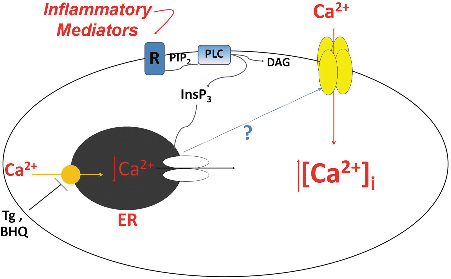
The SOC entry pathway was first described just over 20 years ago by Putney (101). Initially, Putney coined the term “capacitative calcium entry” because he believed that the calcium that entered the cell was directly funneled into the endoplasmic reticulum to refill the calcium store. It was later determined that the calcium that entered the cell first traversed the cytosol before entering the endoplasmic reticulum [reviewed in (102)]. Since then, it has come to be appreciated that in addition to serving the purpose of refilling the calcium store, calcium entry through SOC entry channels contributes to multiple cellular functions that require calcium influx (87). It turns out that not all SOC entry channels and currents are the same. One particular SOC entry current, first described in mast cells (41) and subsequently in Jurkat human leukemic T (147) and rat basophilic leukemia cells (5), is calcium release-activated calcium current (I CRAC). The I CRAC is a highly calcium-selective current that is the prototype SOC entry current and is mediated by the CRAC channel. Endothelial cells also possess a calcium-selective SOC entry current, the I SOC (26, 79, 123). This current is similar, but not identical, to the I CRAC and is likely mediated by a channel different than the CRAC channel. Still, there are other SOC entry currents that are not at all calcium-selective. Since Putney's initial description, SOC entry has been widely and intensely studied in the pursuit to answer two major questions: First, what is the nature of the signal(s) that links store depletion to channel activation? Second, what is the molecular makeup of these channels? To date, the identity of the signal(s) remains unclear. As to the molecular makeup of the channels, only recently has the identity of the CRAC channel been described, whereas still the identity of other SOC entry channels remains unclear.
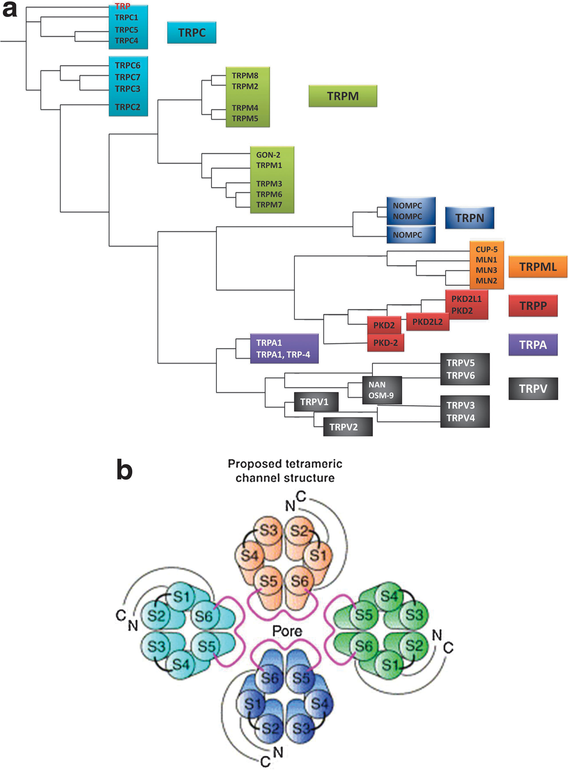
The identity of proteins that make up SOC entry channels has been and still is hotly debated. Currently, they are thought to be made up of mammalian homologs of the transient receptor potential (TRP) protein family and/or orai1. The TRP proteins were the first candidates described for SOC entry channel subunits. Orai1, on the other hand, has just been recently identified and implicated in the molecular makeup of the CRAC channel and possibly other SOC entry channels. TRP proteins were originally identified in the Drosophila melanogaster phototransduction signaling cascade, when it was observed that mutant flies lacking TRP protein expression are blinded by bright, sustained light (19). It was subsequently determined that the TRP protein constitutes a calcium-selective ion channel that, when activated, allows for calcium entry into the cell (36). When expressed in vitro, the Drosophila TRP protein can be activated by store depletion; that is, it acts as a SOC entry channel (124). Homologs of Drosophila TRP proteins have been identified in mammals, Caenorhabditis elegans, and zebrafish, and now TRP proteins constitute a superfamily in which there are seven subfamilies [reviewed in (74, 76)] (Fig. 2a). The mammalian isoforms most closely resembling Drosophila TRP proteins are referred to as the TRPC (canonical) subfamily [reviewed in (91)]. The TRP proteins all exhibit six transmembrane spanning domains (S1–S6), with the pore region residing between S5 and S6, and have cytosolic amino and carboxy termini. Similar to voltage-gated potassium, calcium, and sodium channels, the functional channel is believed to be formed from the coalescence of four subunits (Fig. 2b). The TRPC subfamily members 1, 3, 4, and 5 can exhibit SOC entry channel characteristics, as demonstrated mainly in studies utilizing overexpression systems (33, 65, 66, 95, 96, 126, 129, 134 –136, 142).
In endothelial cells, several studies have looked at the molecular makeup of the endothelial I SOC channel. Two in particular focused on endogenous TRPC proteins and their role I SOC channel function. In the TRPC4 knockout mouse, I SOC in aortic endothelial cells was nearly abolished (29). A second study used small interfering RNA (siRNA) to knockdown TRPC1 and demonstrated a reduced I SOC (11). From these observations, it was determined that the endothelial I SOC channel is comprised of at least TRPC1 and TRPC4 subunits. More specifically, from these data we know that there is at least one TRPC1 subunit and one TRPC4 subunit. However, since the functional channel is likely a tetramer, the question remains as to the identity of the other two subunits. To date, the identity of these other subunits remains to be determined. While there may be more than one TRPC1 or TRPC4 in the functional channel, other TRPC proteins, for example, TRPC3, also represent promising candidates. Another candidate may be orai1, yet the contribution of orai1 to the endothelial I SOC channel structure and function is unknown.
Unlike the TRP proteins, orai1 is a four-transmembrane-spanning domain protein (28). Similar to the TRPs it has cytosolic amino and carboxy termini, and the functional channel is also proposed to be a tetramer. Orai1 was first identified by Feske et al. (28) in 2006 in a study of two patients with severe combined immunodeficiency whose T cells exhibited impaired SOC entry, and orai1 was identified as the mutated protein responsible for the defects in SOC entry. By 2006 another protein involved in SOC entry, stromal interaction molecule (STIM), had also been identified (104) and characterized (138) as a calcium sensor that links store depletion to CRAC channel activation. In their study, Zhang et al. (138) eloquently demonstrated that the endoplasmic reticulum-localized STIM1 translocates to the plasma membrane upon store depletion. They went on to show that when STIM1 translocation can first be detected, I CRAC has also begun to develop, and that the maximum current is observed when the maximal translocation has been achieved. Thus, at this point, both orai1 and STIM1 had been linked to crucial roles in CRAC events. It was not long thereafter that Soboloff et al. (113) drew a connection between orai1 and STIM1. In their study, coexpression of orai1 and STIM1 in human embryonic kidney 293 (HEK293) cells and rat basophilic leukemia cells resulted in greatly enhanced SOC entry and I CRAC, respectively. They went on to propose that orai1 is the actual channel component of SOC entry. Almost at the same time, Prakriya et al. (100) concluded from their study that orai1 is a pore-forming subunit of the CRAC channel. Subsequently, it was determined that orai1 subunits coalesce in a tetramer (69, 92, 141) and further that a tetramer configuration is sufficient to recapitulate the I CRAC in cells stably expressing STIM1 (72).
To date, little is known about the role(s) of orai1 in endothelial SOC entry. Abdullaev et al. (1) examined I CRAC and SOC entry in human umbilical vein endothelial cells (HUVECs). Here they demonstrated that HUVECs exhibit very low amplitude I CRAC, which was abolished by silencing STIM1 or orai1, and which was rescued by ectopic expression of the respective proteins. However, when they inhibited expression of TRPC1 or TRPC4, there was no effect on the I CRAC, collectively suggesting that it is orai1, and not the TRPC proteins, that mediates the endothelial I CRAC. Contrary to these findings, Sundivakkam et al. (115) recently reported that knockdown of orai1 in mouse lung endothelial cells did not prevent calcium entry after thrombin stimulation. However, in mouse lung endothelial cells from TRPC4 knockout mice as well as wild-type cells treated with siRNA to TRPC4, the calcium entry response to thrombin was abolished. Further, in agreement with an earlier study by Brough et al. (11), knockdown of TRPC1 only partially inhibited thrombin-induced calcium entry. Collectively, these results suggest that TRPC4 and TRPC1 play a role in endothelial SOC entry, whereas orai1 does not. Clearly then, a discord exists. On one hand, orai1 appears to play a critical role in endothelial SOC entry; on the other hand, TRPC4 and TRPC1 are the key players. This then leads to the question of why such controversy exists.
Controversy in the SOC entry field is nothing new. Indeed, in the 20 plus years since Putney's initial observation, this field has been hotly debated not only the identity of the channels but also the identity of the signal(s) linking store depletion to channel activation. In addressing the question of which proteins make up SOC entry channels, one must consider that there are a multitude of factors involved in SOC entry as well as in studies thereof. To begin, the proteins that play a role in SOC entry are not just the proteins that make up the pore-forming channel. Indeed, there are likely multiple, very specific protein–protein interactions between channel proteins, regulatory proteins, and even proteins in the endoplasmic reticulum that form an intricate complex that is responsible for overall channel behavior and current properties. However, to date, the molecular players involved in these complexes are only partially resolved. A second factor contributing to the controversy is that there is very likely cell-type specificity in which the molecular makeup of channel complexes varies from one cell type to another. Yet, disparate results have been observed even when comparing studies using the same cell type. This leads to a third confounding factor, the use of overexpression systems. To date, the majority of studies focused on identifying contribution of either TRPC proteins or orai1 to SOC entry have relied on protein expression systems, in which only one type of subunit is overexpressed. However, at least in the case of the TRPC proteins, as they can form either homo- or heteromultimers, one may expect that upon overexpression of a single subunit, the formation of the homomultimer, or a heteromultimer with a different subunit stoichiometry, will be favored. Thus, the channel formed in the overexpression system may or may not faithfully recapitulate the native state. Taking into account these factors, and likely other factors as well, it is not surprising that controversy exists.
To date, the roles of TRPC and orai1 proteins in SOC entry are still being debated. However, should the contributions of these proteins to SOC entry be mutually exclusive? Perhaps not. Indeed, several investigators have already demonstrated a functional synergy between TRPC proteins and orai1. Liao et al. (62 –64) showed that orai1 structurally and functionally interacts with TRPC proteins to mediate SOC entry in HEK cells stably expressing TRPC proteins. Ong et al. (86) examined the interaction of orai1 and TRPC1 in human salivary gland cells and dispersed mouse submandibular gland cells. They observed a dynamic interaction between TRPC1, orai1, and STIM1 that was increased after store depletion, and at least in the human salivary gland cells was important for the activation of SOC entry. Cheng et al. (15) also demonstrated functional interaction of orai1 with TRPC1 and STIM1. In human platelets, Jardin et al. (46) observed increased coimmunoprecipitation between TRPC1 and orai1 upon store depletion. In the presence of an orai1 antibody, SOC entry was decreased and interaction between TRPC1 and STIM1 was interrupted leading the authors to conclude that orai1 functions to link STIM1 to TRPC1. In endothelial cells, interaction of TRPC1 and/or TRPC4 with orai1 has not yet been examined. Indeed, it may be that functional interaction between TRPC proteins and orai1 is a critical determinant of endothelial SOC entry activation and barrier disruption.
SOC Entry Channels and Endothelial Barrier Disruption
The observation that calcium entry through SOC entry channels plays a key role in endothelial barrier disruption was first made in the pulmonary circulation (16, 49). In these and other studies, the plant alkaloid thapsigargin was, and still is, used to activate SOC entry. Thapsigargin is a Ca2+/ATPase inhibitor that causes the endoplasmic reticulum calcium store to become depleted (118, 124). After store depletion, the signal will be initiated to activate SOC entry channels. In pulmonary artery endothelial cells (PAECs), thapsigargin application activates multiple SOC entry channels, including the calcium-selective I SOC channel. PAEC monolayers treated with thapsigargin exhibit disruption of cell–cell and cell–matrix adhesions, an increase in myosin light chain phosphorylation and formation of actin-based stress fibers, leading to formation of interendothelial cell gaps and increased permeability (49, 78, 79) (Fig. 3). In situ, lung permeability increased in a dose-dependent manner after thapsigargin application, with the principal site of fluid leak from large, extra-alveolar vessels (16).

As described above, we know that TRPC1 and TRPC4 contribute subunits to the functional I SOC channel. Importantly, in the TRPC4 knockout mouse thrombin-induced increases in lung permeability were attenuated (119), whereas permeability was increased in TRPC1-overexpressing human dermal microvascular endothelium (88). These observations reflect the critical roles of TRPC1 and TRPC4 and the I SOC channel in endothelial barrier disruption.
Endothelial ROS
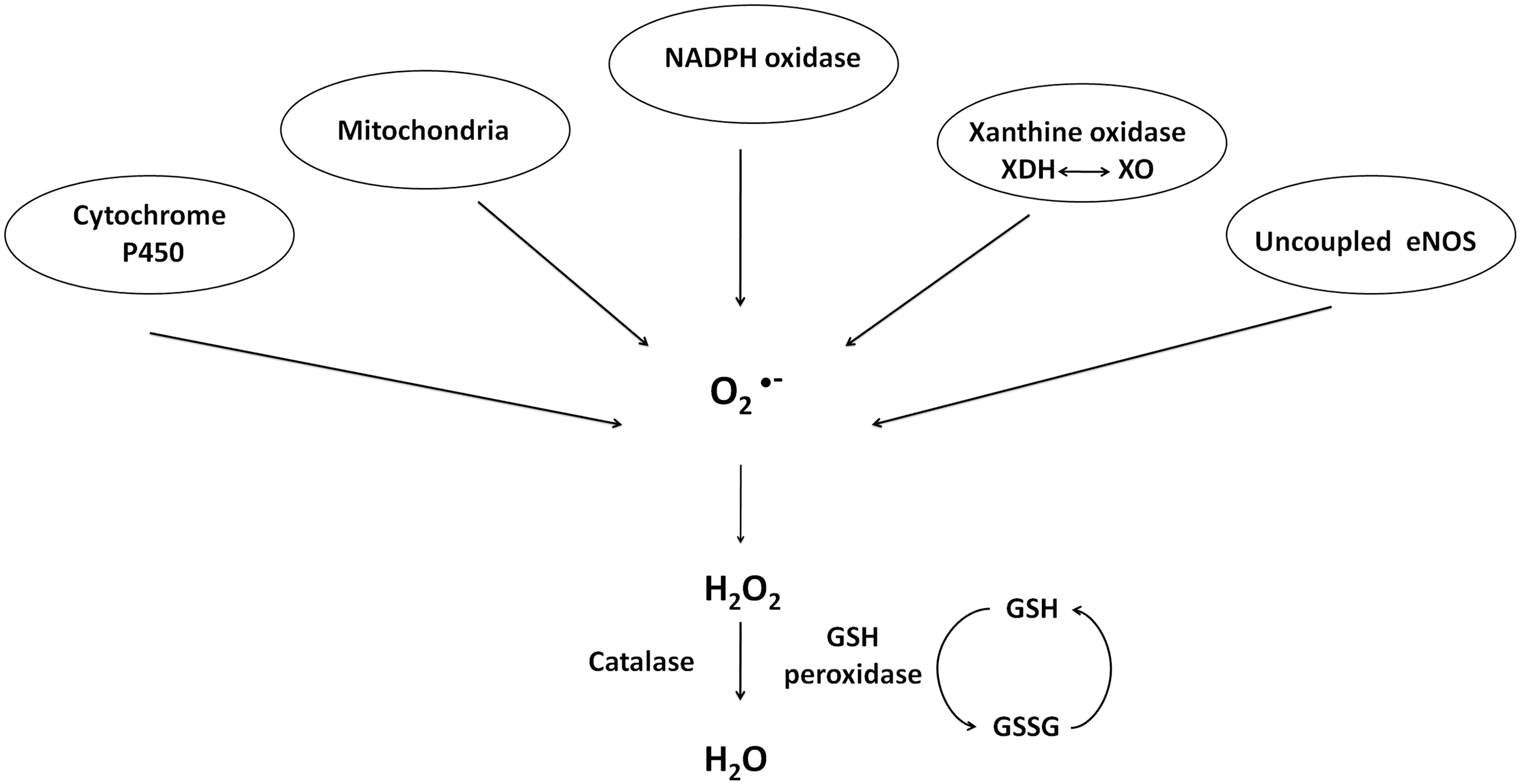
Chemical and biochemical reactions that lead to generation of ROS are complex and numerous, and have been reviewed in detail elsewhere (68, 85). ROS and reactive nitrogen species (RNS) can act in the cell via direct protein oxidation and nitration, respectively, or can be short-lived signaling molecules in specific pathways [reviewed in (73)]. Reactive molecular species include superoxide (
Endothelium synthesizes and responds to ROS. In the 1980s to early and mid-1990s, it was of keen interest to identify specific ROS generated and the generating source. In general, endothelium generates
Xanthine oxidase
Xanthine oxidase is one form of the enzyme xanthine oxidoreductase. The mammalian xanthine oxidoreductase exists in two interconvertable forms, xanthine dehydrogenase and xanthine oxidase [reviewed in (37)]. While either form can reduce molecular oxygen to form
Zweier et al. (145) demonstrated the generation of
NAD(P)H oxidase
Whereas xanthine oxidoreductase function appears to require activation, such as by exposure to anoxia and reoxygenation, nicotinamide adenine dinucleotide (NADH) oxidoreductase does not require exogenous stimulation to produce ROS (75). In bovine coronary artery endothelium, NADH oxidoreductase was shown to be the major synthesizer of
The NADPH oxidase system expressed in endothelial cells is very similar to that of neutrophils. Bayraktutan et al. (8) revealed that in rat coronary microvascular endothelial cells, the heterodimer cytochrome b558, with subunits p22-phox and gp91-phox, which is the principal component of the phagocyte NADH/NADPH oxidase complex, is expressed at both the message and protein levels and is active. While the cDNA isolated in this study exhibited high homology to the phagocyte, and vascular smooth muscle cell, sequences, the authors noted that there are some critical biochemical differences between the endothelial and phagocyte NADPH oxidase systems, including substrate specificity and activation properties. The NADPH oxidase inhibitor diphenyleneiodonium (DPI) was “more effective in inhibiting NADPH- than NADH- evoked activity.” Since DPI can also inhibit other oxidant producing flavoproteins such as xanthine oxidase and NO synthase, specific inhibitors of these compounds were tested and found to have no effect. Thus overall, it was concluded that in coronary microvascular endothelial cells NAD(P)H oxidase is a functional ROS-generating species. Further, whereas the phagocyte system is only functional after specific triggers in initiation of nonspecific host defense pathways, the endothelial system is always active, although to a lesser degree, such that there is continuous production of
Following up on these early studies, Bayraktutan et al. (7) proceeded to determine sequence differences and hence likely structural differences between the neutrophil NADPH oxidase system components p22-phox and gp91-phox and the analogous endothelial NAD(P)H proteins. The p22-phox sequence was observed to exhibit >90% homology between the predicted sequence of rat coronary microvascular endothelial cell protein with neutrophil sequences from pig, mouse, human, and cow. Likewise the endothelial gp91-phox also exhibited a high degree of homology (>90%) with neutrophils of other species. However, two differences were noted. First, in the NADPH binding domain C, a serine to alanine mutation exists at residue 416, which the authors suggested might lead to changes in enzyme activity and substrate specificity, as compared to the neutrophil system. The second difference noted was in potential glycosylation sites. The consensus motif N-X-(S/T), with X being any amino acid residue, occurs four or five times in the phagocyte sequences of murine, human, and bovine, but only three times in the predicted rat endothelial sequence. The authors speculated that the cellular localization of the p22-phox/gp91-phox complex in endothelial cells is influenced by the glycosylation patterns. Indeed, immunofluorescence revealed that endothelial p22-phox and gp91-phox were mainly localized to the perinuclear region with a “more diffuse reticular staining extending toward the cell membrane.” This is in contrast to the phagocyte NADPH oxidase, where in the active neutrophil system, p22-phox and gp91-phox are associated with the plasma membrane [(47, 132) and reviewed in (58)]. Li et al. (60) pursued the observation that the cytochrome b558 subunits, p22-phox and gp91-phox, appear to be localized intracellularly in unstimulated endothelial cells. In their study, the authors used a combination of techniques, including coimmunoprecipitation, subcellular fractionation, and confocal microscopy, to determine the localization of p22-phox and gp91-phox as well as the cytosolic subunits p47-phox, p67-phox, and p40-phox. From their observations they determined that in the resting state these subunits interact in a preformed cytosolic complex that is associated with the cytoskeletal microtubule system. Importantly, this preformed complex is functional and thus capable of generating ROS.
Collectively, from these studies of endothelial xanthine oxidase and NAD(P)H oxidase, it became clear that endothelium does indeed generate
ROS Regulation of TRPC Proteins in Endothelium
It is well established that ROS can lead to increased [Ca2+]i due to calcium release from intracellular stores as well as calcium entry across the plasma membrane [reviewed in (21)]. Endothelial cells are generally considered to be nonexcitable cells [reviewed in (84)], although pulmonary microvascular endothelial cells express a functional voltage-gated T-type calcium channel (140). Without voltage-gated channels, the principal calcium entry pathways in endothelial cells are through SOC and ROC entry. As TRPC proteins are believed to underlie the molecular makeup of many ROC entry channels and at least some SOC entry channels, it is logical to question whether TRPC proteins are sensitive to regulation by ROS. To date, TRPC7 (currently known as transient receptor potential melastatin 2 [TRPM2]) and TRPC3/TRPC4 have been shown to be regulated by ROS in endothelium.
Transient receptor potential melastatin 2
TRPM2, of the melastatin subfamily, was originally named TRPC7 or long transient receptor potential canonical 2 (LTRPC2) (77). Although no longer considered a member of the TRPC subfamily, as much more is known about the mechanism underlying oxidant-induced channel activation for this channel as compared to other TRPC channels, we felt it appropriate to include it in this section. The TRPM subfamily only exhibits limited homology to other TRP proteins, and functional channels comprised of members of this subfamily are believed to be homotetramers (81).
Early work by two groups, Sano et al. (107) and Perraud et al. (93), examined activation of LTRPC2 by intracellular pyrimidine nucleotides based on the fact that LTRPC2 possesses a Nudix motif. The term “Nudix” was originally coined by Bessman et al. (9) to describe a motif found in proteins that is important in the hydrolysis of a nucleoside diphosphate linked to some other moiety, X (i.e., Nudix). The Nudix motif was originally described in the MutT protein of Escherichia coli, and as such was also referred to as a MutT motif, and later was also found in the MutX protein of Streptococcus pneumonia; proteins bearing the Nudix motif contain a conserved signature sequence that imparts nucleoside-triphosphate pyrophosphohydrolase activity [reviewed in (9)]. Perraud et al. (93) observed that in LTRPC2-expressing HEK293 cells, 100 μM adenosine 5′-diphosphoribose (ADPR) activated LTRPC2, but the same concentration of other closely related compounds, including NAD, NAD+, cyclic ADPR (cADPR), ATP, or ADP, did not (Fig. 5). Similarly, Sano et al. (107) showed that LTRPC2 expressed in HEK293 cells was activated in a dose-dependent manner by ADPR, as well as by 1 mM β-NAD. Using an inside-out excised patch configuration, both groups further determined that these intracellular pyrimidine nucleotides act directly, without the aid of cytoplasmic or membrane components, to activate LTRPC2 yielding a nonselective cation current permeable to calcium.

Subsequent studies used H2O2 to induce oxidant stress. Wehage et al. (130) reasoned that under oxidant stress NAD is elevated, and NAD is metabolized to ADPR, a known activator of LTRPC2. In their patch-clamp studies, 0.3 mM ADPR in the patch pipette or 5 mM H2O2 in the bath were able to activate a cation current in LTRPC2 expressing HEK293 cells. Unlike the results of Sano et al. (107), however, 1 mM NAD was not able to activate a current, which the authors attribute to potential experimental differences. An experiment in nature then led to more mechanistic insight into channel activation. Neutrophil granulocytes express a LTRPC2 deletion mutant (Δ1292-1325). As this mutant was activated by H2O2 but not by ADPR, the authors concluded that the mechanism of ADPR-induced LTRPC2 activation is independent of H2O2. In the same year, Hara et al. (35) performed an in depth study in which calcium entry was measured, using the Fura/2 approach, in LTRPC2-expressing HEK293 cells treated with H2O2. In support of a role for the MutT or Nudix motif, upon deletion of this sequence no calcium entry was detected. Further, the idea that channel activation is mediated by direct interaction between β-NAD+ and LTRPC2 was confirmed again using inside-out patches. In a whole-cell configuration, H2O2 application induced a unitary LTRPC2 current; however, when a patch was isolated in the inside-out configuration, the current disappeared and only was restored by β-NAD+ (not H2O2). Further, 32P-labeled β-NAD+ strongly bound to a glutathione S-transferase (GST)—LTRPC2 carboxy terminus fusion protein.
If H2O2-mediated activation of LTRPC2 requires conversion of H2O2 to NAD+ or ADPR, then OH• that is upstream of ADPR formation (Fig. 5) should also be able to activate the current. Along this train of thought, Ishii et al. (44) demonstrated the ability of intracellularly generated OH• to activate LTRPC2 (by now more commonly referred to as TRPM2) in both TRPM2-expressing HEK293 cells and in the rat β-cell line RIN-5F. Kolisek et al. (53) studied the cADPR part of the pathway and demonstrated that at concentrations >100 μM, cADPR was sufficient to gate TRPM2, but at lower concentrations of 10 μM, it was only able to act in an ancillary way to potentiate ADPR channel gating. In a study by Buelow et al. (12) it was demonstrated that the enzyme responsible for converting NAD+ to pADPR, poly(ADP-ribose) polymerase (PARP) isoform 1 was critical for TRPM2 currents and Ca2+ entry in DT40 B cells. From these studies it is clear that H2O2 and many of its metabolites are capable of activating TRPM2. However, the question of which species directly interact with and gate the channel and which species act indirectly as second messengers persists. Indeed, in a recent study, Naziroğlu and Luckhoff (81) measured single-channel conductances in TRPM2-expressing Chinese hamster ovary cells. Here they added H2O2 to the bath solution, or ADPR or NAD+ in the patch pipette. Using any of the three agonists in the whole-cell patch mode they observed a nonselective current. H2O2 in the bath led to current activation typically after a 2–5 min delay and wash-out did not terminate channel activity, suggesting that the H2O2-induced current did not rely on generation of a soluble factor that could be washed away in the inside-out patch configuration. However, the authors recognized that since the channels were first activated by H2O2 in the whole-cell configuration, it could be that H2O2 is not directly acting on the channel but is initiating a cascade of events that culminate in channel activation. To determine whether this was the case, they began in the cell-attached mode and treated cells with H2O2 for several minutes after which the inside-out patch configuration was established, and they observed that H2O2 activated the channels. However, in inside-out patches from cells that had never been exposed to H2O2, no current was detected until addition of ADPR. They thus concluded that both H2O2 and ADPR are sufficient to activate TRPM2, but that they act independently in their gating mechanisms. This is in support of observations of Wehage et al. (130) and Kraft et al. (56) where a carboxy terminal-truncated TRPM2 maintained H2O2 gating but lost ADPR gating. Finally, Perraud et al. (94) proposed that oxidative/nitrosative stress activates a biochemical pathway within the mitochondria, which results in the production and release of ADPR from the mitochondria, after which ADPR acts as diffusible second messenger to activate TRPM2.
Even though there currently are still some uncertainties as to which compounds act directly and which act indirectly to gate the TRPM2 channel, it is clear that TRPM2 does indeed act as a cellular oxidant sensor. What then are the downstream physiological effects of channel activation? Hara et al. (35) determined that activation of LTRPC2 leads to increased susceptibility to cell death and reduced cell survival. More recently, Hecquet et al. (38) described a role for TRPM2 in endothelial permeability. In this study TRPM2 was activated in human PAECs by exposure to H2O2 or an ADPR analog (3-deaza-cADP-ribose). Studies were performed to demonstrate activation of calcium entry by H2O2 and to determine whether this agonist causes release of calcium from intracellular stores. They observed that at H2O2 concentrations <300 μM, calcium was not released from intracellular stores, indicating that the calcium entry that occurred was not through SOC entry channels. Endothelial barrier integrity was monitored by measuring transendothelial electrical resistance. With H2O2 they observed a dose-dependent decrease in resistance with the maximal effect achieved at 300 μM H2O2. Both TRPM2 siRNA and TRPM2 blocking antibody inhibited H2O2-induced calcium entry and also reduced the decrease in transendothelial resistance.
TRPC3/4 channel
Early observations by Balzer et al. (6) revealed that in the presence of oxidants, channels comprised, at least in part, of TRPC3 were activated and contributed to endothelial cell depolarization. Groschner et al. (34) pursued the idea that TRPC3 activation was somehow sensitive to, or regulated by, oxidant stress. They proceeded to describe a mechanism whereby TRPC3 channel function, likely in combination with TRPC4, is activated following “oxidant-induced disruption of cholesterol-rich lipid rafts.” The proposed mechanism provided insight into how TRPC channel function might be intimately coupled to cellular redox status. To test their hypothesis, oxidative stress was induced in TRPC3-expressing HEK293 cells by treatment with tert-butylhydroperoxide or cholesterol oxidase. Caveolin1, which is a marker of caveolae, moved from the membrane into the cytosol, which was interpreted as disruption of caveolae. TRPC3, on the other hand, remained localized to the membrane and importantly was activated upon oxidative stress. As TRPC3 activation is regulated by phospholipase C (PLC), and activated PLC hydrolyzes membrane-bound PIP2, the authors next looked at membrane localization of PIP2. Using a PIP2-binding GFP fusion protein (GFP-PH-PLCδ) they observed that after tert-butylhydroperoxide application, GFP-PH-PLCδ lost its membrane association; however, they did not see the same effect after cholesterol oxidase treatment, leading them to conclude that the mechanism underlying oxidative stress-mediated TRPC3 activation does not involve PIP2 hydrolysis. The authors proposed a potential mechanism of cross-talk between TRPC channels and signaling molecules that reside in caveolae (or lipid rafts) and further speculated that membrane cholesterol oxidation might actually be the signaling molecule that activates TRPC3.
The observation that oxidative stress leads to TRPC3 activation leads to two questions. First, as TRPC channels are most likely tetramers, the question arises as to whether this particular channel is a TRPC3 homomer or heteromer, and if it is a heteromer, what are the identities of the other subunits? Second is the question of the mechanism underlying redox regulation of the TRPC3 containing channel. Poteser et al. (99) resolved, in part, the first question. As in their earlier study they demonstrated that TRPC3-expressing HEK293 cells possessed a redox-sensitive current activated by treatment with tert-butylhydroperoxide or cholesterol oxidase. The current–voltage plot revealed a linear relation with reversal potentials <0 mV, suggesting activation of a nonselective cation current. Additionally, native porcine aortic endothelial cells exhibited a similar current. While the aortic endothelial cells were shown to express several TRPC homologs, including TRPC1, TRPC3, TRPC4, and TRPC6, only TRPC3 and TRPC4 were resolved at the plasma membrane; further through immunoprecipitation and Förster Resonance Energy Transfer experiments, it was shown that TRPC4 interacts with TRPC3. The authors thus concluded that TRPC3 and TRPC4 contribute subunits to the redox-sensitive channel. It is still unclear as to the identity of the other two subunits. It could be any combination of TRPC3 and TRPC4, or it may be that another TRPC homolog is present. In the current study, even though TRPC1 was resolved in aortic endothelial cells and it appeared to interact, though weakly, with TRPC3, it was not detected at the plasma membrane. However, the authors did not rule out a contribution from TRPC1, as they recognized that Strübing et al. (114) had previously identified a channel complex comprised of TRPC1 plus TRPC4 or TPRC5 plus TRPC3 or TRPC6 in mammalian embryonic brain. Nonetheless, to date, the subunit stoichiometry of the endothelial TRPC3/TRPC4 redox-sensitive channel remains unresolved. Further the mechanism by which oxidants lead to channel activation and the specific downstream function(s) of this channel have not yet been explored.
Redox Regulation of SOC Entry in Endothelial Cells
Activation of SOC entry, in general, and I SOC in particular, is an important step leading to interendothelial cell gap formation and barrier disruption. ROS can also contribute to mechanisms underlying endothelial barrier disruption [reviewed in (10)]. In this section we review studies that specifically examined redox regulation of SOC entry in endothelial cells.
Elliott et al. (23) performed early experiments looking at the effect of oxidant stress on agonist-induced calcium entry in vascular endothelial cells. In this study, bovine aortic endothelial cells were subjected to oxidative stress using tert-butylhydroperoxide and then were stimulated with the agonist bradykinin. Earlier studies (18, 111) had already established that bradykinin stimulates calcium release from the endoplasmic reticulum followed by entry across the plasma membrane, that is, SOC entry. Using 0.4 mM tert-butylhydroperoxide, the authors observed, using the fluorescent calcium indicator Fura/2, that after 30 min incubation the bradykinin-induced calcium entry was inhibited by over 50%. Importantly, they also tested for effects of tert-butylhydroperoxide on calcium release from the endoplasmic reticulum and observed no change in release after 30 min incubation, overall indicating that the tert-butylhydroperoxide-induced oxidant stress inhibits calcium entry in bovine aortic endothelial cells. These observations were supported in a follow-up study (25) whereby uptake of 45Ca2+ was measured in calf PAECs treated with 0.4 mM tert-butylhydroperoxide followed by exposure to bradykinin. In line with results from the previous study, after tert-butylhydroperoxide treatment, uptake of 45Ca2+ into the cells was decreased. It was not until 1993 that Elliott and Doan (22) specifically focused on the effect of oxidant stress on SOC entry in endothelial cells. Here they used the Ca2+/ATPase inhibitors thapsigargin or 2,5-di-t-butylhydroquinone (BHQ) to directly activate SOC entry. Application of either thapsigargin or BHQ after 30-min and 1-h incubation with 0.4 mM tert-butylhydroperoxide resulted in reduced calcium entry with no changes in the basal calcium levels or calcium release. As these results mirror those obtained using the Gq agonist bradykinin, the authors concluded that the mechanism underlying oxidant-mediated inhibition of calcium entry does not involve decreased synthesis or increased metabolism of InsP3. Instead they proposed that it is likely that the oxidants directly inhibit the calcium entry channels located on the plasma membrane or interfere with the link between the calcium entry channels and the calcium store fill state.
Wesson and Elliott (131) expanded these early studies by exogenously adding xanthine oxidase in the presence of its substrate hypoxanthine to cultured calf PAECs. Xanthine oxidase caused increased basal calcium levels and inhibited bradykinin-induced calcium release from the endoplasmic reticulum as well as subsequent entry. However, when the substrate concentration was decreased, they observed that xanthine oxidase specifically inhibited calcium entry without decreasing calcium release. To determine the molecular species underlying this inhibitory effect on calcium signaling, they used superoxide dismutase (SOD) and catalase as inhibitors of
In two follow-up studies, Koliwad et al. (54, 55) identified a potential oxidation target. It was known from an earlier study (24) that cytosolic sodium increased after tert-butylhydroperoxide treatment of endothelial cells. In the first study (55) it was determined that treatment of calf PAECs with tert-butylhydroperoxide led to activation of a current that was equally selective for sodium and potassium. Interestingly, the channel underlying this current also permits calcium entry, which the authors suggested could explain the increased basal calcium after tert-butylhydroperoxide exposure. Second, they observed membrane depolarization, likely due to activation of this nonselective cation channel. As SOC entry is inhibited by membrane depolarization (20, 110), the authors proposed that the mechanism underlying tert-butylhydroperoxide-induced inhibition of SOC entry is due to opening of the nonselective cation channel leading to membrane depolarization. In the second study patch-clamp electrophysiology was performed to identify the molecular species important for activating this nonselective channel. Based on the earlier study (39) that identified GSSG as a critical mediator of SOC entry inhibition, Koliwad et al. (54) examined whether GSSG could activate the nonselective current. Using excised membrane patches, internal but not external GSSG did indeed activate this current. Additionally, internal tert-butylhydroperoxide and reduced GSH were unable to activate the current leading the authors to conclude that under oxidant stress endogenous GSSG mediates opening of a nonselective cation channel activation and membrane depolarization. While GSSG washout did not reverse channel activity, the addition of GSH, to increase the [GSH]:[GSSG] ratio, or dithiothreitol did result in reversal. These observations further suggested that GSSG acts via covalent sulfhydryl oxidation or at least binds with high affinity to some internal receptor.
While these early studies revealed that oxidative stress, when induced by 0.4 mM tert-butylhydroperoxide, leads to inhibition of SOC entry, it appears that not all oxidative species act in the same manner. Favre et al. (27) were interested in the effects of NO and S-nitrosylation on SOC entry. Using membrane-permeant NO donors they demonstrated that calcium entry was activated by NO and that calcium store depletion augments the entry response. Calcium entry was also triggered by alkylating agents that react with thiol groups, to mimic S-nitrosylation, and this entry was also enhanced by store depletion. From these observations, the authors concluded that channel S-nitrosylation activates calcium entry that is also activated by store depletion. In a follow-up study, Ma et al. (67) probed the relationship between S-nitrosylation and SOC entry. Here they examined differences between SOC entry activated by store depletion and calcium entry activated by an NO donor. In an ion selectivity experiment, they observed that with barium as a charge carrier, there were differences in cation entry between the two groups. Further, there were also small differences in sensitivity to lanthanum when used to block entry. Collectively, these results suggested that the NO-mediated calcium entry mechanism was distinct for store-mediated calcium entry. In the next study, van Rossum et al. (125) compared calcium entry mediated by store depletion, that is, SOC entry, with that induced by S-nitrosylation and observed that calcium entry induced by an NO donor was insensitive to the InP3 receptor blocker 2-aminoethoxydiphenyl borate (2-APB), thus reinforcing the earlier results indicating that NO-mediated entry is via a mechanism distinct from SOC entry. More recently, Yoshida et al. (137) described activation of TRP proteins, including TRPC1, TRPC4, and TRPC5, by NO. They went on to demonstrate that in TRPC5, two cysteine residues, Cys553 and Cys558, are S-nitrosylated by NO. Interestingly, and perhaps importantly, the other TRP proteins that are activated by NO also have conserved cysteine residues, leading to the idea that the other TRP homologs are also S-nitrosylated. While still unknown at this time, it is interesting to consider whether S-nitrosylation of TRPC1 and TRPC4, when configured in the I SOC channel arrangement, leads to channel activation.
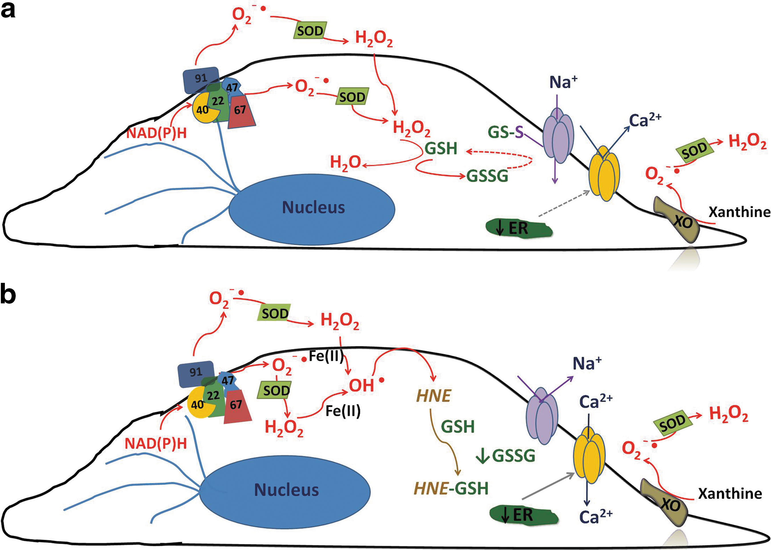
It is clear that endothelial SOC entry is sensitive to redox regulation, at least in part through an indirect mechanism in which a nonselective cation channel is activated by GSSG oxidation leading to membrane depolarization and inhibition of SOC entry. What is less clear is whether there is also direct redox regulation of SOC entry channels and especially whether the endothelial I
SOC channel is directly or indirectly redox regulated. As activation of the endothelial I
SOC is an important step leading to endothelial barrier disruption (133), and conditions of oxidative stress have been shown to lead to endothelial barrier disruption [reviewed in (10)], it is important for future studies to determine whether I
SOC function is regulated by cellular redox status as well. Based on our current understanding of the field, we propose a model describing how I
SOC-induced endothelial barrier disruption can be influenced by cellular redox status. To begin, in an unstimulated state, NAD(P)H oxidase is localized within the cytosol in a perinuclear manner (7). As it is constitutively active (75),
Summary
Activation of the endothelial I SOC channel is an important step leading to intercellular gap formation and endothelial barrier disruption. Likewise, oxidative stress can trigger endothelial barrier disruption. To date it is unclear whether there is a link between I SOC channel function and oxidative stress. Indeed, it is conceivable that the I SOC channel is indirectly regulated by cellular redox status through a nonselective cation channel that is covalently modified by GSSG. Alternatively, the I SOC channel may itself be a direct target of GSSG or some other ROS species. Along this line, we searched for the Nudix motif in TRPC1 and TRPC4, two subunits that contribute to the molecular makeup of the endothelial I SOC channel, but did not find one (unpublished data). However, this does not rule out direct oxidative modification by other species such as RNS, in a manner similar to what has been observed for NO modification of TRPC5. Overall, we have much to learn about redox regulation of SOC entry channels, in general, and the endothelial I SOC channel, in particular. Indeed, we need to better understand the relationship between cellular redox status and I SOC channel function to more fully appreciate the downstream physiological and pathophysiological effects.
Footnotes
Acknowledgments
The author would like to thank Ms. Sidra Rasool for her help in preparing figures for this article. Funding is provided by NIH 5R00HL089361-03.