
Abstract
The integrity and function of many vertebrate organs depend on cellular barriers that are mainly formed by intercellular protein complexes of the plasma membrane. These cell–cell contacts, tight junctions (TJs), exhibit the most apical localization in the lateral membrane; they regulate the permeability of the paracellular space between opposing epithelial and endothelial cells. This Forum reviews the currently available data on the influence of oxidative stress and the effects of antioxidative mechanisms on TJ proteins and on tissue barrier functions inseparably linked to these proteins. The contributions are focused on the most important transmembranal and membrane-associated TJ proteins and on tissue barriers characterized by predominant involvement of the TJs, and alterations at the molecular and functional levels induced by redox signaling are also discussed. This Forum demonstrates that cell barriers are highly sensitive to oxidative stress but also respond to antioxidative intervention. However, our knowledge of the molecular basis of the specific mechanisms responsible for functional disturbances remains limited and needs further investigations. Antioxid. Redox Signal. 15, 1163–1166.
The early morphological characterization of the TJ has been extensively underpinned by its molecular analysis during the last two decades (Fig. 1). Zonula occludens (ZO)-1, a membrane-associated protein with a scaffolding function in TJ, was identified in 1988 (3), followed by ZO-2 (17) and ZO-3 (18). Occludin, a member of the marvel protein family, was the first transmembranal (tetraspanning) protein, discovered in 1993 as a constituent of the TJ (13). Two further members of the TJ-associated marvel proteins are now known: tricellulin, described for the first time in 2005 (19), and marvelD3, discovered as a part of the TJ in 2009 (31). The function of the TJ, particularly with respect to tightness and paracellular permeability, is decisively determined by the expression of different representatives of the tetraspan claudin protein family, which comprises 24 members (21). Junctional adhesion molecules (JAMs) belong to the immunoglobulin superfamily and contain a single transmembranal domain (25); JAMs are not only expressed in TJ-forming cells but also in circulating cells, such as leukocytes or platelets. In addition, numerous additional proteins have been described, which are involved in regulating the TJ as well as in facilitating the structural requirements for creating a functional cell–cell contact (9).

Tetraspan TJ proteins serve two main purposes. First, they control the paracellular permeability for ions, larger solutes (29), and water (30), either by sealing the junction [e.g., claudin-1 (12), claudin-5 (28) and tricellulin (22)] or by forming channels across the TJ [such as claudin-2 (2)]. A second central function of the TJ is the formation of a physical barrier to prevent intramembrane diffusion of lipids and proteins and thus to maintain the asymmetric distribution of membrane constituents, resulting in cell polarity (26). This function seems to be independent of the formation of protein strands, which, in turn, are inseparably linked to the establishment of the paracellular barrier (33).
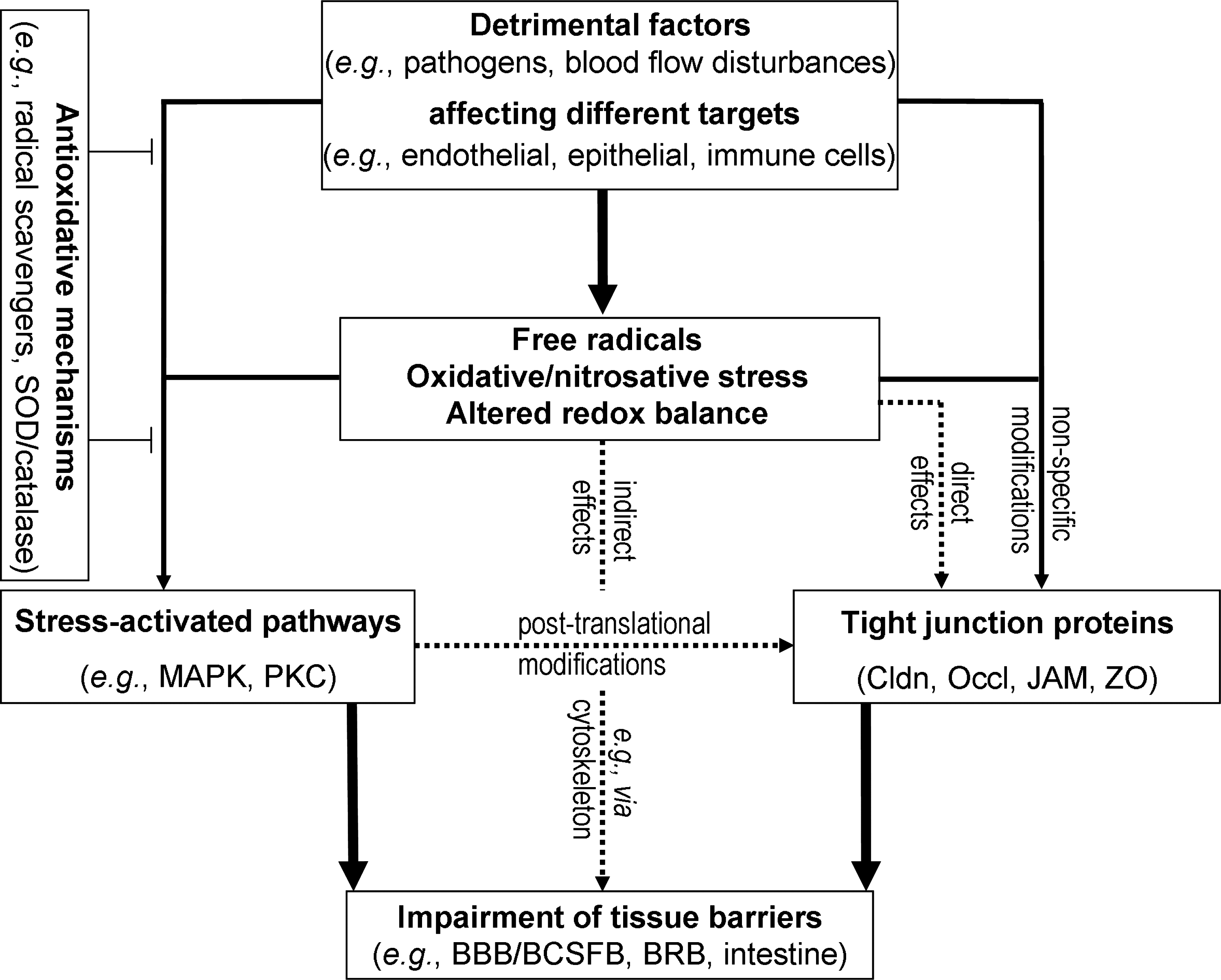
It is obvious that impairment of the TJ may have dramatic consequences for the functions of the respective organs. Oxidative stress and imbalance of the redox equilibrium are important factors that can induce disturbances in the barrier properties of epithelial and endothelial cells (Fig. 2). The Forum “Tight Junctions” is aimed at summarizing recent knowledge on the influence of redox processes on the structure and function of TJ. This includes both the effect of oxidative stress and that of antioxidative treatment. Some of the contributions will focus on TJ proteins, such as claudins (which form the backbone of the TJ), on occludin (a unique marker protein of TJ), on JAMs (that contribute to the regulation of paracellular permeability), and on ZO proteins (that recruit other TJ proteins) (Fig. 1). On the other hand, the Forum will cover endothelial and epithelial tissue barriers exposed to pathological events and pharmacological manipulations, which are accompanied by oxidative stress and involve antioxidative effects, respectively. Redox-related changes may have major functional consequences, as shown for barriers in the central nervous system or in the gastrointestinal tract. Specific pathologies will be discussed, such as hypoxia-related disturbances, inflammatory processes, or metabolic diseases, to illustrate the effect of alterations in the redox status on selected tissue barriers. In this respect, major pathways involve both conventional and novel protein kinase C isoenzymes, which are highly sensitive to the cellular redox state, as this modulates the cellular levels of Ca2+ and acylglycerol, their activators (4). Mitogen-activated protein kinase-related pathways are another key element in the redox-dependent signal transduction to TJ proteins (14) (Fig. 2).
The work of Overgaard et al. (27) summarizes recent developments in the field of claudins directly controlling the barrier function of the TJ. The authors present current progress in identifying the structural elements of claudins that regulate their function and the current understanding of how oxidant stress may affect claudin expression and function. Blasig et al. (6) review the structure, function, and regulation of occludin-like proteins, that is, of occludin itself and, as far as known, of tricellulin and marvelD3. It is pointed out that occludin can oligomerize and modulate TJ functions indirectly, in a redox-dependent manner. In particular, the novel concept developed is that redox regulation of the TJ is a principle function of occludin. The review of Gonzalez-Mariscal et al. (15) is devoted to ZO proteins and oxidative stress. Based on a detailed characterization of the domain structure, the manifold protein–protein interactions involving ZO proteins are analyzed, and functional and evolutionary aspects are covered. The effects of oxidative stress are discussed, with special attention given to stress-related pathologies that affect ZO proteins. The protein family of the JAMs is presented by Bazzoni (5), focusing on their role in the regulation of immune responses as well as on their involvement in various pathological situations, such as inflammation, vascular diseases, and tumorigenesis. The detailed analysis of the function of JAMs is supplemented by information on experimental approaches for the investigation of these proteins.
The second part of the Forum deals with epithelial and endothelial barriers. John et al. (20) summarize the current understanding of the alterations in epithelial barrier function and modulation of TJ proteins in inflammatory diseases of the intestine. Their review includes current therapeutic strategies and new agents that may potentially act at the level of TJ and help in the treatment of various intestinal inflammatory diseases. Up-to-date knowledge of the endothelial blood–retinal barrier during diabetes is provided by Frey and Antonetti (11). The consequences of oxidative stress are discussed with respect to TJ regulation and vascular permeability, and great importance is attached to the role of altered expression of cytokines and growth factors. The review of Coisne et al. (8) deals with the similarities and the differences with regard to the cellular and molecular characteristics of the functional and dysfunctional TJ complexes at the endothelial blood–brain barrier and the epithelial blood–cerebrospinal fluid barrier. The main focus is on neuroinflammation and on the role of reactive oxygen species. The breakdown of the blood–brain barrier is also discussed by Lehner et al. (24), with special consideration of the role of matrix metalloproteinases activated by reactive oxygen species. The authors compare various in vivo and in vitro blood–brain barrier models applied to study the cellular response of cerebral vascular cells to oxidative damage and discuss the therapeutic potential of antioxidants as potential neuroprotective agents.
Original data are included in some of the manuscripts. Lehner et al. (24) present new data on a connection between the nuclear expression of ZO-2 and the transcription of matrix metalloproteinases, which could point to a novel aspect in the adaptation of the blood–brain barrier to oxidative stress. The review of Overgaard et al. (27) also presents unpublished results by applying a recently established technique for analyzing claudin–claudin interactions. In the original contribution by Carrano et al. (7), it is demonstrated that accumulation of β-amyloid, a pathogenic factor in Alzheimer's disease, leads to a marked decrease in expression of TJ proteins in brain capillaries. Addition of β-amyloid to cultivated endothelial cells resulted in its binding to the receptor for advanced glycation endproducts and in generation of reactive oxygen species, with the final consequence of impaired integrity of the blood–brain barrier.
The reviews and the original contribution in this Forum illustrate the progress in the understanding of the architecture and characteristics of TJs, which are essential elements of tissue barriers. Current insight, although far from being comprehensive, justifies a closer look at special features of barrier-associated processes, particularly if these features are indeed related to oxidative stress and its consequences for tissue barriers and hence for organ functions. This Forum clearly demonstrates that TJ proteins and tissue barriers formed by TJ are highly sensitive to alterations in the redox status of the cells. It is shown that the redox sensitivity is caused either directly via specific protein–protein interactions or indirectly via redox-dependent signal transduction pathways modulating the TJ proteins. These mechanisms do not only play a role in regulating paracellular permeability but are also relevant for the function of cell–cell contacts in general.
Footnotes
Acknowledgments
The editors of this Forum express their gratitude to the members of the DFG Research Unit 721/2 “Molecular structure and function of the tight junction” coordinated by Michael Fromm, Charité Berlin (Germany), for very helpful discussions and suggestions with respect to this Forum. The support by the European research project JUSTBRAIN is highly appreciated.