




Abstract
On November 15, 2011, the Japanese Emperor bestowed the Medal with Purple Ribbon on Professor Hideo Utsumi for contributions to redox biology. Professor Utsumi was awarded Ph.D. in Pharmaceutical Sciences from University of Tokyo in 1976, and started his professional career as Assistant Professor at Teikyo University. He visited Cologne University as fellow during 1978–1980. In 1982, he moved to Showa University as Associate Professor. In 1994, he moved to Kyushu University as Professor. During 2008–2010, he served as vice president of Kyushu University. From 2007 to now he serves as the Director of Innovation Center for Medical Redox Navigation. Beginning 2010 he serves as the Executive Director of the Center for Product Evaluation, Pharmaceuticals and Medical Devices Agency. Professor Utsumi was the first to develop in vivo electron spin resonance (ESR; also known as electron paramagnetic resonance) imaging system in Japan and commercialized it to promote redox research. Over 30 in vivo ESR systems are currently used in Japan today. A compact or high-resolution Overhauser-enhanced MRI system has been developed by his group and will be available next year. His translational research activities have uniquely covered instrumentation, organic synthesis, and disease model applications. He synthesized many redox-sensitive compounds, and collaborated with clinicians to understand mechanisms underlying disease systems caused by redox imbalance using his compounds as tools. Thus, Professor Hideo Utsumi contributed a novel technology to investigate in vivo redox status in disease models. This technology platform has immense potential for bedside application to humans. Antioxid. Redox Signal. 16, 463–467.
Introduction
Professor Hideo Utsumi was born in Shimizu, Japan, on February 5, 1947. Professor Utsumi started his scientific research in pharmaceutical sciences: “Free Radical Formed During the Reaction of L-ascorbic Acid with Hydrazine and Isoniazide” as graduation dissertation, followed by “Spin-labeling Studies on Bio-membrane Dynamics” as Ph.D. thesis in Pharmaceutical Sciences from University of Tokyo in 1976. He started his professional career as an Assistant Professor at Teikyo University during 1976–1982. He continued his research on the relation of membrane fluidity with bio-functions by using spin-labeling technique until 1986. He stayed at Institute of Physiological Chemistry, Medical School, Cologne University, as visiting fellow of Alexander von Humboldt Foundation, Germany, to extend his research to 13C nuclear magnetic resonance technique, supervised by Professor Wilhelm Stoffel during 1978–1980. In 1982, he transferred to Showa University as an Associate Professor. He moved to Kyushu University in 1994 to accept a professorship. He was vice president of Kyushu University during 2008–2010, and director of Innovation Center for Medical Redox Navigation (ICMRN) from 2007 to this day. On November 15, 2011, the Japanese Emperor bestowed the Medal with Purple Ribbon on Professor Utsumi for contributions to redox biology.
Professor Hideo Utsumi is bestowed the Medal with Purple Ribbon on November 15, 2011, for contributions to redox biology.

It has been found that a variety of redox reactions play a significant role in maintenance of homeostasis of organism. Aerobic organism produces energy from oxygen metabolism and regulates redox reactions; however, under redox-disorder state, the organism would be suffered from excessive radical oxygen and researchers point out that this disruption can be potential risk of cancers, lifestyle diseases, cardiac diseases, etc.
Before 1980s, most researches in redox-related disease focused to measure the end product of redox reaction as a standard technique, such as malondialdehyde formation, using cultured cells and extracted tissues. However, in the course of disease propagation, kinetics and dynamics of redox disorder changes and the measurement of the end product did not offer sufficient information on mechanisms of redox-related diseases. Researchers tried to establish a methodology to measure redox reaction in living animal in real time, such as fluorescence probes or magnetic resonance technique. Professor Utsumi was interested in investigating the mechanism of oxidative diseases and cure effect of pharmaceutical drug in treatment of oxidative diseases in living animals. However, there was no standard methodology to measure the in vivo redox status in a living animal in 1980s.
In 1975 Feldman et al. reported the first in vivo electron spin resonance (ESR) experiments in rats using aminoxyl radicals as the contrast reagent (1). Based on the experience of spin-labeling, Professor Utsumi realized that the nitroxyl radical is sensitive to biological redox and that small-molecular-weight aminoxyl compounds could be diagnostic drug for redox status in vivo.
Professor Utsumi started development of ESR apparatus for small animals using a low-frequency microwave source in collaboration with JEOL, Ltd. The apparatus was commercialized through JEOL, Ltd. Thus, he has pursued to establish a technology/methodology to measure free radical/redox reaction in vivo in his scientific life. His research included multiple scientific areas, including development of analytical devices for in vivo redox status, organic synthesis of redox-sensitive spin-label, and investigation of mechanisms of oxidative disease in animal.
In Vivo Electron Spin Resonance Imaging and Overhauser-Enhanced MRI Scanner Were Developed for Imaging Redox Status in Living Animal
Professor Utsumi started developing in vivo ESR system in 1980s, since he believed magnetic resonance approach is one of the most useful techniques to investigate the redox status deep inside biological samples, while other techniques, for example, fluorescence technique, are for the research on the surface of biological samples.
The first in vivo ESR imaging system was developed for mouse-sized biological sample using 1-GHz-frequency source. Then, he developed a 300-MHz ESR system for the research of rat-sized biological samples. This 300-MHz ESR system was modified and combined with clinical magnetic resonance imaging (MRI) scanner; thereby, image of redox metabolism is to be superimposed on to MRI images (3), while the spatial resolution of ESR image was not sufficient due to the wide linewidth. The development of the system was a key step to expand in vivo redox research in Japan. Professor Utsumi published many articles on mechanisms of oxidative diseases using in vivo ESR spectroscopy or imaging technique (5), and the technology/methodology was widely used in universities and pharmaceutical companies.
In 1988, Lurie et al. demonstrated proton electron double-resonance imaging for free radical imaging (2), which is the same technique as Overhauser-enhanced MRI (OMRI). OMRI is an indirect free radical imaging technique based on Overhauser effect. Since OMRI utilizes MRI apparatus, Professor Utsumi expected to achieve much higher spatial resolution for free radical imaging in OMRI than in ESR. Gyromagnetic ratio of electron spin is ∼660 times larger than that of proton, and ensuring a good microwave penetration to biological samples, excitation of electron resonance needs to be carried out at around 5 to 20 mTesla. Professor Utsumi invented a new approach for field cycling by moving the sample between ESR magnetic field and MRI magnetic field and is the first to develop a high-field OMRI system over 1 Tesla of MRI field.
Overview of high-field Overhauser-enhanced MRI scanner that Professor Utsumi developed. The system consists of electron spin resonance, magnetic resonance imaging (MRI) magnets, and transportation device.

Nitroxyl Radicals as a Diagnosis Drug for Oxidative Diseases
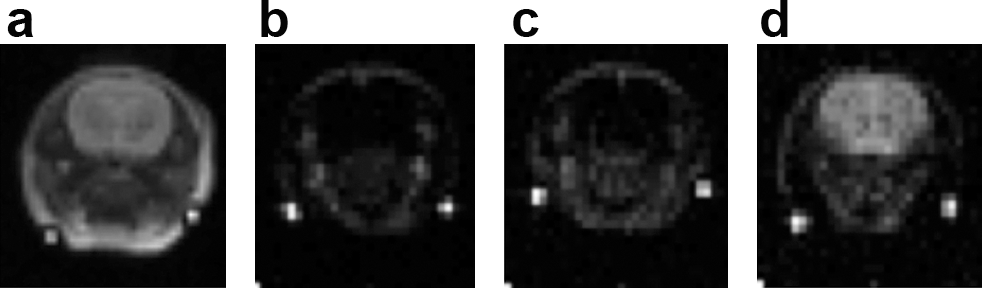
Nitroxyl radicals are paramagnetic compounds that are stable in solution. The nitroxyl radicals are reduced or oxidized to lose their paramagnetic characteristics by redox reactions with redox active compounds and enzymes, such as ascorbate, glutathione, and superoxide. Professor Utsumi synthesized many nitroxyl radicals that are accumulated in specific organs, or are specific to free radical species overproduced in oxidative diseases. One of such compounds is 3-methoxycarbonyl-2,2,5,5-tetramethylpyrrolidine-N-oxyl radical, which is highly accumulated in brain region, after passing through the blood–brain barrier (6), which is used in brain researches such as middle cerebral artery occlusion models (9) (Fig. 1).
Multi-OMRI Imaging Sequence Enabled Redox Molecular Imaging
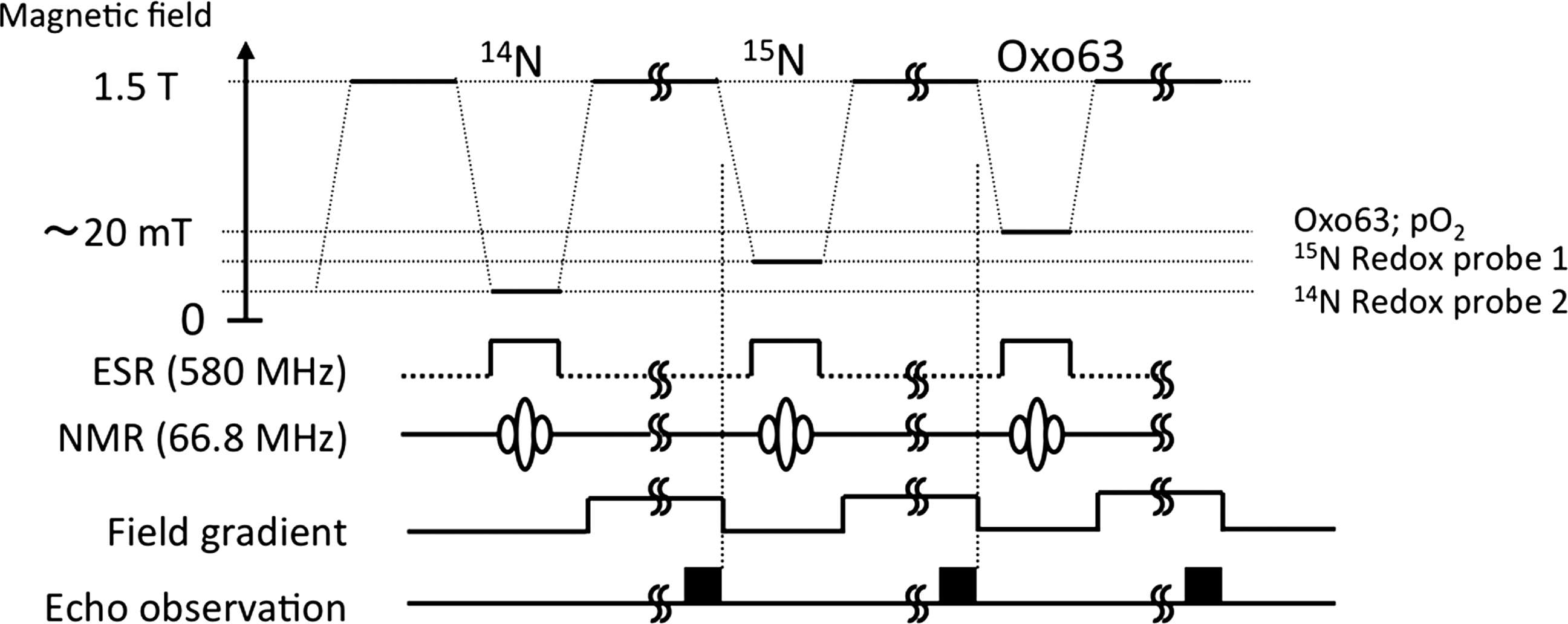
Professor Utsumi found the possibility of OMRI for simultaneous-imaging plural radicals, and developed OMRI acquisition sequence for multiple nitroxyl compounds with 14N or 15N nuclei (7). Since 14N and 15N nitroxyl radicals have different resonance frequencies, both molecules can be simultaneously imaged using a new sequence of OMRI in animals after administration of 14N and 15N nitroxyl radicals. By combining these compounds with different tissue or reaction specificities, the mechanisms of oxidative diseases were analyzed from in vivo ESR or OMRI images (Fig. 2).
Mechanisms of Oxidative Diseases Have Been Investigated with ESR/OMRI In Vivo
The paramagnetism of nitroxyl radicals decays depending on redox reactions. Professor Utsumi vigorously applied the nitroxyl compound to oxidative disease models using ESR system, to prove that nitroxyl radical metabolism should monitor oxidative condition in animal and that the approach gives an insight of disease mechanisms of in vivo oxidative diseases.
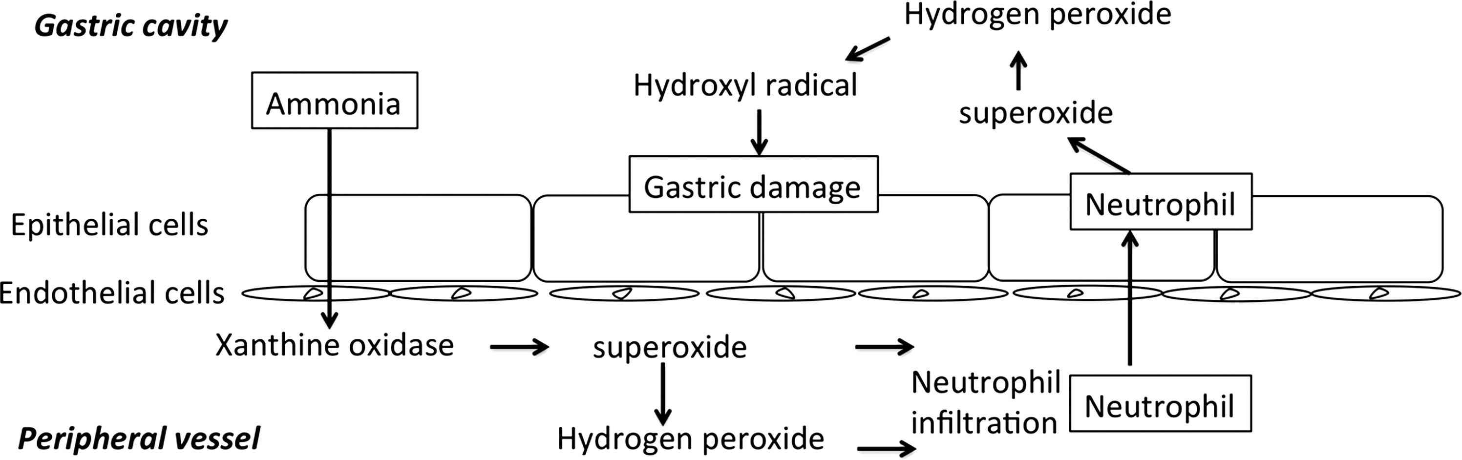
The mechanism of oxidative stress can be investigated by applying various free radical scavengers. Co-administration of specific radical scavengers with nitroxyl radical has served to elucidate the role of specific components of oxidative stress. One of the early studies to elucidate oxidative mechanism was performed in gastric ulcer model caused by indomethacin, a Non-Steroidal Anti-Inflammatory Drugs (8), NH4OH, or water immersion stress. In the case of NH4OH model, nitroxyl reduction was enhanced after intragastric administration of the compound. Co-administration of hydroxyl radical scavengers, mannitol or catalase, suppressed the enhanced reduction of nitroxyl radical in a dose-dependent manner. (Fig. 3)
Professor Utsumi applied this technology/methodology in collaboration with medical doctors in Kyushu University Medical School, analyzing mechanisms of oxidative diseases in vivo, such as streptozotocin-induced diabetic rat (4) and transient middle cerebral artery occlusion in rat. Thereby, the technology/methodology using nitroxyl compounds and magnetic resonance techniques is regarded as a standard approache to investigate redox-related diseases in vivo in Japan.
Professor Utsumi has contributed to the community of redox biology. He was President of Pharmaceutical Society of Japan (PSJ) during 2007 and Society of Electron Spin Sciences and Technology (SEST) during 2005–2007. He was awarded Society Prizes from PSJ, SEST, and Sociefy of Free Radical Research Japan. He hosted the international exchange program “Center for Magnetic Resonance Molecular Imaging of In Vivo Redox System” between universities in United Kingdom and United States during 2007–2008, which was expanded to include universities in Germany, Australia, and China during 2009–2011, supported by Japan Society for the Promotion of Science. Seventeen international meetings were held among these universities and 31 Japanese young researchers and Ph.D. students experienced short-term collaboration research in the U.K., Australia, or U.S. laboratories.
Currently, Professor Utsumi is Director of ICMRN from 2007 to this day. ICMRN was established at Kyushu University with a proposal of “Formulation of Advanced Collaborative Medical Innovation Center” as Special Coordination Funds for Promoting Science and Technology, since Professor Utsumi's achievements were regarded as one of the best pioneering researches on molecular imaging for biological redox disorder and biological redox navigation.
Gathering a collaborative wisdom and creativity in medical science, pharmaceutical science, agricultural science, and engineering from industry and academia, Professor Utsumi directs the national project: to develop biological redox measurement, imaging system and biological redox-sensitive probe; to analyze metabolic changes in redox-related diseases; to clarify redox concerns with diseases; to promote consistent early diagnosis, medical treatment, and development of therapeutic agents for related redox diseases; and to formulate a network system to connect these research achievements to community as well as district hospital.
Recent photo of Professor Utsumi, acting as Executive Director/Director of Center for Product Evaluation, Pharmaceuticals and Medical Devices Agency, Japan.
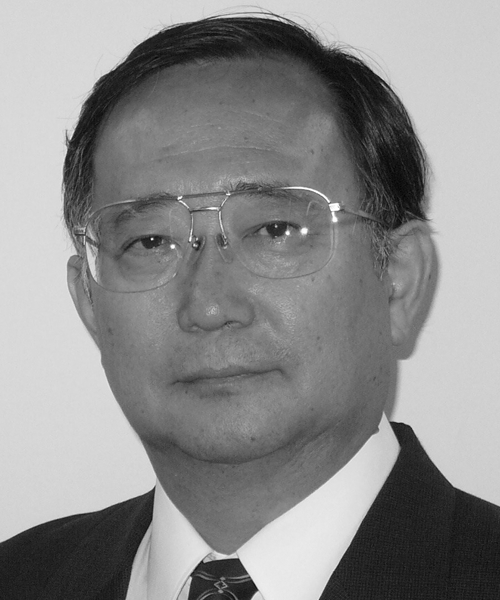
Professor Utsumi is also Executive Director/Director of Center for Product Evaluation, Pharmaceuticals and Medical Devices Agency, Japan (PMDA), since 2010. PMDA is a Japanese regulatory agency and its mission is similar to that of Food and Drug Administration. Professor Utsumi is responsible for scientific reviews of pharmaceuticals and medical devices for marketing.
Footnotes
Acknowledgments
Professor Hideo Utsumi thanks Professors Takao Kwan, Shoshichi Nojima, Akira Hamada, and Wilhelm Stoffel for the supervision. Professor Utsumi also thanks collaborators from JEOL, Ltd., for the collaborations to the research.