
Abstract
Introduction
Innovation
This is the first study to examine the subcellular sites (mitochondrial or extramitochondrial) that modulate cytosolic superoxide in skeletal muscle at rest and during contractions. Our data indicate that mitochondrial sources do not contribute to cytosolic superoxide, but nicotinamide adenine dinucleotide phosphate oxidase is a major source at rest and during contractile activity in skeletal muscle. These results are biologically and potentially clinically relevant and have widespread implications for the understanding of diverse scientific areas, including the responses of muscle to exercise training, age-related loss of muscle mass and function, as well as inflammatory or degenerative muscle diseases, such as the muscular dystrophies that are associated with increased levels of oxidative damage and muscle weakness.
Monitoring of dihydroethidium (DHE) oxidation may provide a reliable technique to monitor superoxide within single skeletal muscle fibers and recent data showed that contractile activity induced a significant increase in DHE oxidation due to increased superoxide within the fiber cytoplasm (60). Other studies that have examined superoxide in the mitochondrial matrix (4) or mitochondrial redox potential (44) indicate that mitochondria may not be the major source for the increased fiber ROS generation observed during contractile activity.
Skeletal muscle mitochondria produce superoxide from both complexes I and III of the electron transport chain (47). Superoxide generated at complex I appears to be released into the mitochondrial matrix, but complex III releases superoxide to both sides of the inner mitochondrial membrane (IMM)—the matrix and the mitochondrial intermembrane space (MIS) (47). Studies that have investigated mitochondrial superoxide formation or changes in mitochondrial redox potential have used MitoSOX Red or mito-roGFP constructs, but these probes accumulate in the mitochondrial matrix and do not respond to changes in superoxide or the redox potential in the MIS. Although superoxide is a relatively membrane-impermeable anion (54), studies of isolated mitochondria indicate that channels in the outer mitochondrial membrane (OMM) can facilitate diffusion of superoxide from the MIS to the cytosol (11, 27). Thus, the increase in cytoplasmic superoxide observed during contractile activity might be related to superoxide released across the OMM, although this possibility does not appear to have been examined in skeletal muscle.
Additional extramitochondrial sites for superoxide production within skeletal muscle have been proposed, including reduced nicotinamide adenine dinucleotide phosphate (NADPH) oxidase enzymes (30, 45), enzymes of the phospholipase A2 family (26, 49), and xanthine oxidase (25), but their contribution during contractile activity has not been fully evaluated.
The aims of the current study were therefore to: (i) identify the primary source(s) that contribute(s) to cytosolic superoxide in mature skeletal muscle fibers at rest and following a period of contractile activity, (ii) determine the effect of increasing frequency of contractions on fiber NO and superoxide, and (iii) define whether mitochondria within single muscle fibers release superoxide from the MIS or mitochondrial matrix to the cytoplasm of skeletal muscles at rest and following contractions.
Results
Single skeletal muscle fibers loaded with 4-amino-5-methylamino-2′,7′-difluorofluorescein diacetate and dihydroethidium
Figure 1A(i), B(i), and C(i) shows bright-field images of single muscle fibers displaying good morphology and well-defined striations along the sarcolemma. 4-Amino-5-methylamino-2′,7′-difluorofluorescein (DAF-FM) fluorescence from a fiber at rest appears homogeneous throughout the fiber, indicating no subcellular localization of this probe (Fig. 1A[ii], [iii]). The unreacted form of dihydroethidium (DHE) appeared to accumulate in the cytosol (Fig. 1B[ii], [iii]) but, upon reaction with superoxide, the oxidized form of DHE indicated by ethidium (E+) fluorescence was predominantly localized to nuclei, shown by the colocalization of 4′,6-diamidino-2-phenylindole dihydrochloride (DAPI) with E+ fluorescence (Fig. 1C[iv]).
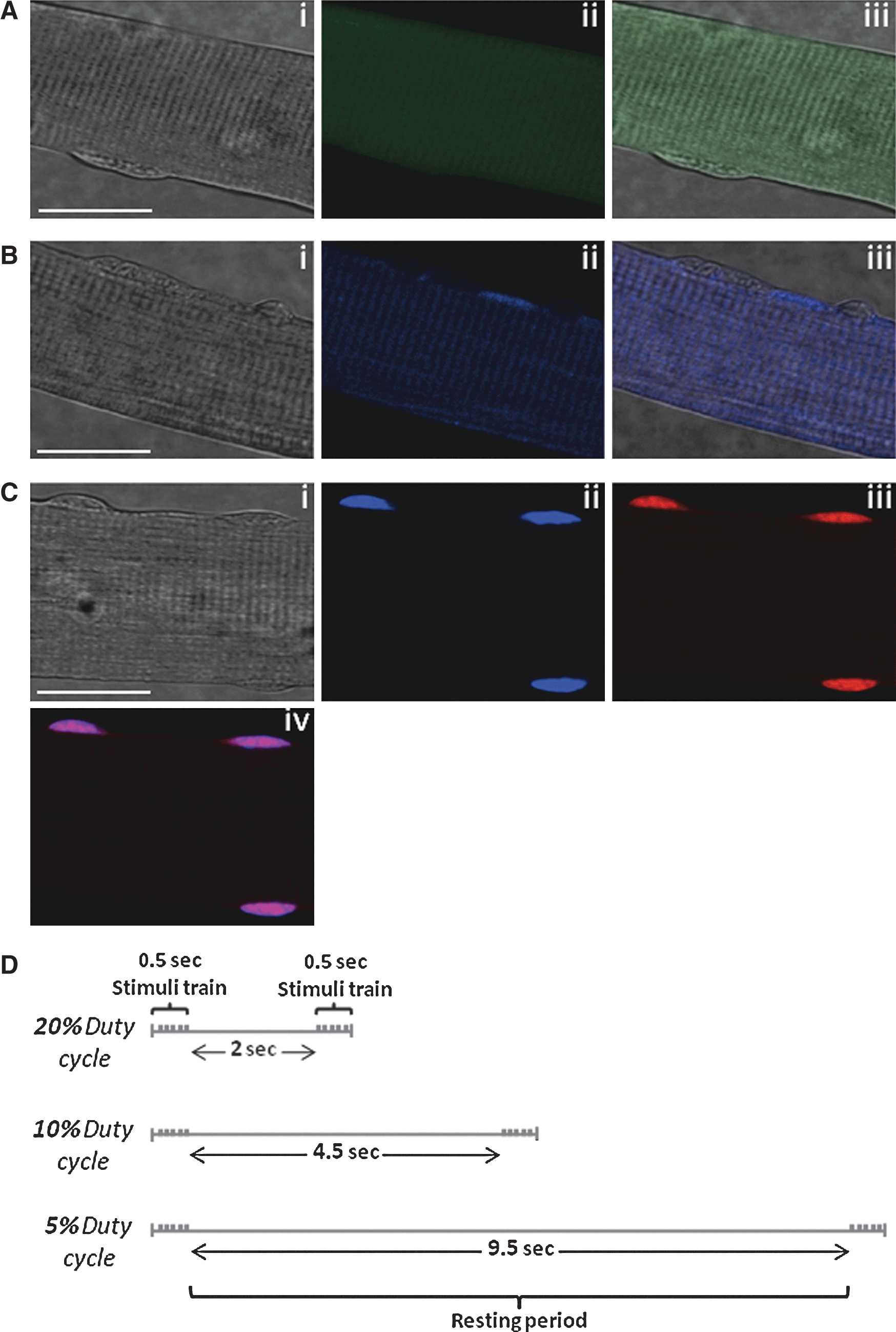
To determine the effect of increasing intensity of contractions on superoxide and NO activities, single muscle fibers were subjected to three different contractile activity protocols that are shown schematically in Figure 1D.
Intracellular NO and superoxide in single skeletal muscle fibers following contractile activity protocols of different intensities
Resting noncontracted flexor digitorum brevis (FDB) fibers loaded with 4-amino-5-methylamino-2′,7′-difluorofluorescein diacetate (DAF-FM DA) showed no significant changes in the rate of change in fluorescence over the time course of the study (Fig. 2A). Fibers subjected to the Moderate and Low contraction protocols showed a significant increase in the rate of change in DAF-FM fluorescence by a mean of ∼75% following contractions indicating an increase in NO production (Fig. 2A). DHE oxidation was observed to increase in an intensity-dependent manner in DHE-loaded fibers following muscle contractions (Fig. 2B) with the group of fibers subjected to the High, Moderate, and Low contraction protocols showing a 5.8-, 4.1-, and 2.8-fold increase relative to resting fibers (Fig. 2C).

Contribution of mitochondria to cytosolic superoxide in single skeletal muscle fibers at rest and during contractile activity
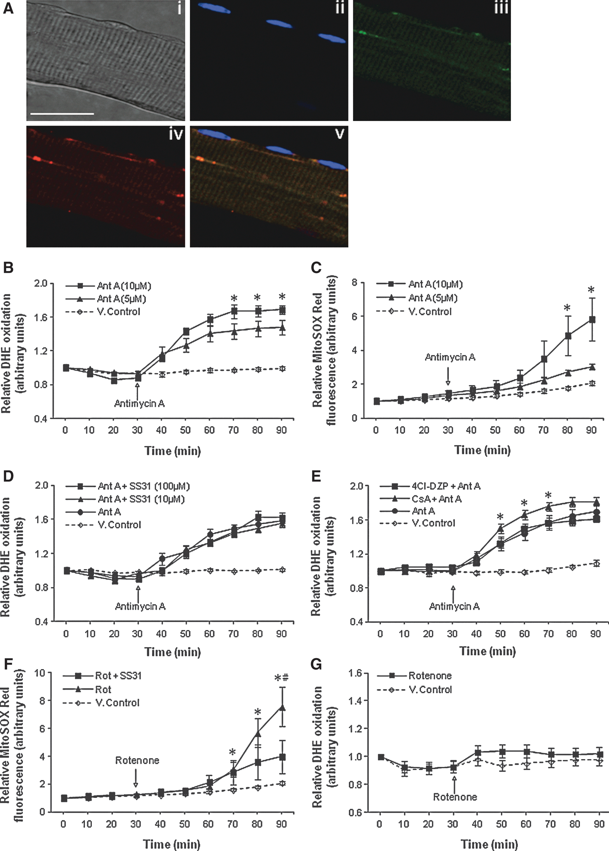
Changes in superoxide in the mitochondrial matrix were monitored using MitoSOX Red. This probe selectively accumulated in the mitochondria of single muscle fibers as shown by the colocalization of MitoTracker Green FM with MitoSOX Red fluorescence. The colocalization coefficients were as follows: Pearson's correlation (R r)=0.51, Manders's overlap (R)=0.78, Manders's colocalization coefficient for channel 1 (M red)=0.91, and Manders's colocalization coefficient for channel 2 (M green)=0.99 (Fig. 3A[v]). MitoSOX Red fluorescence was not detectable in the nuclei of muscle cells (Fig. 3A[v]).
Skeletal muscle mitochondria release superoxide to the cytosol of fibers following treatment with antimycin A
To examine mitochondria as a potential source of superoxide detected in the cytosol, fibers were treated with the electron transport chain inhibitors—Antimycin A (Ant A) and rotenone (Rot). Treatment of fibers with either 5 or 10 μM Ant A at 30 min induced a dose-dependent increase in DHE oxidation compared with control untreated fibers, indicating release of superoxide from mitochondria to the cytosol (Fig. 3B). MitoSOX Red–loaded fibers also showed a dose-dependent increase in fluorescence following addition of Ant A, indicating an increase in superoxide in the mitochondrial matrix (Fig. 3C).
Release of superoxide from mitochondria following treatment with Ant A does not occur through the mitochondrial permeability transition pore or the innermembrane anion channel
Previous reports have shown that superoxide can be released from the mitochondrial matrix through the mitochondrial permeability transition pore (mPTP) (1) and the innermembrane anion channel (iMAC) (41). To identify whether either of these two channels contributed to the extramitochondrial superoxide release seen following treatment with Ant A, fibers were pretreated with the SS-31 peptide, a mitochondrial-targeted antioxidant peptide that accumulates on the IMM and can scavenge superoxide to form tyrosine hydroperoxide (15, 41). Treatment with SS-31 at either 10 or 100 μM had no effect on the Ant A–stimulated increase in DHE oxidation (Fig. 3D). The potential role of the mPTP and iMAC in release of superoxide was further examined using inhibitors of the two potential pores/channels (cyclosporin A [CsA] and 4′-chlorodiazepam [4Cl-DZP]) but neither was found to reduce DHE oxidation in Ant A–treated fibers (Fig. 3E). Together these data suggest that superoxide released following treatment with Ant A did not derive from the mitochondrial matrix. The role of matrix superoxide was further examined by treatment of fibers with the complex I inhibitor Rot. This treatment increased MitoSOX Red oxidation, indicating increased superoxide generation in the matrix (Fig. 3F), but had no effect on DHE oxidation (Fig. 3G).
Channels of the OMM mediate the diffusion of superoxide from the MIS to the cytosol of skeletal muscle fibers
To identify channels that might play a role in the diffusion of superoxide from the MIS to the cytosol of single muscle fibers, mitochondrial and cytosolic fractions from gastrocnemius (GTN) muscles were prepared. Figure 4A illustrates the relative purity of the mitochondrial and cytosolic fractions obtained and Figure 4B illustrates the protein contents of the three voltage-dependent anion channel (VDAC) isoforms. In the presence of dextran sulfate (DS), a VDAC inhibitor, the increase in DHE oxidation observed following treatment with Ant A at 30 min was partially inhibited over the next 20 min, but reached a similar level by the end of the experiment compared with values from fibers loaded with Ant A only (Fig. 4C). No effect on baseline DHE oxidation was observed following treatment of fibers with DS alone at 30 min (Fig. 4D). Potential effects of inhibition of VDAC by 4,4′-diisothiocyano-2,2′-disulfonic acid stilbene could not be assessed since this agent was found to affect fiber viability at IC50 values of 0.2 mM (data not shown in detail). The protein expression of the Bax channel in fibers is shown in Figure 4E. Following treatment with the Bax channel blocker (Bax CB), fibers showed a slightly lower rate of increase in DHE oxidation (by ∼8%) during the 40–50 min period following addition of Ant A (Fig. 4F). Fibers pretreated with the Bax CB and VDAC inhibitor (DS) together showed a further reduction in fluorescence, with the relative increase in DHE oxidation being statistically lower (∼13%) during the 20–50 min period following Ant A treatment compared with fibers treated with Ant A only (Fig. 4F). The increase in DHE oxidation in the Bax CB and DS group was lower over the 30–90 min time course compared with the Ant A–treated fibers (Fig. 4F) but this did not occur in fibers treated with DS only (Fig. 4C), suggesting that both channels might play a synergistic role in mediating the diffusion of superoxide from the MIS across the OMM. Inhibition or blocking of these two OMM channels did not completely prevent extramitochondrial superoxide release following addition of Ant A (Fig. 4F). To ensure that data were not influenced by any confounding direct interactions between Ant A and DHE, unrelated to effects of Ant A on the electron transport chain, control experiments were conducted in a cell-free medium but these showed no effect of Ant A on DHE or MitoSOX Red oxidation (data not shown in detail).

The effect of VDAC (DS) and Bax (Bax CB) inhibitors was assessed in fibers following a 10-min contraction period but no differences in DHE oxidation were observed between the contracted groups (Fig. 4G).
Identifying the cytosolic sources of superoxide production in single skeletal muscle fibers at rest and during contractile activity
Effect of inhibition of calcium-independent phospholipase A2 enzyme and NADPH oxidase(s) on cytosolic superoxide
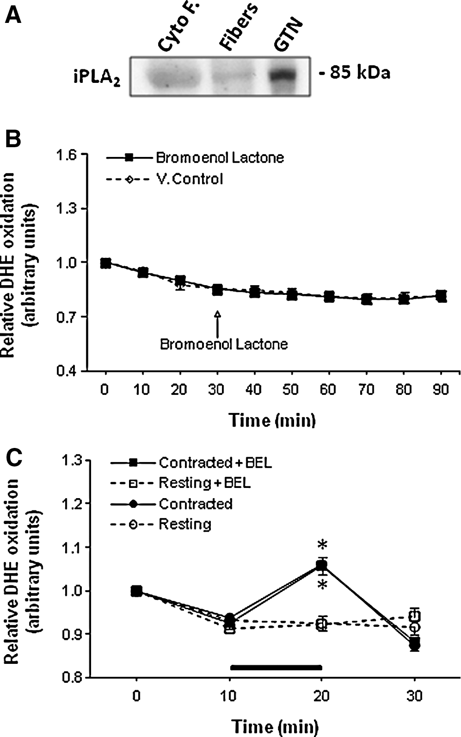
The presence of calcium-independent phospholipase A2 enzyme (iPLA2) in the cytosolic compartment of skeletal muscles was confirmed by Western blotting (Fig. 5A) but no effect of the selective iPLA2 inhibitor, bromoenol lactone (BEL), on DHE oxidation was observed either in quiescent (Fig. 5B) or contracted fibers (Fig. 5C).
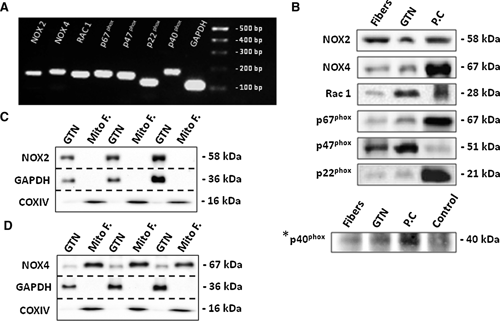
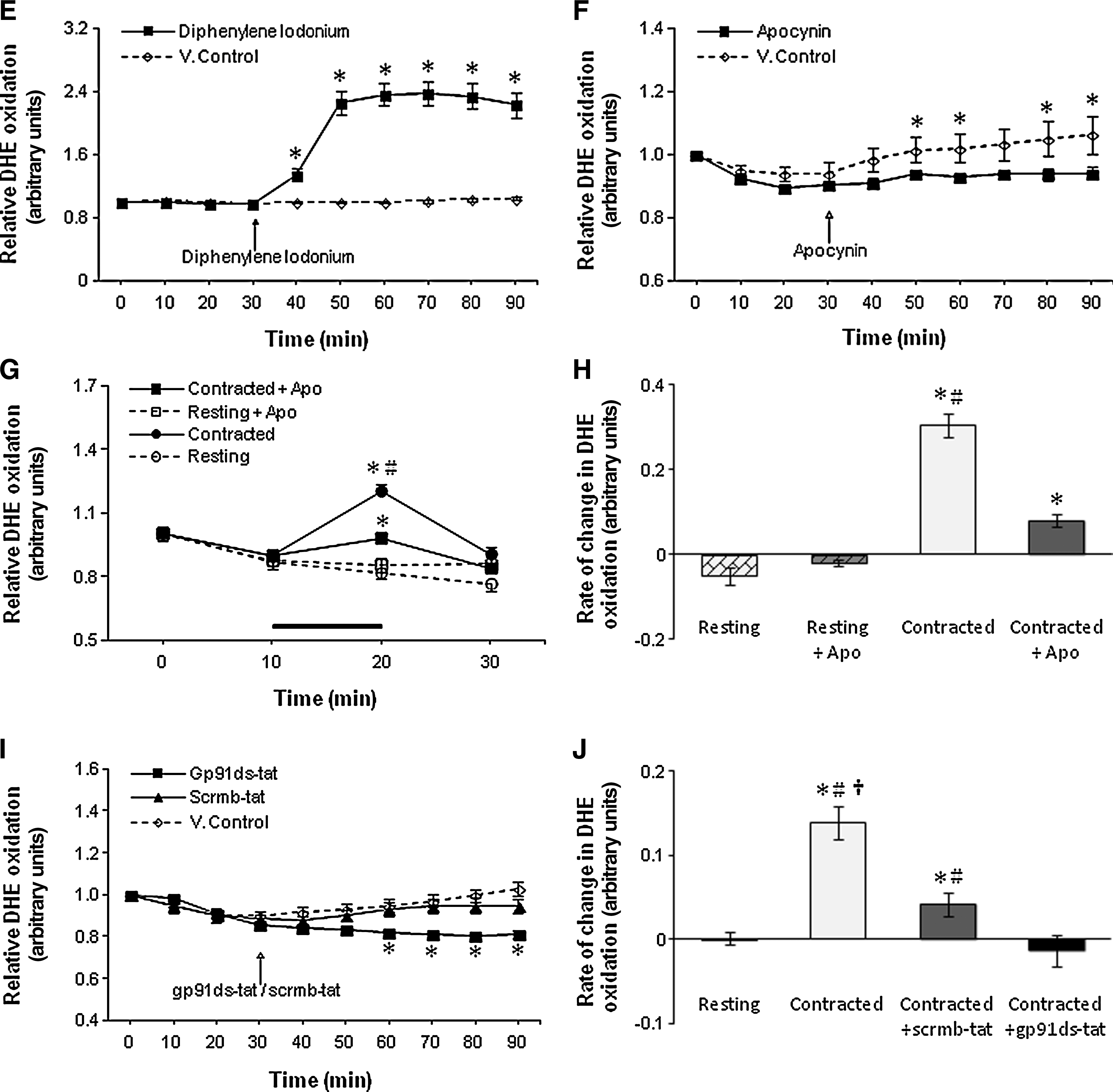
The mRNA and protein expression of NADPH oxidase subunits in single skeletal muscle fibers of the FDB muscle is shown in Figure 6A and B. Further experiments were also undertaken to investigate whether the NOX4 or NOX2 subunits were present in mitochondrial fractions from GTN muscles and demonstrated the presence of NOX4 in this fraction (Fig. 6C, D). The role of NADPH oxidase complexes in producing superoxide under resting conditions was initially assessed using the nonspecific flavoprotein inhibitor diphenyleneiodonium chloride (DPI) (Fig. 6E). Resting muscle fibers treated with DPI at 30 min of the experimental period showed a rapid increase in fluorescence compared with control nontreated fibers (Fig. 6E). In addition, DPI was also observed to prevent the fibers from contracting following field stimulation. An alternative NADPH oxidase inhibitor, 4-(2-aminoethyl)-benzenesulfonyl fluoride inhibitor, was also found to inhibit muscle contractions (data not shown in detail). Fibers were therefore incubated in the presence of apocynin (APO), a nonspecific NADPH oxidase inhibitor. Quiescent muscle fibers showed a reduction in fluorescence by ∼10% over the 50–90 min time course compared with vehicle-treated fibers (Fig. 6F). APO was also found to decrease DHE oxidation following a period of contractile activity by ∼70% compared with untreated fibers (Fig. 6G, H). Recent data have suggested that APO can also act as a scavenger for reaction products of hydrogen peroxide (H2O2), in vascular cells that lack myeloperoxidase (MPO) or produce low amounts of ROS (29), and hence the potential inhibitory effects of gp91ds-tat, a specific peptide inhibitor of NADPH oxidases, were also examined (Fig. 6I, J). Resting fibers showed a decline in fluorescence by a mean of 18% compared with vehicle-treated fibers and fibers treated with the control peptide scrambled-tat (Fig. 6I). Treatment of fibers with gp91ds-tat also prevented the increase in DHE oxidation in response to a 10-min period of contractions (Fig. 6J), but the scrambled-tat-treated fibers also showed a lower rate of increase in DHE oxidation than the vehicle-treated control group (Fig. 6J).
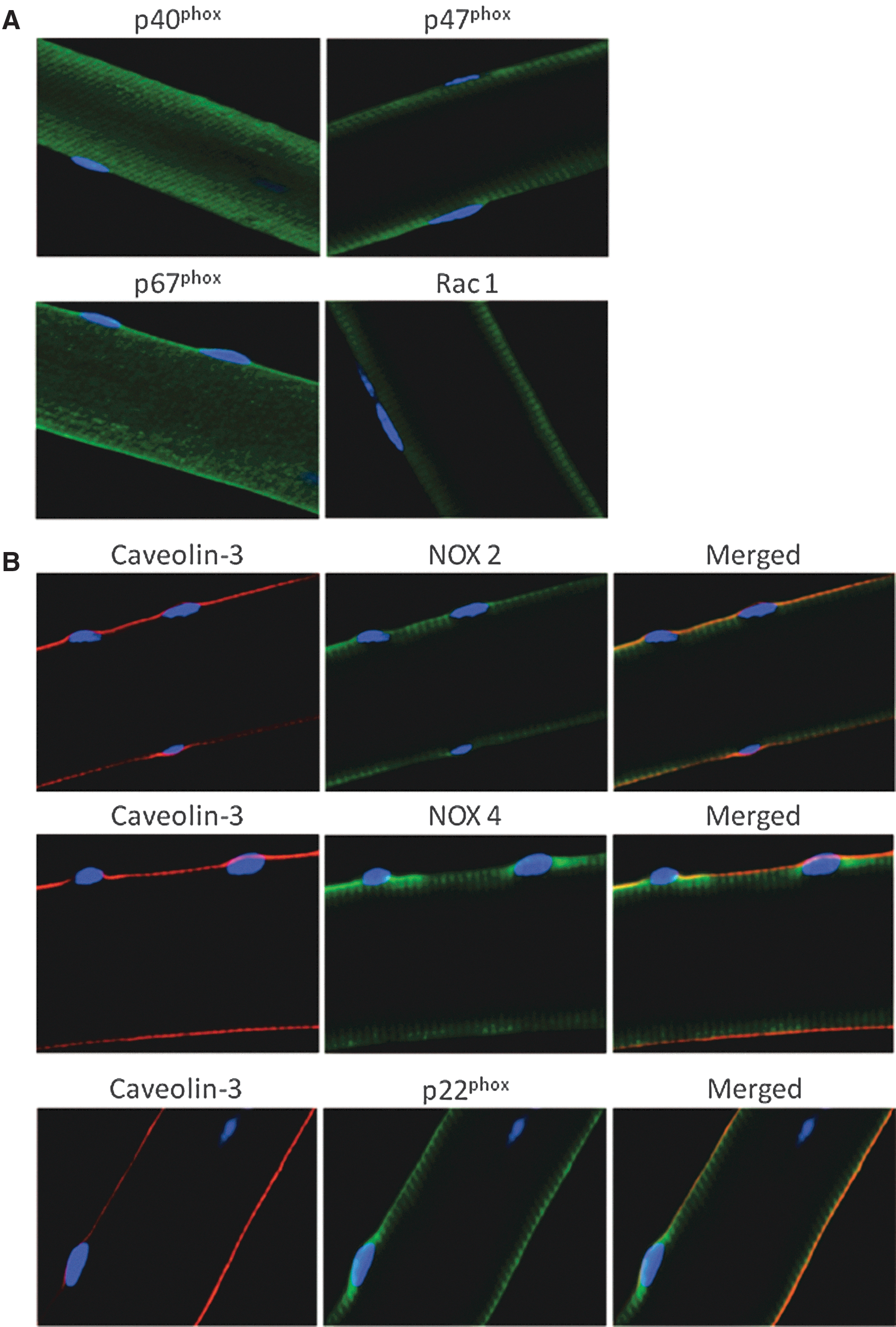
NADPH oxidase subunits are located on the plasma membrane and transverse tubules of single skeletal muscle fibers
To determine the cellular location of NADPH oxidase subunits, immunocytochemistry of single isolated fibers from the FDB muscle was undertaken (Fig. 7A–D). The regulatory subunits (p40phox, p47phox, p67phox, and Rac1) were found to be localized on, or in close proximity to the, sarcolemma, but immunofluorescence from p40phox and p67phox was also observed in the cytosolic compartment of the muscle fibers (Fig. 7A). The catalytic subunits (NOX2 and NOX4) and the small membrane-bound integral p22phox subunit were localized to the plasma membrane of the muscle fibers, as indicated by colocalization (yellow staining) with caveolin-3, the muscle-specific caveolin isoform, present in sarcolemmal caveolae (Fig. 7B). A striated pattern of staining for all three subunits was also observed in close proximity to the sarcolemma, potentially due to expression in the transverse tubules (T-tubules) (Fig. 7B). To examine this possibility, the membrane-bound subunits of the NADPH oxidase complex from single muscle fibers were co-immunostained with the α1s subunit of the dihydropyridine receptor (Fig. 7C). Confocal images showed a high degree of colocalization (yellow staining), strongly suggesting that the NADPH oxidase components NOX2 (R r=0.60, R=0.67, M red=0.85, M green=0.96), NOX4 (R r=0.98, R=0.98, M red=0.98, M green=1), and p22phox (R r=0.92, R=0.97, M red=1, M green=0.92) were also expressed on the T-tubule membrane in skeletal muscle fibers (Fig. 7C).
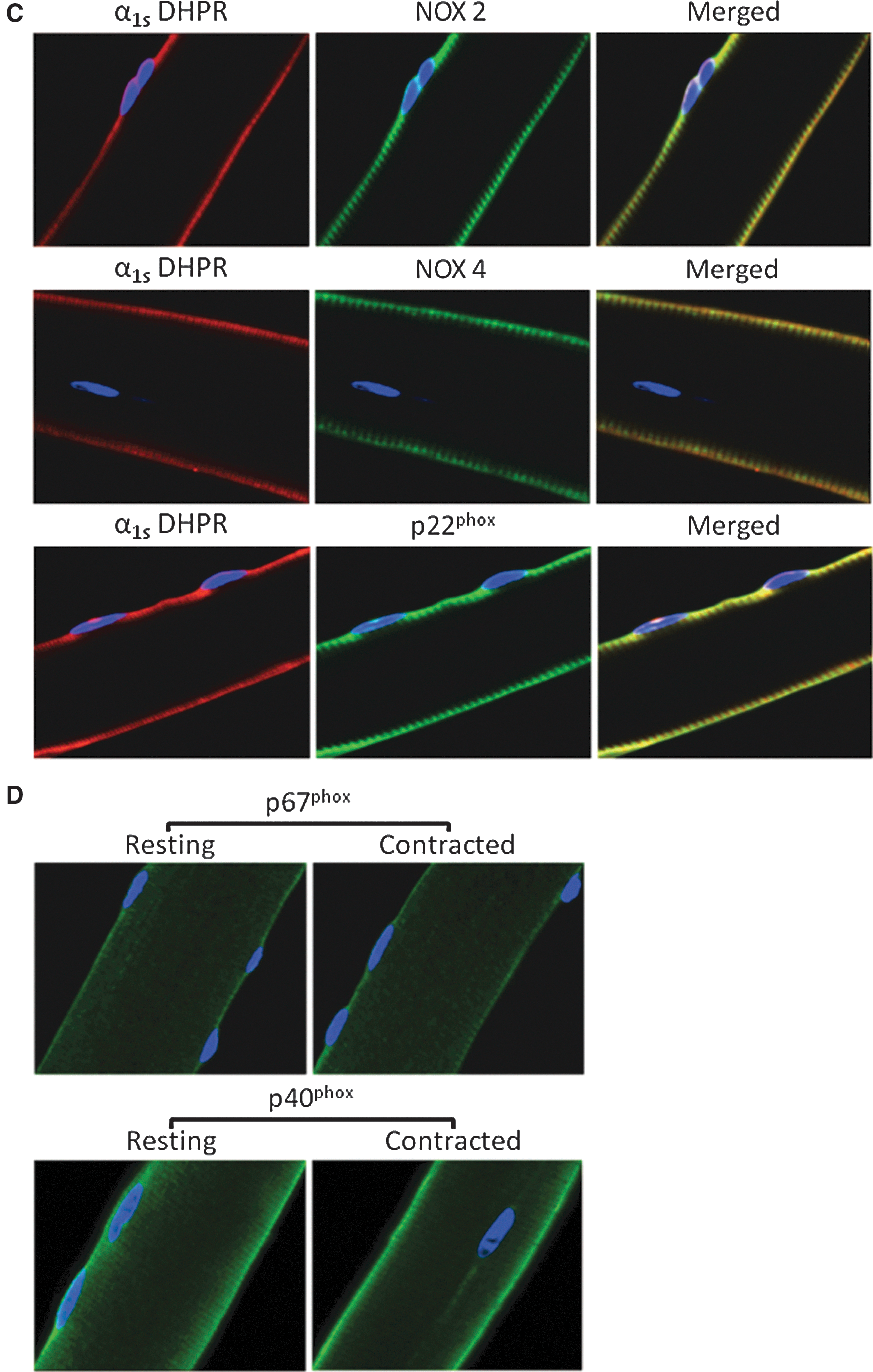
The effect of contractions on p40phox and p67phox translocation in single skeletal muscle fibers
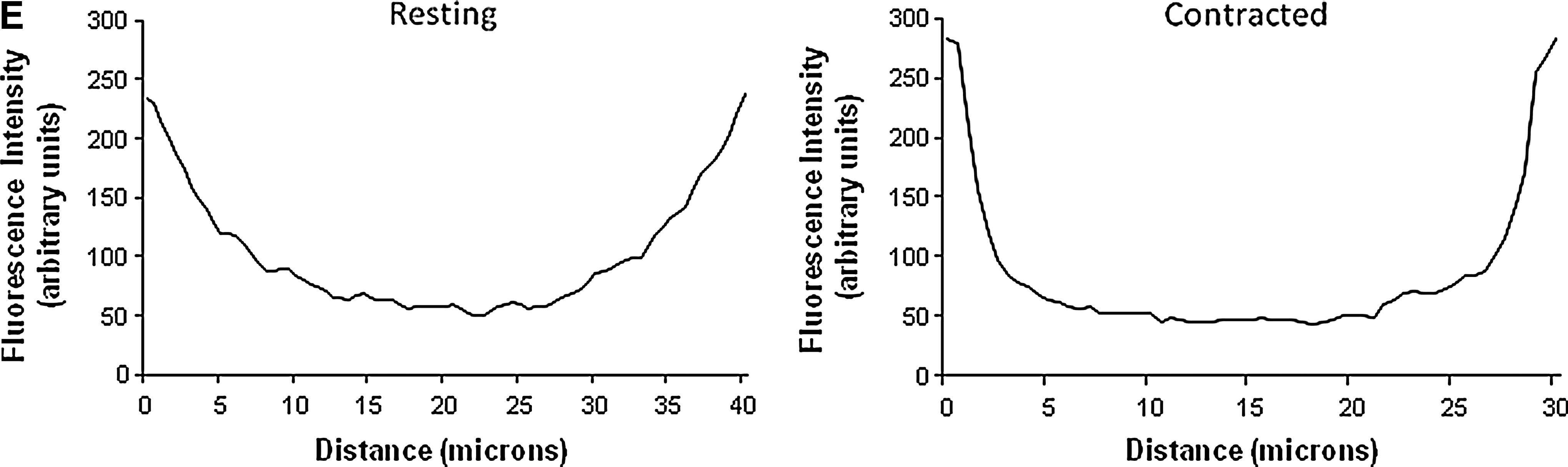
The effect of contractions on p40phox and p67phox protein translocation is shown in Figure 7D. Fluorescent distribution analysis showed no evidence for contraction-induced translocation of the p67phox protein (not shown in detail), but fluorescent distribution analysis for p40phox (Fig. 7E) showed an increased proportion of the fiber fluorescence at the sarcolemma and a relative reduction in the cytosolic compartment of contracted fibers compared with quiescent fibers. These data suggest that NADPH oxidase activation in skeletal muscle fibers during contractions may involve translocation of p40phox to the plasma membrane.
Discussion
Methodological considerations
We have previously utilized electron-spin resonance spectroscopy (53), fluorescence-based microscopic assays (51, 55, 60), microdialysis (12), and high-performance liquid chromatography techniques (53, 60) to assess intracellular and extracellular changes in RONS produced by skeletal muscle. In the current study, we utilized fluorescence imaging microscopy to allow monitoring of real-time changes in radical species in single isolated muscle fibers and to examine potential subcellular pathways that are involved in the regulation of cytosolic superoxide content at rest and during contractile activity. Within subcellular compartments of skeletal muscle, superoxide will be rapidly dismutated to H2O2 or undergo chemical reactions to generate products, such as peroxynitrite [see ref. (60) for a discussion], but superoxide per se can be detected in subcellular regions, such as the mitochondria or cytoplasm, due to its poor membrane permeability. Superoxide has also previously been claimed to directly induce changes in muscle function and degeneration during aging (60).
Assessment of E+ fluorescence following MitoSOX Red and DHE loading as a measure of superoxide anion radical in cellular compartments has been criticized and recent studies have identified 2-hydroxyethidium (2-OH-E+) as a specific product of the reaction of DHE and MitoSOX Red with superoxide (71). We have previously evaluated the use of DHE oxidation and showed that E+ and 2-OH-E+ followed the same pattern of change in resting and contracted fibers from wild-type mice and mice lacking SOD1 (60). Previous work also showed that the anticipated increase in DHE oxidation following contractions was completely abolished following loading of fibers with the superoxide scavengers Tiron or Tempol (60). Thus, we argue that the technique used in this study, based on monitoring E+ fluorescence from single muscle cells, is capable of detecting changes in superoxide production at rest but also in response to a physiological stimulus such as contractile activity.
NO and superoxide production following contractile activity protocols of different intensities
The increase in RONS activity during exercise appears to be in major part due to generation by contracting skeletal muscle (32, 54) and one objective of the present study was to determine the effect of increasing frequency of contractions on muscle fiber NO and superoxide activity. We previously showed that contractile activity induces an increase in intracellular DAF-FM fluorescence in mouse skeletal muscle fibers due to increased NO generation (55). In the present study an increase in DAF-FM fluorescence was seen from fibers subjected to all contraction protocols, but the increase reached statistical significance only for the Moderate and Low contraction protocols.
For DHE-loaded fibers, those subjected to the High contraction protocol exhibited the largest increase in DHE oxidation and those subjected to the Low protocol had the least. This finding is in agreement with a previous study that demonstrated that intracellular ROS generation in primary myotubes was higher following an intense electrical stimulation protocol than that with a moderate stimulation protocol (61). The increase in DHE oxidation immediately postcontractions was followed by a reduction in fluorescence at 10 min following the contractile activity period. Early studies investigating the electrochemical reduction of E+ reported DHE as the final product (31) and the reduction of E+ to DHE has been reported in cell free (6) and cellular systems (23). Work from our group (unpublished data) has also shown that the reduction of E+ at 10 min postcontractions is related to an increase in DHE as assessed by changes in blue fluorescence.
Contribution of mitochondria to cytosolic superoxide in single skeletal muscle fibers
We reasoned that the isolated single mature skeletal muscle fiber approach would provide a valid model to study the role of mitochondria in regulating changes in cytosolic superoxide. These fibers contain an intact mature mitochondrial matrix in situ and retain excitability to electrical stimuli, hence facilitating the study of ROS generation following contractions in real time. The current approach indicated that the rapid increase in superoxide observed following contractions likely originated from nonmitochondrial sources since the change in DHE oxidation reflects reaction with superoxide within the cytosolic compartment of the muscle cells (33). Superoxide is a membrane-impermeable anion, but recent data have indicated that superoxide can diffuse out of isolated mitochondria through the mPTP (1), the iMAC (15), and channels located on the OMM (11, 27).
Treatment of fibers with the electron transport chain inhibitors Ant A and Rot increased the oxidation of matrix-localized MitoSOX Red, but only Ant A caused an increase in cytosolic DHE oxidation, supporting the possibility that only superoxide within the MIS can contribute to cytosolic changes. The lack of any role for matrix superoxide in oxidation of DHE in the cytoplasm was further supported by the lack of any effect of SS-31 peptide that scavenged superoxide within the mitochondrial matrix as shown by the reduction of MitoSOX Red fluorescence following addition of Rot. These data provide evidence that superoxide generated by complex III and released into the matrix does not escape from intact mitochondria in muscle fibers. These findings are consistent with previous reports that failed to show any extramitochondrial superoxide release from mitochondria in response to the addition of Rot (47). Inhibitors of iMAC or mPTP were also ineffective in reducing cytosolic DHE oxidation in Ant A–treated fibers, further indicating a lack of a role for these channels in release of superoxide from the mitochondrial matrix to the cytosol of muscle fibers.
A role for OMM channels in release of superoxide from the MIS to the cytoplasm was supported by the inhibitory effects of VDAC and Bax inhibitors on DHE oxidation following Ant A treatment, and inhibition of VDACs resulted in a greater reduction in DHE oxidation than that of Bax inhibition, potentially because VDACs are the major channels of the OMM responsible for the passage of proteins and solutes between the OMM and cytoplasm (42). Neither VDAC nor Bax inhibition fully prevented the extramitochondrial superoxide release induced by Ant A, suggesting that either of the inhibitors failed to completely close the respective channels or other channels, such that peptide-sensitive channel or the translocase of outer membrane (11) might also mediate the release of superoxide across the OMM.
Despite the effects of VDAC and Bax channel inhibitors seen on DHE oxidation in Ant A–treated fibers, no similar effects were seen following contractions, indicating that the increase in cytosolic superoxide during contractile activity does not derive from mitochondria. DHE oxidation following contractions increased by ∼20%, whereas the change in fluorescence in response to Ant A increased by ∼90%. Thus, the observed increase in superoxide following addition of Ant A may be considered “nonphysiological” compared with that seen following a period of contractile activity. Hence, we propose that intact mitochondria do not release superoxide to the cytoplasm of skeletal muscle fibers at rest or in response to contractile activity, but can release superoxide under conditions where mitochondrial superoxide production is grossly excessive.
Contribution of iPLA2 enzymes to cytosolic superoxide in skeletal muscle fibers
Recent work (26) demonstrated that iPLA2 enzyme can modulate cytosolic oxidant activity in skeletal muscle cells indicated by a reduction in 2′,7′-dichlorodihydrofluorescin oxidation (a general indicator of ROS activities) following inhibition of iPLA2 (26). In the present study no effects of iPLA2 inhibitors on DHE oxidation were seen. The previous study (26) used BEL at 10 μM whereas in the present study 0.6 μM was used since preliminary studies indicated that higher concentrations affected the viability of muscle fibers (data not shown in detail). BEL at 0.6 μM has previously been shown to inhibit iPLA2 activity by 100% (2). Thus, current data do not support the possibility that cytosolic superoxide levels at rest or during contractions are modulated by the activity of the iPLA2 enzymes although the higher concentrations of inhibitor used by other researchers could not be examined in the model. Zuo et al. demonstrated that PLA2 enzymes in skeletal muscle do not directly produce superoxide but can regulate the activity of the lipoxygenases by modulating the release of arachidonic acid (72).
Expression of NADPH oxidase and its contribution to cytosolic superoxide in skeletal muscle fibers
Skeletal muscles have previously been shown to express NADPH oxidase(s) (22, 30, 45, 67), but little information is available regarding the role and regulation of this complex in generation of superoxide in muscles. Our initial results show mRNA and protein expression of NADPH oxidase subunits in single isolated fibers from the FDB muscle. Others have identified various NADPH oxidase subunits in mouse and rabbit skeletal muscles (30, 67), but this appears to be the first report to demonstrate the expression of NADPH oxidase components in a pure muscle fiber preparation devoid of all nonmyogenic cells.
Inhibitor studies support a role for NADPH oxidase in contributing to cytosolic superoxide production. Resting fibers treated with APO and gp91ds-tat inhibitors showed a decrease in DHE oxidation compared with control fibers, suggesting that the complex is active and modulates superoxide generation under resting conditions. Similarly, fibers subjected to contractile activity showed a significantly lower increase in DHE oxidation following treatment with APO whereas gp91ds-tat treatment abolished the increase in response to contractions. Scrambled-tat-pretreated fibers also showed a decrease in fluorescence following contractions, but some previous studies have also observed this (57). Gp91ds-tat appeared to be more effective than APO, which can be attributed to the different inhibitory properties of the compounds.
Recent evidence suggests that APO might require activation by peroxidases (69) and can also act as an antioxidant for peroxide-dependent ROS in cellular systems that lack MPO, or produce low amounts of ROS (29). The proposed inhibitory mechanism of action of APO on NADPH oxidases is to prevent the translocation of p47phox and p67phox to the membrane-located catalytic subunits (46) whereas gp91ds-tat peptide, the most potent inhibitor (IC50: 3 μM), has been reported to selectively inhibit NOX2 oxidase (14), although early studies suggested that the peptide additionally inhibited NOX1 and NOX4 homologues (9, 57) by preventing their interaction with p47phox (57). The inhibitory effects of both APO and gp91ds-tat imply that p47phox is a critical component of the NADPH oxidase complex and can regulate the activity of the enzyme.
Treatment of resting fibers with the nonspecific inhibitor DPI showed a paradoxical increase in DHE oxidation. The mechanism by which DPI induced increased DHE oxidation in the current study is unclear, but DPI has been reported to inhibit flavoenzymes in addition to NADPH oxidases, such as NOS, xanthine oxidase, and NADPH-cytochrome P450 reductase (5, 39, 52), and hence it can affect cellular processes unrelated to NADPH oxidase. Data on the regulation of intracellular ROS by DPI are controversial, with both inhibitory and stimulatory actions of DPI being reported. Some studies indicate that DPI can stimulate the production of ROS (37, 58) and reactive nitrogen species (5), affecting cellular redox status indicated by increased levels of lipid peroxidation (58), DNA damage (39, 52), increased glutathione disulfide (58), apoptosis (5, 39, 52), and increased nitration of tyrosine residues in cellular proteins (5). Some previous studies have also reported an increased oxidation of ROS-sensitive probes, including dihydrorhodamine 123 (5) and 2′,7′-dichlorodihydrofluorescein diacetate (5, 52), following addition of DPI. Potential mechanisms through which DPI might stimulate RONS production have been proposed, including increased mitochondrial superoxide production via inhibition of complex I (37, 38). Formation of phenyl radicals during NADPH oxidase inhibition by DPI has also been reported (39, 50).
The data presented imply that NOX2 is likely to be the predominant oxidase system contributing to cytosolic superoxide generation in muscle fibers, since most recent data indicate that the gp91ds-tat peptide selectively inhibits the assembly of NOX2 oxidase (14). Immunocytochemistry of single isolated fibers from the FDB muscle revealed that NOX isoforms and subunits were localized on, or in close proximity to, the plasma membrane of muscle fibers although p40phox and p67phox were also present in the cytosolic compartment of quiescent muscle fibers. Further experiments supported previous findings (22, 30) of apparent localization of these subunits to the T-tubules. In addition, NOX4 was found to be present in skeletal muscle mitochondria. Recent reports have shown that NOX4 localized to cardiac (3, 15, 36) and liver (7) mitochondria. NOX4 has been predicted to localize on the IMM (7) and produce H2O2 (66) but recent reports have shown that it can also regulate changes in superoxide within the mitochondrial matrix (3, 7, 15, 36). These findings, however, have not been examined in skeletal muscle; thus, future studies are warranted to examine the role of NOX4 in skeletal muscle mitochondria.
In phagocytic cells, NADPH oxidase activation requires the translocation of the cytosolic regulatory subunits to cytochrome b 558, the catalytic core of the enzyme (20, 35). Fluorescent distribution analysis showed no evidence for translocation of the p67phox protein following contractions but p40phox-immunostained fibers showed an increased proportion of fluorescence at the membrane and a reduction in the cytoplasmic compartment following contractions, which suggests that NADPH oxidase activation in single skeletal muscle fibers from young mice in response to contractions may involve translocation of p40phox to the plasma membrane. Although the role of p40phox has been controversial, studies in phagocytes indicate that on stimulation p40phox translocates to cytochrome b558 (20) and functions as a positive regulator of the superoxide-producing phagocyte oxidase by enhancing recruitment of p67phox and p47phox to the membrane (35). In addition, lack of p40phox in homozygotic knockout mice reduced superoxide production in both in vivo and in vitro models (21) and decreased the expression of p67phox (21, 64).
Physiological implications
Previous studies indicated that myotube depolarization induced superoxide generation through activation of NADPH oxidase (22) and data presented here support this general proposal. The presence of NADPH oxidase that is localized to the sarcolemma and T-tubules together with the effects of increasing frequency of contractions on superoxide suggest that repeated membrane depolarization may regulate the activity of skeletal muscle NADPH oxidase. A potential physiological role for this process was proposed by Hidalgo et al. who suggested that the superoxide generated by NADPH oxidase can stimulate calcium release from the sarcoplasmic reticulum through oxidative modification of the ryanodine receptor (30).
There is also emerging evidence of interplay between ROS generated by NADPH oxidases and that by mitochondria; for instance, NADPH oxidase generated superoxide triggering mitochondrial ROS formation. Studies in vascular tissue have shown that NOX-derived cytosolic ROS can trigger mitochondrial ROS formation (10) by opening the mitochondrial ATP-sensitive potassium channels, leading to changes in the mitochondrial membrane potential (18). Conversely, increased mitochondrial H2O2 release has been reported to activate NADPH oxidase via a protein kinase C–dependent pathway, resulting in increased cytosolic superoxide production (16, 18). The concept that NADPH oxidase activation can trigger mitochondrial ROS formation and vice verse is also supported by in vivo pharmacological (70) and transgenic approaches (68) that have shown that mitochondrial ROS can regulate the expression of the NADPH oxidase components. The location of NOX4 in skeletal muscle mitochondria that is reported in the current work also supports the concept of interplay between the NADPH oxidases and mitochondrial ROS sources, although it is unknown whether this cross-talk plays any role in skeletal muscle at rest or during contractile activity.
In conclusion, these data are the first to report the relative contribution of potential mitochondrial and cytosolic sources to superoxide production within the cytosolic compartment of single muscle fibers both at rest and during contractile activity. Data indicate that contractile activity increases superoxide content in an intensity-dependent manner. Experiments with isolated mature skeletal muscle fibers support the conclusion that mitochondrial sources do not contribute to cytosolic superoxide either at rest or following contractile activity. In contrast, under conditions where mitochondrial superoxide production is grossly excessive, channels of the OMM, such as VDAC and Bax, appear to be able to release superoxide from the MIS to the cytosol of skeletal muscle fibers. Inhibition of NADPH oxidase activity induced a significant decrease in DHE oxidation at rest and following contractions. Our data are therefore consistent with the proposal that NADPH oxidase is the major contributor to superoxide production both at rest and during contractile activity in skeletal muscle.
Materials and Methods
Mice
Four- to eight-month-old, female C57Bl/6 mice were used in this study. All experiments were performed in accordance with United Kingdom Home Office guidelines under the United Kingdom Animals (Scientific Procedures) Act 1986. Mice were sacrificed by cervical dislocation and the FDB muscles were rapidly removed for isolation of single muscle fibers (see paragraph “Isolation of single mature skeletal muscle fibers”). Other muscles and tissues were harvested and stored at −70°C until analysis.
Chemicals and reagents
Unless stated otherwise, all chemicals used in this study were obtained from Sigma Chemical Company (Dorset, United Kingdom).
Isolation of single mature skeletal muscle fibers
Single muscle fibers were isolated from the FDB muscles of mice as previously described (51, 55). Experiments were only performed on fibers that displayed excellent morphology and exhibited clear striations along the sarcolemma (e.g., see Fig. 1).
Use of DAF-FM DA to monitor intracellular NO in single isolated fibers
To monitor changes in NO (Fig. 1A), fibers were loaded with DAF-FM DA as previously described (55).
Use of DHE to monitor cytosolic superoxide changes in isolated fibers
Fibers were loaded with 5 μM DHE (Invitrogen, Paisley, United Kingdom), in a similar manner to DAF-FM DA. DHE is a noncharged fluorescent probe specifically sensitive to superoxide. Unreacted DHE displays “blue” fluorescence (71) in the cytoplasm of single muscle fibers (Fig. 1B), but upon oxidation, the oxidized form of DHE, namely, the E+ cations, accumulate in the nucleus and intercalate with the negatively charged DNA phosphate backbone, producing E+ fluorescence (Fig. 1C). Application of the technique to the study of single isolated mature skeletal muscle fibers has been recently reported by Sakellariou et al. (60).
Use of MitoSOX Red to monitor mitochondrial superoxide changes in isolated fibers
Fibers were loaded with 250 nM MitoSOX Red (Invitrogen), using the same protocol as for DAF-FM DA and DHE. MitoSOX Red is a derivative of DHE designed for highly selective detection of superoxide in mitochondria and exhibits fluorescence (MitoSOX Red fluorescence) on oxidation and subsequent binding to mitochondrial DNA (Fig. 3A) (59).
Microscopy and fluorescent imaging
The imaging system consisted of a Zeiss Axiovert 200M epifluorescence microscope equipped with 500/20 excitation and 535/30 emission filter set for the detection of DAF-FM fluorescence and a 510/60 nm excitation and 590 nm emission filter set for E+ and MitoSOX Red fluorescence (Carl Zeiss GmbH, Welwyn Garden City, United Kingdom). Using a×20 objective, fluorescence images were captured with a computer-controlled Zeiss MRm charged-coupled device camera (Carl Zeiss GmbH) and analyzed with the Axiovision 4.0 image capture and analysis software (Carl Zeiss GmbH, Welwyn Garden City, United Kingdom). All experiments were carried out at 25°C.
Contractile activity protocols
Contractions in single isolated muscle fibers were induced by electrical field stimulation using established techniques (51, 55, 60). Following loading, fibers remained at rest for 10 min and were then exposed to trains of bipolar square wave pulses of 2 ms in duration for 0.5 s at 50 Hz and 30 V/well. The duty cycle (i.e., proportion of time that the fiber was stimulated) was varied to modify the intensity of the contraction protocol. The High contraction protocol consisted of a 2.5-s stimulation cycle (20% duty cycle), the Moderate protocol consisted of a 5-s stimulation cycle (10% duty cycle), and the Low protocol consisted of a 10-s stimulation cycle (5% duty cycle) (Fig. 1D). The Moderate intensity protocol has been used in previous studies and shown to increase RONS activities in single isolated muscle fibers (51, 55, 60). A video of contracting FDB fibers was presented in previous work by Palomero et al. (51).
Use of pharmacological agents to identify sources of superoxide
Mitochondrial superoxide production was induced by treatment of fibers with Rot (250 nM) or Ant A (5 or 10 μM). The mitochondria-targeted SS-31 peptide (10 or 100 μM) was obtained from W.M. Keck Fdn. Biotechnology Resource Laboratory at Yale (New Haven, CT). CsA (0.5 μM), 4Cl-DZP (12 μM), DS Mr 6500–10,000 (DS; 0.1 mM), and the Bax CB [(±)-1-(3,6-dibromocarbazol-9-yl)-3-piper azin-1-yl-propan-2-ol; 5 μM; Merck Chemicals Ltd, Nottingham, United Kingdom] were used to assess the role of specific pathways in superoxide release from mitochondria. The contribution of NADPH oxidase enzymes was assessed using the NADPH oxidase inhibitors: APO (0.5 mM), DPI (10 μM; Enzo Life Sciences, Exeter, United Kingdom), gp91ds-tat, and the control peptide scrambled-tat (5 μM; Anaspec, Fremont). The iPLA2 was blocked by the selective inhibitor BEL (0.6 μM; Enzo Life Sciences). Incubation of fibers with inhibitors was commenced at 30 min prior to the start of fluorescence measurements, with the exception of the SS-31 peptide, which was added 4 h prior to measurements. Cell viability following treatments was determined by assessment of propidium iodide exclusion and control experiments were undertaken to examine any potential effects of the drugs on muscle contractions (data not shown in detail). Contractile activity was monitored and fibers that did not visibly contract during the entire contractile activity period were excluded.
RNA isolation and real-time–polymerase chain reaction analysis
Single skeletal muscle fibers from the FDB muscle were isolated, washed with cold phosphate-buffered saline (PBS), and frozen in liquid nitrogen. RNA from single muscle cells was extracted using Tri Reagent (Qiagen, Sussex, United Kingdom). All RNA samples were DNase-treated and purified using the RNeasy MinElute cleanup-kit (Qiagen). Purified RNA was utilized to generate first-strand cDNA using the iScript cDNA synthesis kit (Bio-Rad, Hertfordshire, United Kingdom). Primers for real-time polymerase chain reaction (PCR) analyses were designed (Table 1) and the optimal annealing temperature for each primer set was determined by using an annealing temperature gradient between 55°C and 62°C. Real-time PCR reactions were performed on an iCycler Detection System (Bio-Rad) using iQ SYBR Green Supermix (Bio-Rad). Specificity of the PCR products was determined by melt curve analysis and agarose gel electrophoresis.
NADPH, nicotinamide adenine dinucleotide phosphate; RT-PCR, real-time–polymerase chain reaction.
Western blotting of muscle proteins
Protein extracts (50 μg/sample) were separated using a standard protocol for Western blots (60). Peroxidase activity was detected using an ECL kit (Amersham International, Cardiff, United Kingdom), and band intensities were analyzed using Quantity One Software (Bio-Rad). Mitochondrial and cytosolic subcellular fractions were obtained from the same GTN muscle as previously described (13).
Immunoprecipitation of p40phox
Single isolated muscle fibers from the FDB muscle, powdered frozen muscle tissue from GTN, and heart tissue were homogenized and lysed in CelLytic-M Mammalian Cell Lysis/Extraction Reagent. Protein extracts (∼1 mg protein/sample) were incubated with 2 μg of anti-p40phox antibody (Santa Cruz Biotechnology, Middlesex, United Kingdom) overnight at 4°C. The mixture was added to 30 μl of protein G-Sepharose beads and was further incubated for 2 h at 4°C. Beads were centrifuged and washed five times with immunoprecipitation buffer and bound proteins were eluted by boiling in loading buffer (National Diagnostics, Hessle, United Kingdom) before being resolved by electrophoresis and identified by Western blotting.
Immunocytochemistry of NADPH oxidase subunits in single isolated muscle fibers
Single skeletal muscle fibers from the FDB muscle were isolated and plated on culture dishes. Cells were rinsed with warm PBS and immediately fixed in 4% paraformaldehyde for 20 min at room temperature. After three washes with PBS, fibers were permeabilized and blocked in PBS containing 0.1% Triton X-100 and 1% bovine serum albumin (BSA). After 10 h of incubation, fibers were washed with PBS and incubated overnight at 4°C with primary antibodies diluted in PBS and 1% BSA. Fibers were washed with PBS plus 1% BSA three times for 5 min, followed by incubation with appropriate secondary antibodies (Alexa Fluor 488 and 532; Invitrogen; diluted 1:800) for 1.5 h at room temperature. Fluorescence images were obtained using a C1 confocal laser scanning microscope (Nikon Instruments Europe BV, Surrey, United Kingdom) equipped with a 405 nm excitation diode laser, a 488 nm excitation argon laser, and a 543 nm excitation helium-neon laser. Emission fluorescence was detected through a set of 450/35, 515/30, and 605/15 emission filters. Using a×60 objective, fluorescence images were captured and analyzed with the EZC1 V.3.9 (12bit) acquisition software. To quantify subcellular (cytosolic and membrane) fluorescent distribution and the degree of colocalization of proteins and fluorescent probes in muscle fibers, NIH Image J software was used. The colocalization coefficients were as follows: Pearson's correlation (R r) coefficient, Manders's overlap (R) coefficient, and Manders's colocalization coefficient for each image; channel 1 (M red) and channel 2 (M green) were calculated over the entire confocal image.
Statistical analyses
Data are presented as mean±standard error of the mean for each experiment. For multiple comparisons at any time point, analysis was done by one-way analysis of variance followed by the post hoc LSD test. Single comparisons between two experimental conditions at a single time point were undertaken using the unpaired Student's t test. Comparisons between data from individual fibers at different time points were undertaken using Student's paired t test. Data were analyzed using SPSS 18 and p-values of<0.05 were considered statistically significant.
Footnotes
Acknowledgments
The authors thank Dr. M.B. Reid (University of Kentucky Center for Muscle Biology) for the kind gift of the SS-31 peptide. Generous financial support from the Wellcome Trust (Grant No. 073263/Z/03), U.S. National Institute on Aging (Grant No. AG-20591), and the Alexander S. Onassis Public Benefit foundation (Greece) is gratefully acknowledged.
Author Disclosure Statement
No competing financial interests exist.