
Abstract
Introduction
The formation of Dsb in the periplasm of most γ-Proteobacteria is facilitated by two processes: (i) oxidative disulfide folding, and (ii) disulfide isomerization. The most extensively studied example of oxidative disulfide folding is that of the DsbAB system of Escherichia coli K-12, where periplasmic thiols are oxidized by the soluble protein DsbA, and the electrons are shuttled to the electron transport chain by the membrane-bound DsbB (4). Misfolded disulfides are then corrected by the soluble disulfide isomerase DsbC, driven by reducing power from the integral membrane protein DsbD [reviewed in (14)]. However, a wide diversity of oxidizing machineries exists among bacteria (15), exemplified by the existence of three functionally distinct DsbA proteins in Neisseria meningitidis (52).
Innovation
Many bacterial pathogens overcome the biocidal action of copper during infection. The scsABCD locus of Salmonella encodes four proteins with thioredoxin-like motifs that restore tolerance to copper in copper-sensitive mutants of Escherichia coli. Here we have determined the structural and biochemical properties of the soluble, periplasmically located ScsC protein. We also demonstrate that ScsC contributes to the survival of Salmonella during conditions of copper stress. Our findings have implications on the mechanism by which Salmonella circumvents the toxic effects of redox-active copper (II) ions. Scs proteins are encoded by several pathogens, suggesting this system may constitute a broad protective mechanism.
In addition to the oxidative DsbAB system, S. Typhimurium also encodes an alternative DsbLI redox pair similar to that of Uropathogenic E. coli (UPEC) strain CFT073 (57), as well as a plasmid-encoded DsbA-like protein called SrgA (6). S. Typhimurium DsbA (StDsbA) is required for disulfide folding of the P-ring flagellar protein FlgI (58), DsbL is likely to be involved in the assembly of arylsulfate sulfotransferase (AssT) (18), and SrgA is necessary for the assembly of the major structural subunit of the plasmid-encoded fimbriae, PefA (6). In addition to these TRX-like proteins, the scs (
Human protein disulfide isomerase has two CGHC motifs.
The current work describes the structural and biochemical characterization of the soluble, periplasmically located, S. Typhimurium ScsC (StScsC) protein, and provides evidence that the scs cluster has a role in protection against the toxic effects of redox-active copper (II) ions.
Results
Three-dimensional architecture of ScsC: ScsC is a soluble TRX-like protein
StScsC was crystallized in the monoclinic space group P21, the crystals diffracted to 2.04 Å and contained four molecules in the asymmetric unit. The structure was solved by molecular replacement using the TRX-like protein from Silicibacter pomeroyi (PDB: 3GYK) as a model. After subsequent building and refinement, the structure of StScsC corresponding to residues 9–186 of the processed peptide (178 residues) was refined to a final R
factor of 19.9 (R
free=25.3) (Supplementary Table S1; Supplementary Data are available online at
The architecture of StScsC comprises all structural hallmarks that characterize canonical thiol oxidases like StDsbA [PDB: 3L9S (25)]; it consists of a TRX domain (residues 38–75 form the βαβ motif and residues 141 to 189 form the connecting helix and the ββα motif) and a long helical insertion (residues 75–140) (Fig. 1A, B). Despite the overall structural similarity, superposition of these two proteins is poor with an r.m.s.d. value of 3.3 Å for 128 of the 189 Cα aligned. This poor match is primarily due to the different topology of StScsC, where the core β-strands (β2–β5) are structurally equivalent to those of StDsbA, but the strand β1 is hydrogen bonded to the opposite edge of the central β-sheet in the TRX domain (Figs. 1B & 2A). Another notable discrepancy between StScsC and StDsbA resides in the helical domain inserted in the TRX fold. In StDsbA, this region consists of a three helical bundle (α2–α4), plus an additional helix (α5) and an extension of the connecting helix α6, while in StScsC, a deletion in this section results in the loss of the α5 helix, which is substituted by a comparatively short loop (Fig. 1A, B). Additionally, superposition of StScsC with StDsbA (Fig. 2A) highlights a shorter loop linking β5 to α7 and a shorter α7 helix (by 1.5 turns) in StScsC compared to StDsbA. Moreover, unlike StDsbA, the StScsC β5 strand and β5-α7 loop map near the active site, a position that is stabilized by hydrogen bonds between Ile166 (O and NH) and Thr159 (NH and O) and Gly168 (NH) and cis-Pro157 (Supplementary Fig. S1). The latter modifications in StScsC alter the size and shape of the groove adjacent to the active site, which in prototypical DsbAs is defined as the hydrophobic peptide-binding groove, which binds to the redox partner DsbB (31). Other minor differences between StScsC and StDsbA include a less pronounced kink in α1 of StScsC, and longer loops linking α2, α3, and α4 in StDsbA.

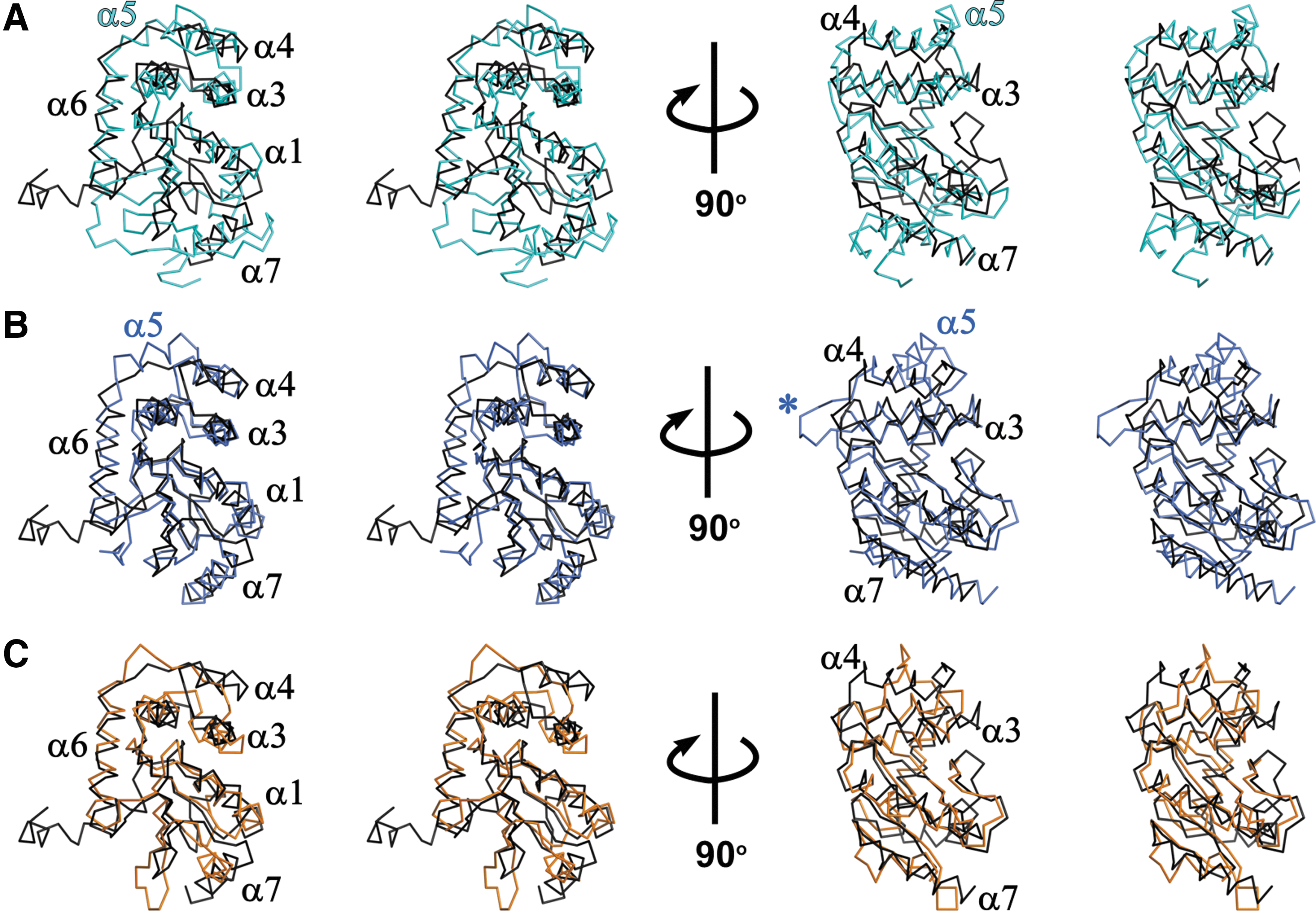
The Dali search (29) identified 3GYK, BdbD (PDB code 3GHA), and DsbG (PDB code 1V58) as the closest structural homologues to StScsC. 3GYK and StScsC exhibit high structural similarity (r.m.s.d. value of 1.5 Å over 165 Cα aligned); this protein is a DsbA-like homologue from Silicibacter pomeroyi although no functional characterization or publication is available. Structural superposition of StScsC with Bacillus subtilis DsbA protein BsBdbD (11) (r.m.s.d. value of 2.0 Å for 148 Cα aligned) highlights features of StScsC that deviate from this close structural homologue. For example, although BsBdbD and StScsC share a similar topology (β1 interacts with β3 instead of β5), the α5 helix is present in the helical domain of BsBdbD (Fig. 1B, 2B). Rotation of these structures by 90o also shows a longer loop connecting BsBdbD α3 and α4, which protrudes into the catalytic site (Fig. 2B). StScsC also shows structural similarity with DsbG, a disulfide isomerase from E. coli (23), which has also been shown to control cysteine sulfenylation in the oxidizing periplasm (13). EcDsbG is a homodimer, where each monomer incorporates a dimerization domain and a TRX domain linked by a connecting helix. StScsC superimposes unexpectedly well with the TRX domain of EcDsbG (2.3 Å for 134 Cα aligned, Fig. 2C). Furthermore, the lack of α5 in StScsC appears to emulate the shorter insertion present in this reductase, which only contains two helices and a long loop embedded in the TRX fold (23).
Analysis of surface electrostatics of StScsC
The electrostatic surface of StScsC partially resembles that of the thiol oxidase StDsbA, displaying a hydrophobic peptide-binding groove running along the protein surface and a hydrophobic patch, both adjacent to the CPYC catalytic motif (Fig. 3A). However, compared with StDsbA (Fig. 3B), the hydrophobic groove in StScsC is truncated. Moreover, next to the hydrophobic patch, StScsC displays a positively charged ridge above the exposed cysteine, comprising Lys52 on α1, Lys78 on the loop between β3 and α2 and Arg111 in the α3-α4 loop that overhangs the entrance to the active site cysteines (Fig. 3A). This cluster of positive residues surrounding the catalytic site is likely to stabilize the thiolate form of the nucleophilic cysteine Cys48. Interestingly, EcDsbG also has a positively charged ridge above the active site, with Lys139 on the loop between β3 and α2 directly above the exposed cysteine, although not as close as the Lys78 of StScsC. Lys168 (loop between α3 and α6) and Lys113 (α1) provide the additional positive charge to the right of Lys139 of EcDsbG (Fig. 3C).
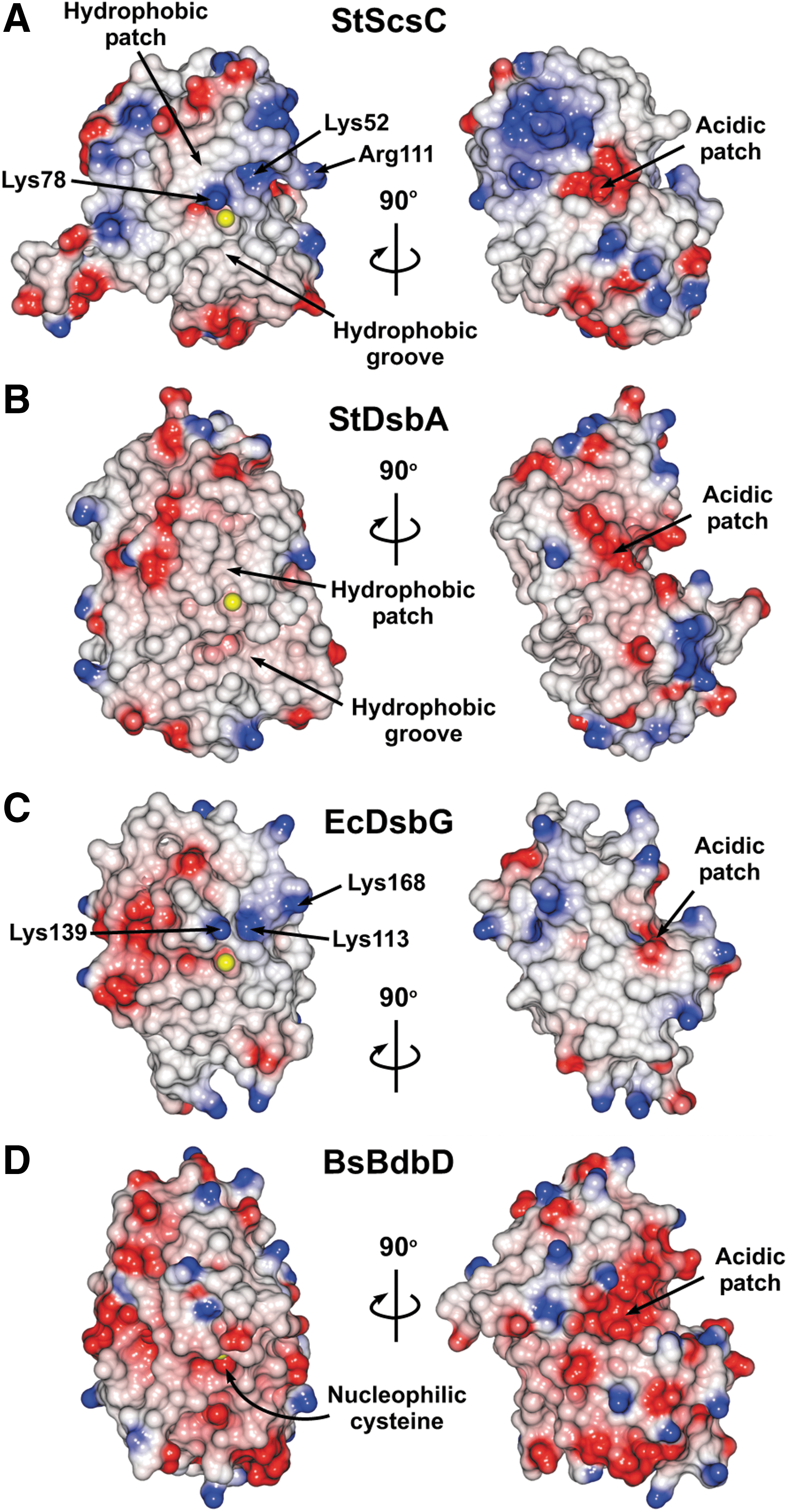
Although BsBdbD is the closest structural homologue for StScsC, these two proteins are not closely related (24% sequence identity), which results in discrepancies in their surface properties. Overall, BsBdbD displays a more acidic surface, primarily in areas surrounding the catalytic CPSC motif (Fig. 3A–D). The long loop between α3 and α4 is lined with negatively charged residues that map on top of the active site (11) (Fig. 3D). Despite their different surface properties, both StScsC and BdBdbD have acidic patches in the groove between the TRX and the helical domain, which map at the opposite side of their active sites, (Fig. 3, right panels). This acidic patch is conserved in other DsbA homologues although its exact role remains unclear (40).
The StScsC redox active site
The catalytic active site of StScsC resembles that of the disulfide isomerase EcDsbG rather than the oxidase StDsbA, it consists of a 48CPYC51 motif located at the N-terminus of helix α1 and a cis-Pro loop (156Thr-157 cisPro) in the α6-β4 connecting loop (Fig. 4A). StScsC was crystallized in its reduced form, as cleavage of the His-tag with tobacco etch virus (TEV) protease before crystallization involved incubation with 1 mM dithiothreitol (DTT). As shown in Figure 4B, the StScsC cysteine sulfur atoms are separated by a distance of 3.2 Å, which clearly indicates that these residues remained in their free thiol/thiolate forms upon crystallization. Several interactions may stabilize the reduced form of this protein; the nucleophilic N-terminal Cys48 Sγ is within hydrogen bond distance of the main chain nitrogen of the second cysteine (3.5 Å) and the side chain hydroxyl of Thr156 (3.5 Å). Furthermore, the protruding lysine residue (Lys78) forms a positively charged lip that overhangs the access route to the Cys48 residue. The close proximity between Lys78 and Cys48 could promote stabilization of the Cys48 thiolate, following a conformational rearrangement in this region; the Cδ atom of Lys78 is 3.9 Å away from the Cα atom of Cys48, but the Nζ of Lys78 is 6.6 Å from the Sγ of Cys48, so a conformational rearrangement would be required for this interaction to provide a significant stabilizing effect (Fig. 4A). The catalytic sites of BsBdbD and EcDsbG also reveal hydrogen bonds between the Sγ of the first cysteine and two acceptor groups: the second cysteine mainchain NH and the hydroxyl group of the Thr preceding the cis-Pro (Fig. 4C). The residues corresponding to StScsC Lys78 in BsBdbD and EcDsbG are His103 and Lys139, respectively. In BsBdbD, the Nδ2 of His103 is 4.8 Å away from the Sγ of Cys69, although a histidine residue is less likely to be cationic at physiological pH compared to a lysine residue, and is therefore less able to stabilize a cysteine thiolate. In EcDsbG, Lys139 generates a basic patch near the active site, which may also contribute to stabilizing the thiolate form of the cysteine (Fig. 4C).

Biochemical characterization of StScsC
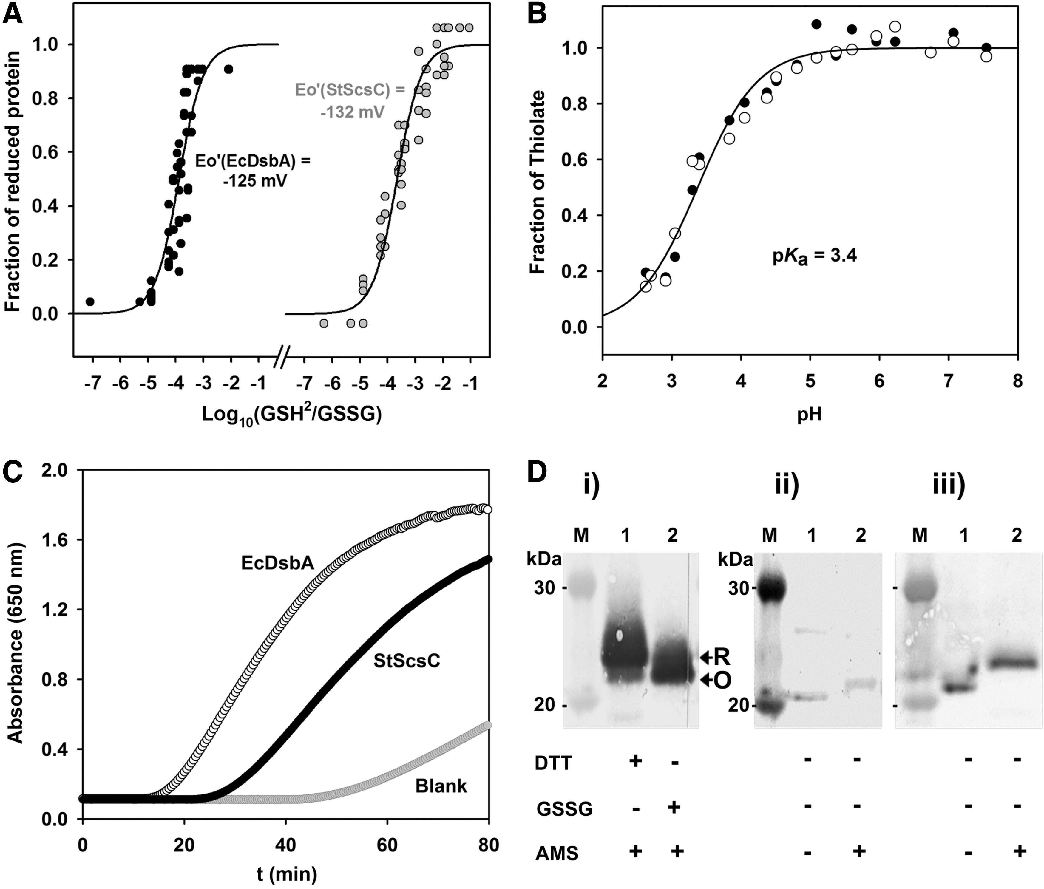
The biochemical properties of StScsC were characterized to gain further insights into its potential function. The oxidizing/reducing power of StScsC was quantified by measuring the equilibrium constant (K eq) for the disulfide exchange reaction with glutathione via monitoring changes in NMR spectra (Fig. 5A). The measured constants for StScsC and DsbA protein of E. coli (EcDsbA) were 21.7±2.6×10⁻⁵ M and 12.9±2.1×10⁻⁵ M, respectively. These K eq values convert into intrinsic redox potential values of −132 mV and −125 mV, respectively (Table 1). The redox midpoint of our EcDsbA-positive control is in close agreement with previously measured values for this protein (3). In addition, control spectra were recorded for the fully oxidized protein (using 5 mM oxidized glutathione [GSSG]) and for the fully reduced protein (using 5 mM DTT), providing confidence for the choice of peaks used to monitor the redox transition.
The first of the two cysteines in the
Given the redox potential exhibited by StScsC is similar to that of disulfide isomerases (Table 1) and its low pK a, which is also common in highly oxidizing proteins, we investigated the in vitro oxidoreductase activity using the classic insulin reduction assay: most TRX-like oxidoreductases, including DsbC and DsbA, catalyze DTT-induced insulin reduction. StScsC catalyzed the reduction of insulin more slowly than EcDsbA, and showed an activity ∼71% of the maximal rate of EcDsbA (Fig. 5C). To further understand the function of StScsC, we also investigated its ability to catalyze Dsb isomerization using the scrambled RNaseA (scRNaseA) refolding assay (27). Under our conditions, StScsC did not show any activity in this assay (Supplementary Fig. S2). In addition, to experimentally verify that StScsC is able to perform disulfide reduction in the native host, the in vivo redox status was measured using the thiol modification reagent 4-acetamino-4-acetamido-4′-maleimidylstilbene-2,2′-disulfonic acid (AMS) (Fig. 5D). Alkylation assays clearly indicate that StScsC is predominantly in the reduced state in the periplasm of S. Typhimurium.
Loss of StScsC elicits copper sensitivity in S. Typhimurium
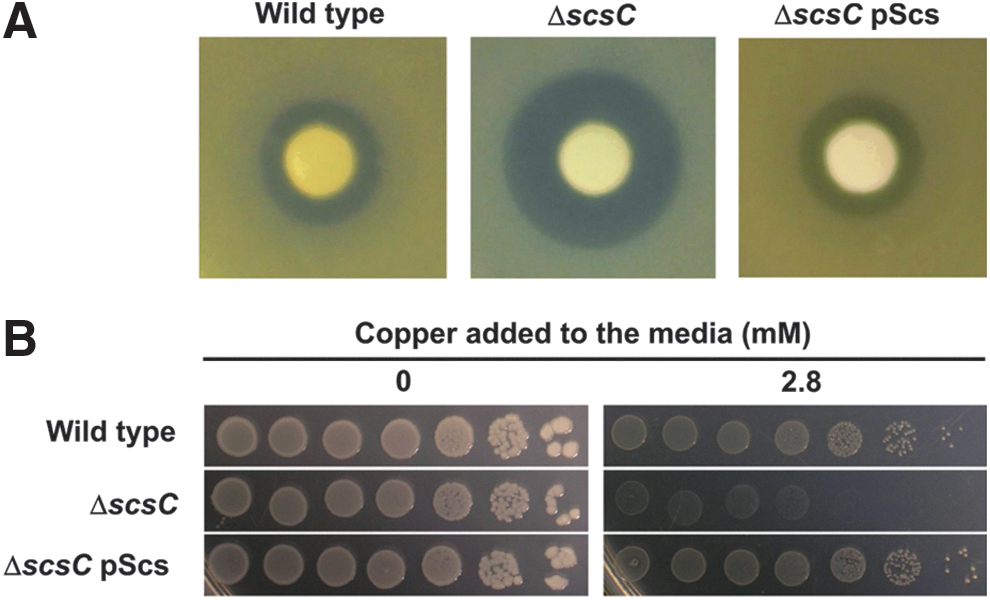
To examine the role of StScsC in S. Typhimurium, we constructed a scsC mutant in SL1344 (referred to as SL1344scsC). Loss of StScsC expression in SL1344scsC was confirmed by Western blot analysis with a StScsC-specific antibody (data not shown). Wild-type SL1344 and SL1344scsC were assessed for copper sensitivity using disk diffusion assays. In these assays, SL1344scsC displayed enhanced sensitivity to copper, as evidenced by the larger zones of growth inhibition observed in the presence of copper (Fig. 6A). Reintroduction of the scs genes on plasmid pScs restored the wild-type phenotype, confirming a role for StScsC in resistance to copper. To confirm this role for StScsC in S. Typhimurium and to more accurately assess the concentration of copper that inhibited growth of SL1344scsC, wild-type SL1344 and SL1344scsC strains were assessed for copper sensitivity by spotting a series of 10-fold dilutions of overnight cultures onto agar plates containing a range of different copper concentrations. In these assays, wild-type SL1344 and SL1344scsC displayed significantly different sensitivity to 2.8 mM copper (Fig. 6B). Reintroduction of the scs genes on plasmid pScs restored the wild-type phenotype, confirming a role for StScsC in resistance to copper.
Discussion
The formation of Dsb is an essential process for the correct folding of many periplasmic and secreted proteins. Bacteria exhibit a diverse array of disulfide folding machineries (15), particularly the Gram-negative bacteria, which encode a variety of systems that have evolved to catalyze the oxidative folding of virulence proteins required for infection (18, 38). A striking example of this is S. Typhimurium, which in addition to the oxidative DsbAB system also encodes an alternative DsbLI disulfide oxidation system similar to that of UPEC strain CFT073 (18, 57), as well as a plasmid-encoded DsbA-like protein called SrgA (6). In addition to these TRX-like proteins, the scs (
To date, little is known about the specific function of the Scs additional redox pathway in bacteria. A recent work on the C. crecentus Scs system showed that the membrane protein CcScsB and the soluble CcScsC form a redox pair similar to the DsbC/DsbD system. CcScsB mediates the reduction of dimeric CsScsC which, in turn, catalyzes the folding of proteins containing multiple cysteines via an isomerization mechanism (9). StScsC and CcScsC exhibit 24% sequence identity (Supplementary Fig. S3). Notable sequence differences between these two proteins include a different catalytic motif (CPYC vs. CGYC), the absence in StScsC of the long CcScsC N-terminal extension (which appears to mediate dimerization), and a deletion in CcScsC thirty residues upstream of the conserved cis-Proline present in these TRX-like proteins. To gain a better understanding of the Scs machinery in S. Typhimurium, we pursued a comprehensive structural and biochemical characterization of StScsC.
Structural characterization of StScsC revealed a canonical TRX domain with an inserted helical domain, characteristic of disulfide oxidoreductases like StDsbA or EcDsbA (25, 40). Despite this, StScsC has structural properties intermediate between DsbA-like thiol oxidases and disulfide isomerases. Indeed, a Dali search identified a DsbA homologue (BsBdbD) and an isomerase (EcDsbG) as close structural homologues for StScsC. Structural comparisons with those homologues revealed interesting peculiarities of StScsC. For example, a striking difference between StScsC and archetypal DsbA-like proteins is that the antiparallel β-strands in the TRX domain differ with respect to their topological arrangement: the β1 strand forms a β-sheet with β3 rather than β5 (Fig. 1). While this topology is uncommon in DsbA-like proteins, [only observed in few cases, including Wolbachia pipientis DsbA1 (WpDsbA1 (37)] and BsBdbD), this arrangement is present in the TRX domain of disulfide isomerases like EcDsbG and EcDsbC (23, 41). Moreover, a deletion in the helical domain of StScsC, which is not conserved in its homologue CcScsC (Fig. S2), results in StScsC lacking α5, one of the four usual alpha helices present in DsbA-like proteins like StDsbA or BsBdbD. Thus, the StScsC helical domain is somewhat reminiscent of the isomerase EcDsbG, which has a helical insertion containing two helices and a long loop substituting α4 and α5.
StScsC retains some surface features of the canonical StDsbA or EcDsbA, including a hydrophobic patch, shown to be important for substrate binding (40, 46), and a comparatively small hydrophobic groove under the active site, which in EcDsbA interacts with the partner protein EcDsbB and is key for reoxidation (31). The groove truncation in StScsC may compromise its ability to interact with StDsbB, maintaining StScsC separate from the S. Typhimurium StDsbA/StDsbB oxidative pathway. Furthermore, unlike archetypal DsbAs, the surface of StScsC also incorporates positively charged residues that generate basic protrusions flanking the 48CPYC51 active site (Fig. 3A). The latter surface properties resemble the lysines found adjacent to the also conserved 109CPYC112 active site of EcDsbG (Fig. 3C) and may contribute to stabilizing the thiolate form of the nucleophilic N-terminal cysteine.
StScsC differs from EcDsbG in that this protein is monomeric. Disulfide isomerases, EcDsbC and EcDsbG, contain extended N-terminal domains that allow dimerization, which is essential for their isomerase activity (50, 51). StScsC has an N-terminal helix that projects into the solvent (Fig. 1B, black, initial eight residues were not modeled due to weak electron density). This N-terminal helical domain is shorter than the one predicted in the C. crescentus homologue CcScsC (Supplementary Fig. S3) which is proposed to promote dimerization (9). Thus, unlike CcScsC or EcDsbG, StScsC purifies as a monomer. Furthermore, a protein interfaces, surfaces, and assemblies analysis (62) of StScsC crystal packing revealed no indication of dimer formation.
The structural analysis of ScsC revealed significant similarities to the TRX family proteins catalyzing both oxidation and isomerization/reduction. The redox properties of StScsC are similar to those of the archetypal disulfide-oxidizing DsbA and disulfide-reducing DsbG (Table 1). Furthermore, the pK
a of the exposed nucleophilic cysteine of StScsC is 3.4 (Fig. 5B), a low value that is a common characteristic of Dsb-like proteins like DsbA and DsbG (Table 1). This low pK
a may arise from the sulfur atom of Cys48 being hydrogen bonded to the backbone amide group of Cys51 and the hydroxyl of Thr156 in the cis-Pro loop as well as being flanked by a cluster of positively charged residues (Figs. 3 and 4) all of which contribute to the stabilization of Cys48 in the thiolate form. The low pK
a value of the nucleophilic cysteine for StScsC would favor a reduced disulfide, which is consistent with the relatively oxidizing redox midpoint of −132 mV (this disulfide is relatively easy to reduce). In the case of EcDsbA, a linear relationship between the pK
a of the exposed
Copper (II) is known to drive the oxidation of thiols (53) and since StScsC is required for protection against copper stress, this suggests that StScsC is part of a system in S. Typhimurium that is dedicated to protecting periplasmic proteins against the formation of non-native Dsb induced by this transition metal. The mechanistic role that StScsC plays is still unclear; however, our results indicate that StScsC exists in the reduced state in resting cells, which may indicate that this protein is a disulfide reducing protein. The reason why S. Typhimurium would possess a system dedicated to protection against copper-catalyzed Dsb formation is not entirely clear, but it is noted that copper ions have recently been shown to play an antimicrobial role in the innate immune response and there is evidence that this ion is trafficked to the phagosome in macrophages (28, 60). In macrophage infection studies, we did not observe any difference in the bacterial load when we compared wild-type S. Typhimurium with the scsC mutant (Achard & McEwan, unpublished), but it should be noted that copper tolerance in S. Typhimurium is complex and no single locus may be essential for survival in the macrophage.
Materials and Methods
Production of purified proteins
Native ScScsC was expressed using a method similar to the one descried previously for other S. Typhimurium DsbA-like proteins (32). Briefly, the coding DNA sequence for ScScsC lacking the signal peptide (residues 19–189), was cloned into a pET21a-based LIC vector (for cloning primers, see Supplementary Table S1). The recombinant protein containing an N-terminal hexa-histidine tag was expressed using autoinduction (54), and then purified by Ni2+-affinity chromatography and gel filtration chromatography (in 50 mM HEPES pH 7.0, 150 mM NaCl). Upon removal of the tagged signal peptide by cleavage with TEV protease (in a gel filtration buffer supplemented with 1 mM DTT), uncleaved protein was removed using cobalt affinity (Talon resin) chromatography. Histidine-tagged E. coli DsbA (EcDsbA) was purified as previously described (48).
Crystallization and diffraction data collection
Purified StScsC (in 50 mM HEPES pH 7.0, 150 mM NaCl) was stored in aliquots frozen at −80°C until needed. StScsC was concentrated using a 3K Amicon centrifugal concentrator up to 26 mg·ml−1. High-throughput crystallization experiments were performed with a Mosquito robot using commercial screening kits. Initial condition screening was performed in the hanging-drop format in 96-well plates, StScsC was at 26 mg·ml−1 and the commercial screens JCSG (Molecular Dimensions), Index (Hampton), and PegRx1/PegRx2 (Hampton) were used. Drops were set using the Mosquito® robot (TTP LabTech Ltd.) by mixing 100 nL of protein with 100 nL of well condition. The plates were incubated at 20°C and monitored and scored using the RockImager and Rockmaker system (Formulatrix, Inc.). Crystals were observed in three PegRx conditions (PegRx1 #33, 0.1 M Tris pH 8.0, 28% w/v PEG 4000; PegRx2 #28, 18% v/v 2-Propanol, 0.1 M sodium citrate tribasic dihydrate pH 5.5, 20% w/v PEG 4000; PegRx2 #29, 6% v/v Tacsimate pH 6.0, 0.1 M 2-(N-morpholino)ethanesulfonic acid monohydrate pH 6.0, 25% w/v PEG 4000). Optimization using random and grid screening strategies produced crystals that diffracted to 2.04 Å. These crystals were grown in 96-well plates in gradient screens of 18%–27% (w/v) PEG 4000 and 0.1 M Tris buffer pH 8.2–9.2. A further round of optimization designed to grow larger crystals free of precipitate was performed in 24-well plates using 18%–27% (w/v) PEG 4000 with 0.1 M Tris buffer pH 8.2 with protein at 9 mg·ml−1 using microseeding. The hanging drops were prepared by adding 1 μl of protein (9 mg·ml−1) to 1 μl of well condition and were microseeded within 30 min of setup with a seed stock prepared from small crystals grown in the same condition. Diffraction data were collected on a crystal (∼300×50×50 μm) grown by this method (Supplementary Fig. S4) with a well condition of 22% (w/v) PEG 4000 in 0.1 M Tris buffer pH 8.2. The crystal was cryoprotected by the well solution supplemented with 25% ethylene glycol and flash cooled in the cryostream. Data were collected using a Rigaku FR-E Superbright X-ray generator with Osmic HiRes2 optics and a Rigaku R-Axis IV++ Image Plate detector. CrystalClear 2.0 was used to record the diffraction and indexing and scaling was performed using HKL2000 (45).
Structure determination
The crystal structure of StScsC was solved by molecular replacement using BALBES (39) with a final search model based on the crystal structure of a TRX-like oxidoreductase from Silicibacter pomeroyi (PDB: 3GYK): Model building and refinement were carried out iteratively using COOT (16) and Phenix (2). Structure validation was performed using Molprobity (8). Supplementary Table S1 provides the statistics for the X-ray data collection and final refined model. Superposition of molecules and generation of molecular figures (including electrostatic potential) were carried out using the CCP4 Molecular Graphics program (47).
Bioinformatic analyses
Pairwise sequence alignments and calculation of sequence identities were performed using Biolign (21). Structural alignments were performed using MUSTANG (34), and the secondary structure was assigned using PDBsum.
Determination of the equilibrium constants with glutathione
The redox equilibrium of StScsC with glutathione was determined alongside that of EcDsbA as a positive control, using a recently developed NMR approach (56) (using his-tagged proteins). To determine the reduction potentials, different ratios of [reduced glutathione (GSH)]/[GSSG] were used, where [GSH]+[GSSG] equalled 5 mM. All NMR data were obtained at 298 K using a 14.1 T (600 MHz 1H) Bruker Avance III NMR spectrometer equipped with a QCI-F cryoprobe. All NMR samples were 330 μL within a Shigemi NMR tube and contained 100 μM protein in a 20 mM sodium phosphate buffer at pH 6.5 containing 50 mM sodium chloride and 5% (v/v) deuterium oxide. All buffers had nitrogen bubbled through to ensure the removal of any oxygen. We found that bubbling for 30–60 min was sufficient to remove all effects of dissolved oxygen and the redox equilibrium was complete in all samples within 5 min of equilibration of buffers. All 15N-1H HSQC spectra were acquired with 2048 points in the direct F2 dimension (1H) and 256 points in the F1 dimension (15N). NMR data processing was completed using NMRpipe. NMR backbone assignments have previously been completed for EcDsbA (10) and were used to confirm analysis. Two control spectra were recorded for each protein (Supplementary Fig. S5), the fully oxidized protein (using 5 mM GSSG) and the fully reduced protein (using 5 mM DTT). To determine the reduction potential of each protein, the fraction reduced was calculated for four and six resonances for StScsC and EcDsbA, respectively. The equilibrium constant K eq and redox midpoint potential were determined from standard thermodynamic equations (42). A redox midpoint potential of −240 mV was used for the glutathione couple (49).
Determination of pKa values
The pKa of the nucleophilic cysteine in StScsC was determined by monitoring the specific absorbance of the thiolate anion at 240 nm (43). Measurements were carried out at room temperature in a buffer system consisting of 10 mM K2HPO4, 10 mM boric acid, 10 mM sodium succinate, 1 mM EDTA, and 200 mM KCl, pH 7.5. Oxidized and reduced protein samples (final concentration of 20 μM) were prepared by incubating the proteins with 20 mM GSSG or 10 mM DTT, respectively. Upon removal of the oxidizing and reducing agents using a PD10 column (GE Healthcare), the absorbance of the samples at 240 and 280 nm was recorded as the pH of the protein solution was lowered to 2.5 by the stepwise addition of aliquots of 0.2 M HCl. The pH dependence of the thiolate-specific absorbance signal (S=(A 240/A 280)reduced/(A 240/A 280)oxidized) was fitted according to the Henderson–Hasselbalch equation as previously described (42). The oxidation state of StScsC samples was assessed using the DTNB assay with an extinction coefficient of ɛ412= 13.8 mM −1 cm−1 (17).
Insulin reduction and refolding of scRNaseA assays
The disulfide oxidoreductase activity of StScsC and EcDsbA was determined by measuring the ability to catalyze insulin reduction in the presence of DTT as described previously (37). The in vitro isomerase activity of StScsC, EcDsbC, and EcDsbA was assessed using the refolding of scRNaseA assay essentially as described by Hillson et al. (27).
Bacterial strains, plasmids, and culture conditions
S. Typhimurium SL1344 strains (wild-type and deletion mutants) and E. coli strains (BL21[DE3] and DH5α) were routinely cultured at 37°C on the solid or in liquid Luria Bertani (LB) medium supplemented with the appropriate antibiotics; kanamycin (Km, 50 μg·ml−1), ampicillin (Ap, 100 μg·ml−1 or chloramphenicol (Cm, 30 μg·ml−1). Culture media were supplemented with 1 mM isopropyl β-D-thiogalactopyranoside (IPTG) to induce expression of StScsABCD from plasmid pScs. Plasmid pScs was generated by PCR amplification of the scsABCD operon (primers scsA-DF, scsA-DR) from the chromosome of SL1344 and subsequent cloning into EcoRI-XhoI digested pWSK29 (61). Expression of cloned genes was controlled by the inducible lac promoter. All primers used in this section are shown in Supplementary Table S2.
Construction of S. Typhimurium SL1344 mutants
To delete the scsC gene, a three-step PCR procedure was employed to generate an amplification product containing the Km cassette from pKD4 (12) flanked on both sides by ∼500 bp of DNA sequence homologous to the target gene to be modified. The following sets of primers were used for scsC: scsCP1F, scsCP2R, scsCP5F, and scsCP6R (Supplementary Table S2). These primers were used in combination with the Km cassette amplification primers KanP3 and KanP4 (Supplementary Table S2). The final three-step PCR product for each target gene was gel-purified and cloned into pT7Blue (Merck) according to the manufacturer's instructions, to generate a donor plasmid. SL1344 deletion mutants were constructed using the gene gorging method as previously described (26). Briefly, SL1344 cells were electrotransformed with a donor plasmid [pT7Blue::scsC::kan, and a mutagenesis plasmid (pACBSR, (26)]. Cells containing both plasmids were cultured for 9 h in the LB broth supplemented with 25 μg·ml−1 Cm and 0.2% (w/v) L-arabinose to induce the λ-Red and I-SceI genes on pACBSR. Induction of these genes leads to linearization of the donor plasmid and a double recombination event with the regions flanking scsC, resulting in disruption of the respective gene. Clones sensitive to Ap and Cm (but resistant to Km) were selected and deletion of scsC was confirmed by PCR and subsequent DNA sequencing.
Copper sensitivity assays
Copper sensitivity was assessed via growth on solid media. For disk diffusion assays, strains were grown at 37°C to mid-exponential phase, adjusted to the same cell density and plated using the agar overlay technique on LB supplemented with 1.5% agar. Sterile filter disks soaked with 5 μL of 1 M CuSO4 were placed on the center of each plate and the zones of inhibition were measured after an overnight incubation at 37°C. The data presented are representative of three independent experiments. For colony spot assays, serial 10-fold dilutions of cultures of the same density were prepared and 5 μl of each dilution was spotted on the Tris agar medium supplemented with 1.4–3.6 mM CuSO4 (at 0.2 mM increments) or left untreated. Pictures were taken after 24 h of incubation at 37°C. The data presented are representative of three independent experiments comparing growth in the presence or absence of 2.8 mM CuSO4.
Determination of the in vivo redox state of StScsC
The periplasmic protein fraction of SL1344 and SL1344scsC pScs was prepared by cold osmotic shock. Bacteria grown overnight at 37°C were washed in an ice-cold buffer containing 20% surcrose, 200 mM Tris HCl, pH8, and cells were harvested by centrifugation and resuspended in ice-cold 10 mM Tris HCl, pH8. After centrifugation, the supernatant was retained for analysis. The redox state of ScsC was measured by thiol alkylation using AMS and analysis of the molecular mass of proteins following sodium dodecyl sulfate polyacrylamide gel electrophoresis and Western blotting as previously described (30). ScsC proteins were detected using a StScsC-specific antibody (1:100) and an anti-rabbit IgG coupled with alkaline phosphatase (1:10000; Sigma) as the secondary antibody. Blots were stained with 5-bromo-4-chloro-3-indolyl-phosphate/nitro blue tetrazolium.
Footnotes
Acknowledgments
This work was supported by an Australian Research Council (ARC) grant DP1096395 to M.A.S., A.G.M., and B.H. M.A.S. is supported by an ARC Future Fellowship (FT100100662) and B.H. by a La Trobe Institute for Molecular Science Fellowship, (La Trobe University). The Wellcome Trust supported the reduction potential NMR experiments via Equipment Grant 091163/Z/10/Z to M.J.H. The authors acknowledge the use of the Australian Synchrotron and the UQ ROCX diffraction Facility.
Author Disclosure Statement
No competing financial interests exist.
Abbreviations Used
References
Supplementary Material
Please find the following supplemental material available below.
For Open Access articles published under a Creative Commons License, all supplemental material carries the same license as the article it is associated with.
For non-Open Access articles published, all supplemental material carries a non-exclusive license, and permission requests for re-use of supplemental material or any part of supplemental material shall be sent directly to the copyright owner as specified in the copyright notice associated with the article.