
Abstract
Tumor hypoxia is a well-established biological phenomenon that affects the curability of solid tumors, regardless of treatment modality. Especially for head and neck cancer patients, tumor hypoxia is linked to poor patient outcomes. Given the biological problems associated with tumor hypoxia, the goal for clinicians has been to identify moderately to severely hypoxic tumors for differential treatment strategies. The “gold standard” for detecting and characterizing of tumor hypoxia are the invasive polarographic electrodes. Several less invasive hypoxia assessment techniques have also shown promise for hypoxia assessment. The widespread incorporation of hypoxia information in clinical tumor assessment is severely impeded by several factors, including regulatory hurdles and unclear correlation with potential treatment decisions. There is now an acute need for approved diagnostic technologies for determining the hypoxia status of cancer lesions, as it would enable clinical development of personalized, hypoxia-based therapies, which will ultimately improve outcomes. A number of different techniques for assessing tumor hypoxia have evolved to replace polarographic pO2 measurements for assessing tumor hypoxia. Several of these modalities, either individually or in combination with other imaging techniques, provide functional and physiological information of tumor hypoxia that can significantly improve the course of treatment. The assessment of tumor hypoxia will be valuable to radiation oncologists, surgeons, and biotechnology and pharmaceutical companies who are engaged in developing hypoxia-based therapies or treatment strategies. Antioxid. Redox Signal. 21, 1516–1554.
I. Introduction
T
The detection and assessment of tumor hypoxia now plays a critical role in both the validation and development of hypoxia modification therapies for their eventual adoption into routine clinical practice. Recent clinical trial data appear to demonstrate how patients identified by their hypoxic tumor status have beneficial treatment responses to hypoxia-modified therapies compared with standard therapies, while high-risk patients with hypoxic tumors receiving the control therapies performed poorly. However, despite current research into hypoxia-modification therapies, measurement of tumor hypoxia within lesions is not performed routinely and, consequently, has hindered the development of these therapies. Therefore, there is an acute need to identify tumor hypoxia, to enable appropriate patient classification, and to determine the extent of hypoxia within each lesion, enabling clinicians to make decisions regarding the therapy management for the patient. The hypoxia assessment may aid not only radiation oncologists and surgeons, but also biotechnology and pharmaceutical companies in developing tumor hypoxia therapies or other new treatment strategies for hypoxic tumors.
II. The Clinical Importance of Tumor Hypoxia
A. Pathophysiology of hypoxia
Hypoxia is a pathophysiological property that is defined as a state of depressed oxygen tension. Hypoxia can be present in tissues, including tumors, causing the impairment of cellular or organ functions once critical oxygen levels are breached. Localized tissue hypoxia, as it relates to tumors, can be the result of two general types of oxygen starvation. Hypoxia can be perfusion limited (“acute hypoxia”), caused by a temporary reduction in blood supply. Alternatively, hypoxia can be diffusion limited (“chronic hypoxia”), caused by insufficient vascularization impairing the metabolic needs of the growing tumor. The presence of hypoxia in cancerous tissue was first reported by Thomlinson and Gray, who observed that hypoxic, yet viable, lung carcinoma rods were surrounded by a necrotic core caused by a tissue oxygen gradient (263). All types of solid tumors, especially malignant solid tumors, are subject to hypoxia, often exhibiting oxygenation levels measurably lower than their tissue of origin (Table 1) (7, 27, 124). Recurring tumors often exhibit a higher hypoxic fraction than primary tumors (258, 280). A tumor's hypoxic status cannot be accurately determined anatomically, as the presence of tumor hypoxia is independent of a tumor's size, stage, grade, or histology. Intratumoral oxygenation often disperses heterogeneously, and, therefore, an accurate characterization of tumor hypoxia is possible only from composite measurements.
HPx—the percent frequency of pO2 measurements below a given mmHg value, see Figure 3 for further explanation.
DFS, disease-free survival; HIF, hypoxia-inducible factor; OS, overall survival.
The physiology and biochemistry of hypoxic tumors adapts to oxygen starvation to preserve both tumor growth and propagation. For example, in depressed oxygen environments, hypoxic cells readily revert from aerobic respiration to anaerobic glycolysis, which increases both glucose consumption and the production of pyruvate. These cells can continue to function using this metabolic pathway even in the presence of oxygen (the Warburg effect) (247). This survival mechanism is common among hypoxic tumors, resulting in a twofold increase in glucose uptake (determined in vitro) (29), elevated tissue acidity (44), and an evolutionary selection for a progressively more malignant phenotype (102, 122).
The proteomic and genomic transformations of tumor cells in response to hypoxia lead to permanent alterations in their cellular composition that is regulated predominantly by hypoxia-inducible factors (HIFs). HIFs are a family of heterodimeric transcription factors that are critically up-regulated in response to hypoxia. HIF-1α is a member of this family and it plays an integrative role in the cellular response to hypoxia (245, 274). HIF-1α contains two transactivation domains known as the N-terminal activation domain (N-TAD) and the C-terminal activation domain (C-TAD). C-TAD regulates HIF-1α transcriptional activation under hypoxia, while N-TAD modulates stability of HIF-1α. Under normoxic conditions, amino-acid-specific hydroxylations deactivate HIF-1α activity. Prolyl hydroxylase domain protein (PHD) hydroxylates proline residues on HIF-1α in an oxygen-dependent fashion, enabling von Hippel-Lindau (VHL) tumor suppressor protein to bind HIF-1α for subsequent ubiquitylation and targeted degradation by 26S proteasome (Fig. 1). HIF-1α can also undergo hydroxylation by factor inhibiting HIF1 (FIH-1), which inhibits the recruitment of HIF-1α coactivators and the expression of downstream HIF-1α target genes. However, when intracellular levels of oxygen decrease, the rate of HIF-1α hydroxylation by PHD and FIH-1 also decreases. Consequently, VHL fails to bind to HIF-1α, leading to an accumulation of HIF-1α in the cell. This enables HIF-1α to interact with CREB-binding protein/E1A-binding protein p300 (CBP/p300), a transcriptional co-factor, followed by heterodimerization with HIF-1β. The heterodimer readily binds to the HIF-responsive element (HRE) of DNA, leading to the transcriptional activation and up-regulation of multiple HIF-1α target genes. Other factors affecting HIF-1α activation will be discussed in a subsequent section.

Through the onset of hypoxia, HIF-1α initiates multiple hypoxia-derived molecular processes that drive tumor growth, proliferation, and metastatic potential. Growth and survival mechanisms such as angiogenesis via vascular endothelial growth factor (VEGF), pH regulation via involving carbonic anhydrase IX (CA-IX), metabolism via glucose transporter 1 (GLUT-1), and oxygen management are up-regulated in response to hypoxia (130). Transcription factors that control cell proliferation, cell survival via nuclear factor kappa-light-chain-enhancer of activated B cells (NF-kB), and apoptosis via signal transducer and activator of transcription 3 (STAT3, p53) are also up-regulated in hypoxic tumor cells. The up-regulation of these proteins directly influences the malignancy of hypoxic tumors (102, 275, 276), including a propensity for the development of metastatic disease (23, 255, 297) by propagating cells that have lost their apoptotic potential through decreased p53 expression, increased mutant p53 or bcl-2 protein expression (105, 155), increased angiogenesis (254), and overall increases in proteinase activity (31, 106). Over-expression of several of the protein markers mentioned earlier and linked to hypoxia, each one alone or in conjunction with other markers, has been found to be prognostic for 10-year OS and cancer-specific survival in head and neck patients (161).
B. Hypoxia's negative impact on the effectiveness of curative treatment
1. Hypoxic tumors accumulate and propagate cancer stem cells
Tumor-initiating cells, also known as cancer stem cells (CSC), have phenotypic characteristics that include differentiation, self-renewal, apoptotic resistance, pluripotency, and sufficient motility to initiate new tumor growth at distant sites. Unsurprisingly, the presence of CSC in tumors correlates strongly with both treatment failure and tumor recurrence, despite being a smaller subset of the overall tumor cell population (58). Hypoxic regions within solid tumors harbor CSC in an area known as the hypoxic niche (173), which enriches the CSC population through accumulation and selective propagation. In breast tumors, CSC populations increase within hypoxic zones that are modulated by protein kinase B (Akt)/β-catenin signaling, a pathway which is reported to enable self-renewal of breast CSC (52). Tumor hypoxia reprograms cell dedifferentiation in ductal breast carcinomas that are characterized by a down-regulation of estrogen receptor-α and an increase in the epithelial breast stem cell marker cytokeratin 19, creating a more aggressive, stem cell-like phenotype (118).
2. Hypoxia reduces the effectiveness of radiotherapy
Evidence that depressed oxygen levels compromise the effects of radiation was established more than 75 years ago. Mottram and co-workers observed that poorly oxygenated normal and malignant tissues were resistant to the effects of both X- and γ-irradiation because of a lack of long-lived reactive oxygen radicals (197). Thomlinson and Gray demonstrated that hypoxic tumor cells were thrice more resistant to radiation than well-oxygenated ones, thus forming the basis of understanding that hypoxia impairs the effectiveness of radiotherapy (109, 122). In addition to the physical aspects of radio-resistance imparted by hypoxia, the acquired genetic traits of hypoxic tumor cells during their malignant progression also actively contribute to mechanisms of radioresistance. Hypoxic cells with decreased apoptotic potential and deregulated cell cycle arrest mechanisms still undergo cellular proliferation despite damaged DNA. In addition, the expression of proteins downstream from HIF1-α, such as VEGF and basic fibroblast growth factor (bFGF), were also found to confer radioresistant effects (193, 245). CSC are reported to have reduced amounts of endogenous reactive oxygen species (ROS) relative to both tumorogenic and nontumorogenic cells of the same type, which may prolonged survival of CSC, especially during treatment cycles (66). In addition, mitochondrial dysfunction in a subset of head and neck squamous cell carcinoma CSC is associated with a decrease in levels of endogenous ROS within the cell (98). In the clinical setting, both head and neck and prostate cancer patients with hypoxic tumors undergoing radiotherapy have been reported to be at elevated risk for poor LRC, OS, and biochemical failure (21, 270).
3. Hypoxia increases metastasis risk and reduces the effectiveness of surgery
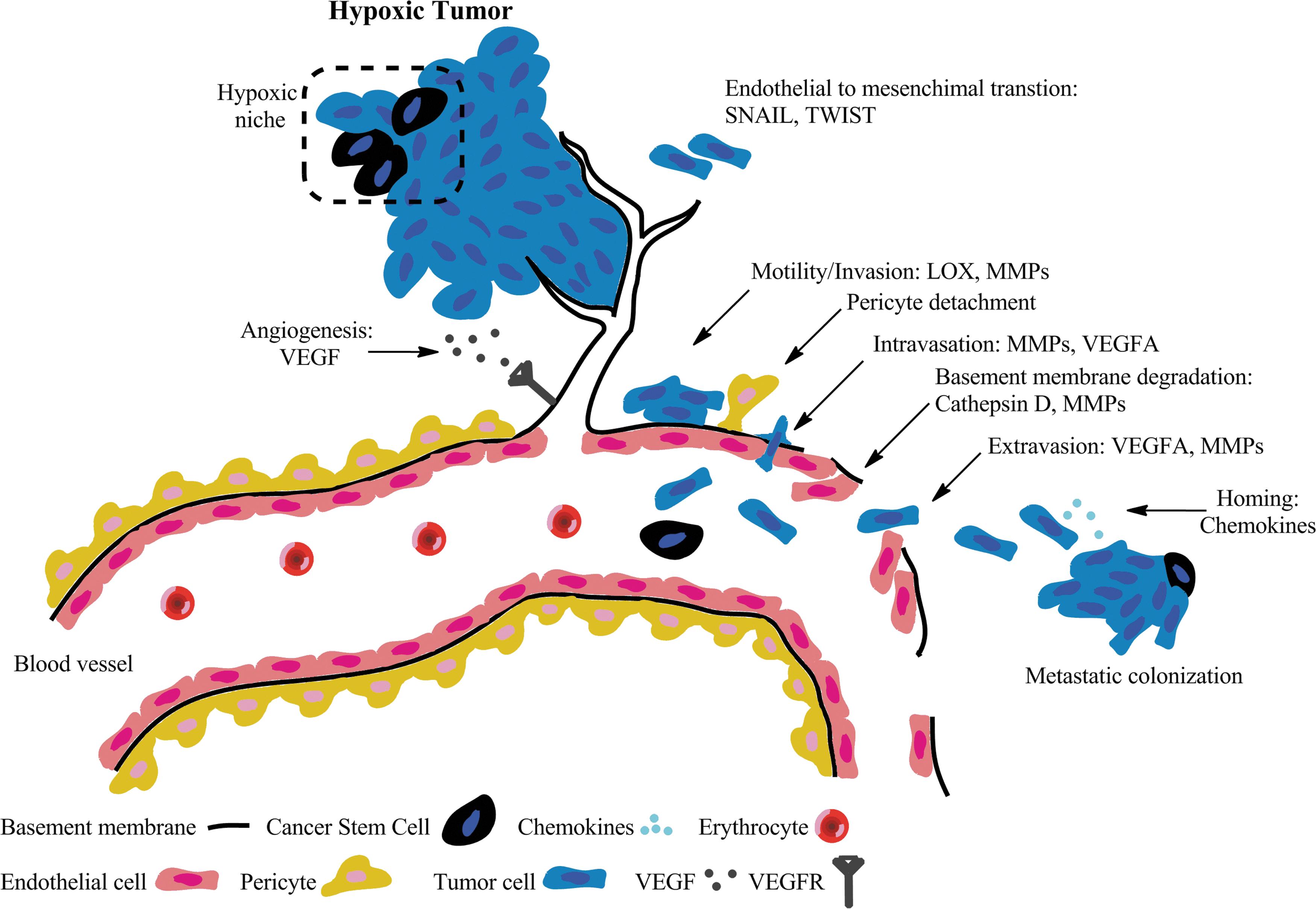
Hypoxia has been linked to the formation of metastatic disease and, thus, provides important prognostic information (255). The downstream expression of hypoxia proteins initiates and supports metastatic spread via tumor cell motility and invasion, intravasation, extravasation, and metastatic colonization (Fig. 2) (179). Hypoxia-mediated activation of snail family zinc finger 1 (SNAIL1) and class A basic helix-loop-helix protein 38 (TWIST) induces epithelial to mesenchymal transition in tumor cells enhancing cell motility by diminishing cell–cell adhesion properties and inducing a loss of cell polarity. Cellular motility is further supported by the expression of lysyl oxidase (LOX), an extracellular matrix remodeling enzyme. Both matrix metalloproteinase (MMP) and cathepsin D compromise the basement membrane, facilitating tumor invasion. Subsequent pericyte detachment from the basement membrane exacerbates vascular structure and function. The overexpression of VEGF leads to leaky vasculature and high vessel permeability, facilitating both intravasation and extravasation of circulating tumor cells and even CSC. Lastly, secreted chemokines facilitate the localization of tumor cells, leading to the formation of metastatic colonies (133).
The effects of hypoxic-mediated metastasis have also been reported in the clinic. In a study of H&NC patients undergoing planned neck dissection after primary radiotherapy treatment, pathology analysis of the neck dissection specimens confirmed the presence of viable tumor from patients having hypoxic lesions, yet no residual disease was found in patients with normoxic lesions (21). In addition, clinical reports have cited an increased propensity for distant metastases in patients with hypoxic soft tissue sarcomas (STS) and cervix cancer undergoing surgery (23, 121). Patients with hypoxic prostate tumors treated by radical prostatectomy were at high risk for biochemical failure over a period of 8 years independent of pathological tumor stage, Gleason score, serum prostate-specific antigen (PSA) concentration, and margin status (281).
4. Hypoxic tumors are resistant to the effects of chemotherapy and chemoradiation
Tumor hypoxia exacerbates chemotherapy resistance through both physiological and genomic mechanisms (257, 260). From a physiological perspective, hypoxia potentiates the growth abnormal vascular networks that support cancer progression. Deregulated vasculature consists of vessels with pathologic size, inconsistent dilation, tortuousness networks, and hyper-permeability. In these conditions, the delivery of agents that are beneficial for cancer treatment is both irregular and inefficient, increasing the immune tolerance of cancer. Hypoxic tumors continually up-regulate angiogenic factors, such as VEGF, to meet metabolic demands; however, this neovascularization fails to support adequate blood supply that further worsens local hypoxia, ensuring a vicious cycle. Prolonged treatment of anti-angiogenic therapies has been known to exacerbate hypoxia, resulting in subsequent treatment failures (32, 101).
On the genomic level, reduced proliferation rates, up-regulation of multidrug resistance, and increased cellular acidification can diminish drug toxicity (44). Consequently, angiogenic factors are up-regulated, furthering the cycle of hypoxia. Hypoxic cells are known to be more resistant toward fluorouracil (251), doxorubicin (92), bleomycin (233), and platinum-based drugs (261) than normoxic cells. Combined therapeutic modalities have also demonstrated a diminished performance in hypoxic tumors (229, 287). Despite the overall improvement in locoregional control (LRC) and OS, treatment strategies, including concurrent chemo-radiotherapy, have had higher failure rates with hypoxic tumors than normoxic ones (214). Patients with hypoxic tumors undergoing chemoradiation therapy often exhibit both poor treatment response (22) and survival rates (201). Hypoxia compensation is, therefore, a critical aspect in treating cancer.
C. Hypoxia is prognostic for poor patient outcomes
Multiple clinical studies have concluded that hypoxia is associated with poor prognosis across multiple tumor types as evidenced by adverse outcomes, including poor OS, DFS, and LRC (Table 1). While there are likely multiple factors influencing a patient's poor outcome and response to therapy, tumor hypoxia is established as one of the strongest prognostic indicators, especially for patients with H&NC. Key clinical studies are highlighted next.
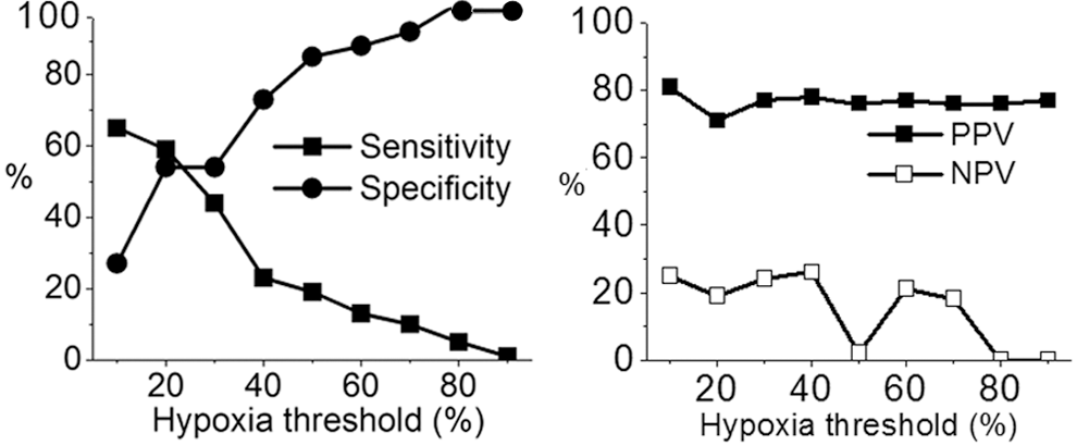
In a study reported by Rudat et al. (Table 2- Study F), for patients with Stage IV H&NC treated with radiation with or without chemotherapy, the hypoxic fraction (HP2.5) was shown to have a predictive value for OS, with the hypoxic subgroup having lower probability of survival (P=0.05). A subsequent analysis by Rudat et al. (236), using pooled data from very advanced H&NC patients with inoperable tumors, also found that tumor oxygenation above the median (HP2.5<9%) was associated with longer survival, confirming the influence of tumor oxygenation on the prognosis of patients with H&NC. The authors also concluded that the sensitivity for predicting patient survival status using a ≤10% threshold for HP2.5 was 75% (1 year survival) and 65% (2-year survival); the corresponding specificity was 41% (1 year survival) and 27% (2 year survival). While the negative predictive value (NPV) was low (25%), the positive predictive value (PPV) was high (81% for 2 years), which is desirable for selecting patients for treatment alternatives. A separate study determined that the NPV of pO2 in predicting pathological clinical response in normoxic patients was 80%, and the PPV to predict persistent disease in hypoxic tumors was 62% (Fig. 3) (22).
DFS, disease free survival; HR, hazard ratio; HSV, hypoxia subvolume; LRC, locoregional control; R, radiation; R+C, radiation plus chemotherapy; w/wo S, with or without surgery.
Nordsmark et al. (201) reported the results of a joint analysis of the data from all of the studies in Table 3, except Study A and Study H, as well as some previously unpublished data. Based on the pooled tumor hypoxia data, Nordsmark concluded that pretreatment tumor hypoxia defined by HP2.5 was a significant prognostic factor for survival after treatment with radiation alone or in combination with surgery, chemotherapy, or a radiation sensitizer (201). Based on the univariate analysis, the relationship between 3 year survival and tumor hypoxia (HP2.5) was found to be significant. In multivariate models, only pretherapy HP2.5 was prognostic for 3 year OS (P=0.006) using a hypoxia threshold of HP2.5>19%. Nordsmark also concluded that a change in HP2.5 from 30% to 40% in two otherwise identical tumors increased the relative risk of death by 13%. The authors concluded that the international, multi-center study firmly established the prognostic significance of hypoxia in H&NC patients after radiotherapy. In conclusion, while there are likely multiple factors influencing a patient's response to therapy, tumor hypoxia has been established as one of the strongest prognostic indicators, and, thus, pretreatment measurements of tumor oxygenation will be useful in the search for therapeutic strategies for overcoming hypoxia in H&NC.
Includes additional patients not reported in Studies E and F.
Presumed to be a subset of patients in Rudat et al. (236).
397 is total number of patients across studies and centers; however, one or more prognostic factor was missing for many patients; the final regression model was based on data from 253 patients.
Tx, treatment; R, radiation; R+C, radiation plus chemotherapy; w/wo S, with or without surgery.
III. Diagnosis of Tumor Hypoxia
Given the numerous treatment complications related to hypoxia, researchers have investigated various ways to assess hypoxia within tumors. Interestingly, the emerging field of hypoxia modification therapies has been largely ongoing without a convenient means for assessing tumor hypoxia, severely restricting the ability to stratify patients based on their tumor's hypoxia status. To continue the development of hypoxia modification therapies, there should be an accurate, composite, and noninvasive means for tumor hypoxia assessment that enables both appropriate patient selection and, ultimately, a change in therapy management.
Methods for assessing tumor hypoxia can be separated into three major groups: methods directly related to assessing the oxygen concentration, methods reporting on the physiologic processes involving oxygen molecules, and methods evaluating the expression of endogenous markers as a response to hypoxia (Fig. 4). Table 4 summarizes the existing methods used to evaluate tissue hypoxia status.

BOLD, blood oxygen level dependent; CA-IX, carbonic anhydrase IX; CT, computed tomography; EPR, electron paramagnetic resonance; PET, positron emission tomography; •, method of hypoxia assessment; +, approved clinical procedure; −, not approved clinical procedure.
Direct methods for detecting tissue hypoxia rely on the explicit interaction of oxygen with a selective sensor providing oxygen concentration data within the vicinity of the probe. Physiologic methods report on processes also directly involving oxygen molecules. While these methods do not directly measure oxygen concentrations in tissues, their response is proportional to the concentration of oxygen. Endogenous markers of hypoxia comprise proteins that are overexpressed in response to diminished oxygen supply. Considerable effort has been invested into the development of simple, reliable, and accurate methods for determining hypoxia using immunohistochemical (IHC) staining (via biopsy or tumor sectioning) or plasma protein assays (7, 283).
A. Direct methods
1. Oxygen electrode—direct pO2 measurement most used in cancer research
The polarographic electrode is an invasive, yet direct method for measuring tissue oxygen concentration based on the electrochemical reduction of oxygen molecules. Between 1988 and 2005, more than 70 research articles quantified data on the oxygenation status of solid tumors (277), and the method is often cited as the “gold standard” for hypoxia determination (93, 253). The procedure is considered safe, and no major adverse effects in using the probes for hypoxia assessment have been reported so far (277). Similar oxygen electrodes are currently used clinically for brain oxygenation assessment during neurocritical procedures.
The oxygen measurements involve inserting an electrode into a tumor or metastatic lymph node and measuring oxygen from several points per needle track in sub-millimeter steps. The more regular the lesion shape results in a more representative sampling of pO2 measurements (100). Typically, more than a hundred measurements are generated over the accessible areas of the lesion, providing a composite overview of the lesion's hypoxia status. The probes are reported to sample a tissue volume of about 50–100 cells (253). The polarographic probes have limited sampling capabilities, accessible only to surface lesions including metastatic lymph nodes. The distribution of pO2 tension between primary tumors and lymph node metastases have shown little difference, suggesting that node measurements can be surrogates for the hypoxic status of the primary tumor, (277) although later positron emission tomography (PET) imaging studies have reported a discordance in uptake between lesions (vide infra) (229).
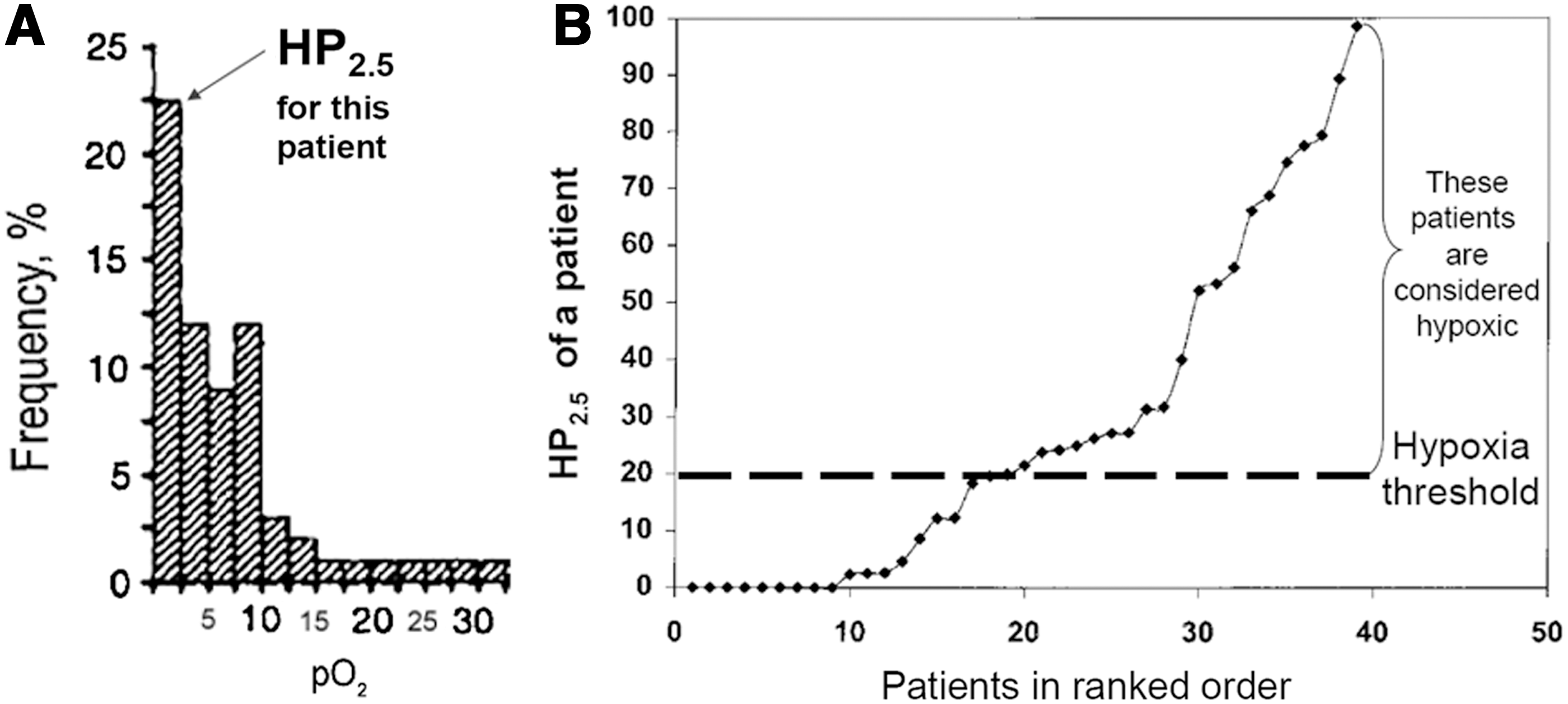
Data obtained from all tracks form a histogram: a graph plotting the oxygen pressure versus the frequency of this particular pressure within the tumor (Fig. 5A) (158). In order to facilitate the assignment of hypoxic versus normoxic tumors across patient populations, several descriptive parameters of the histogram have been reported, including the frequency of measurements below a given mmHg value, referred to as the hypoxic fraction. An example using 2.5 mmHg (HP2.5) as the hypoxic cut-off is shown in Figure 5A. Other descriptive statistical parameters include HP5, HP10, and median pO2. Hypoxia subvolume is another metric for hypoxia quantification defined as the total tumor volume multiplied by the hypoxic fraction. The stratification of patients into hypoxic or normoxic subgroups requires defining a hypoxic threshold. Figure 5B illustrates a defined hypoxia threshold of a large patient population (262). While there is no standard threshold commonly accepted for defining hypoxia, researchers reported using median HP2.5 (236) or HP2.5>19% (108, 201) as hypoxic cut-offs to identify patient groups, as these values are linked to poor treatment outcomes.
Oxygen electrodes have several limitations that impede their routine use in clinical practice. Most notably, the method is highly invasive, making repeated measurements extremely rare. The machine itself requires a technically skilled user, and inter-operator variability can be significant. The construction of three-dimensional (3D) oxygen maps is using electrodes difficult, despite a spatial resolution of 50–100 cells, preventing electrode-based therapy planning. The probe does not discriminate between viable and necrotic tissue; thus, it overestimates hypoxia when necrotic areas are sampled (277). Polarographic electrodes function poorly when patients are administered halogenated anesthetics (such as halothane), giving rise to inaccurate oxygen measurements (64, 248).
2. Phosphorescence quenching—alternative direct pO2 measurement
Phosphorescence quenching relies on the interaction of oxygen molecules with phosphorescent dyes. When illuminated with a short flash of light, the dyes emit their own light and the intensity of this emission decays exponentially at a rate proportional to the local oxygen concentration. Using precalibrated parameters that include the rate of decay in the absence of oxygen and the Stern–Volmer constant, the rate of decay translates into tissue oxygenation. Unlike other oxygen-sensing techniques, the oxygen feedback is independent of tracer concentration, because phosphorescence lifetime is analyzed rather than signal intensity. Another key advantage of this methodology is its high temporal resolution that provides a real-time tissue oxygenation profile which is not easily obtainable with other methods.
The two basic phosphorescent quenching methods employed for the in vivo assessment of oxygen concentration rely on either molecular reporters or physical needle probes. Molecular reporters are largely based on various palladium-containing porphyrins with preclinical investigations starting in 1986 (238, 273). An early in vivo investigation of phosphorescence quenching examined the behavior of an intravenously administered porphyrin solution in a perfused rat liver subjected to either normoxic or anoxic conditions. The phosphorescence quenching in the liver increased fourfold under anoxic conditions with a response time of approximately milliseconds, indicating that the discrimination of oxygen levels was possible using this modality.
Dendrimerized palladium porphyrins, Oxyphor R2 and G2, were later developed for mapping oxygen concentrations in murine tumor models at oxygen levels below 10 mmHg (76) using either basic oxygen contour plots or reconstructed oxygen histograms (301). From a preclinical validation study, the Oxyphor G2 signal differentiated well-oxygenated malignant melanomas (37.8±5.1 mmHg) from both moderately oxygenated renal cell carcinomas (24.8±17.9 mmHg) and hypoxic Lewis lung carcinomas (1.8±1.1 mmHg). The assessment of hypoxia was confirmed by tumor histology, pentafluorinated etanidazole (EF5) binding, and tumor blood flow by contrast-enhanced ultrasound suggesting that Oyxphor G2 provides critical tissue oxygenation information in tumors. Recently, new Oxyphor derivatives were developed to tolerate a variety physiological environments (289). This new generation of the “protected” dendritic probes operate in either albumin-rich (blood plasma) or albumin-free (interstitial space) environments over a range of physiological oxygen concentrations with excellent submillimeter spatial and subsecond temporal resolution (85).
Recently, phosphorescent molecules embedded into a solid support within physical needle probes enable tissue oxygen measurements, albeit invasively. These instruments monitor regional pO2 concentrations in brain tissue (63) and surface accessible tumors (110).
3. Electron paramagnetic resonance
The method requires an injection of an exogenous probe bearing an unpaired electron that is selective in its interaction with oxygen (97). Recently, implantable and metabolically inert paramagnetic lithium phthalocyanine crystals have been utilized as oxygen-sensitive electron paramagnetic resonance (EPR) probes to monitor changes in tissue hypoxia (140). The width of the spectral band corresponding to the probe signal correlates with oxygen concentration. Similar to oxygen electrodes, EPR imaging data provide quantitative pO2 values. With EPR, repeated measurements of absolute pO2 from the same tissue spanning a few minutes to days or even months enables the sensitive detection of fluctuating hypoxia, which is an advantage over other imaging methods.
Oxygen levels determined by EPR imaging closely mirror oxygen levels assessed by Oxylite measurements in fibrosarcoma (FSa) xenografts (79). A significant correlation in the median pO2 levels was observed between the modalities (R=0.64), indicating that EPR imaging provides relevant in vivo oxygen tension data.
Although stand-alone EPR imaging intrinsically provides no anatomical details, recent studies have obtained anatomical magnetic resonance imaging (MRI) imaging data with functional EPR pO2 measurements in a sequential imaging system yielding co-registered composite images of hypoxia (240). This dual imaging modality relies on a resonator tuned to a frequency of 300 MHz, which is optimal for detecting OX63, a paramagnetic oxygen-sensitive triaryl methyl radical used as the in vivo oxygen probe. Both instruments detect the probe's signal, enabling facile image co-registration. Using this dual-modality imaging approach, multiple 3D pO2 imaging maps were generated over 30 min, detecting rapid fluctuations in oxygen tension (187, 295). Rodents bearing SCCVII (murine squamous cell carcinoma) or HT29 (human colorectal carcinoma) tumors subjected to breathing cycles of air/carbogen (95% oxygen:5% carbon dioxide)/air led to observable modulations in intratumoral pO2. Although relatively minor changes in oxygen tension were observed during the air breathing phase (0–12 min, pO2 median=7.2 mmHg), larger changes in oxygen tension were evident during the carbogen breathing phase (12–24 min, pO2 median=13.1 mmHg), consistent with the pharmacologic effect that carbogen breathing reduces tissue hypoxia.
EPR imaging can monitor the effects of radiation and chemoradiation in preclinical tumor models using co-registered MRI and EPR imaging data (80). The curative effect of radiation doses (21.1 to 52 Gy) administered to mice bearing FSa tumors was correlated against several descriptors for pO2 obtained by EPR imaging. Based on survival data, HP10 was most strongly correlated with curative treatment (pseudo-R 2=0.59) and identifying tumors at risk for treatment failure, consistent with clinical data obtained from oxygen-sensitive electrodes. In a follow-up radiation treatment study, HP10>10% and HP10>15% were predictive for treatment failure in FSa tumors and MCa4 (mammary carcinoma) tumors, respectively (81). The hypoxic treatment outcomes cut-offs observed in this study are similar to hypoxic cut-offs observed in human patients.
EPR imaging has been clinically used, but on a limited basis. Several reports disclosed the use of this method in humans, including melanoma and H&NC patients as well as healthy volunteers (288). Clinical development of this technique enabling rapid and absolute pO2 data collection over various timepoints would enhance the clinical development of hypoxia-based therapies.
4. 19F-magnetic resonance spectroscopy
19F-magnetic resonance spectroscopy (MRS) utilizes perfluorinated molecular probes to quantify tissue oxygen concentration with high specificity (147), which is made possible by the linear relationship between local oxygen tension and the spin-lattice relaxation rate of perfluorinated probes (49). This technique was validated preclinically against polarographic pO2 measurements in rodents bearing human glioma xenograft tumors (mean pO2/relaxo vs. median pO2/electrode, R 2=0.95). In a small group of rats bearing human prostrate carcinomas undergoing radiation treatments, the intratumoral oxygenation as assessed by electrodes and MRS revealed a significant correlation (R 2>0.8), suggesting that MRS is a sensitive modality for monitoring changes in oxygen tension as a function of radiation therapy (18). The preclinical results for assessing tumor hypoxia are encouraging, and the ability to translate this technology into the clinical setting requires further validation.
A recent report has disclosed the exploratory use of 19F-MRS to detect hypoxic tissue in cancer patients using SR4554, a trifluorinated 2-nitroimidazole (164). The results indicated that MRS detected SR4554-related signals in various tumor types, including gastrointestinal (GI) stromal tumors, head and neck tumors, and melanomas at doses of 1400 mg/m2, albeit with modest signal intensity. An increase in the number of fluorine atoms would theoretically boost the sensitivity of the method, but further confirmatory testing would be required.
5. Overhauser-enhanced MRI
Overhauser enhanced MRI (OMRI) is a hypoxia imaging technique combining the advantages of MRI with quantitative pO2 measurements, providing both hypoxia and microvascular permeability data noninvasively. By saturating the electron spin of OX63, a paramagnetic oxygen-sensitive triaryl methyl radical contrast agent, water protons in tissue become hyperpolarized via dynamic nuclear polarization. The resultant images reflect both the concentration of the contrast agent and local oxygen concentration. Similar to EPR, this technique provides absolute pO2 data in tissue. Using OMRI, simultaneous vascular and oxygen tension levels were successfully detected in mice bearing implanted hypoxic squamous cell carcinomas (36% HP10 [tumor] vs. 8% HP10 [muscle]) (188). These hypoxic regions displayed increased vessel permeability independent of blood perfusion, suggesting the presence of leaky vasculature and impaired oxygen delivery. Reduced pericyte density in the hyper-perfused regions matched areas of impaired and leaky vasculature caused by hypoxic conditioning. Noninvasive hypoxia imaging with OMRI is a powerful technique which simultaneously provides multiple blood and oxygen parameters that are critical for deducing tissue hypoxia and for enabling the therapeutic discovery of new hypoxia-targeted therapies.
B. Endogenous markers of hypoxia
1. Hypoxia-inducible factor-1α
HIF-1α is an oxygen-regulated transcriptional activator. It potentiates a variety of biochemical processes that are aimed at alleviating the effects of hypoxia (vide supra). It is constantly expressed and degraded via oxygen-dependent oxidation; therefore, its degradation is slowed at low oxygen pressure (103) with elevated protein levels found in many hypoxic tumors (245). HIF-1α is a critical molecule for translating tumor hypoxia into the expression of multiple hypoxia-related targets.
Activating HIF-1α expression in human cancer has ties to both hypoxic and normoxic signaling pathways (Fig. 1) (244). Activation of the phosphoinositide 3 kinase (PI3K)/Akt/mammalian target of rapamycin apoptosis pathway leads to increased gene expression of HIF-1α (127). In addition, mutated variants of phosphatase and tensin homolog (PTEN), VHL, succinate dehydrogenase, or fumarate hydratase are also known to increase HIF-1α transcription (296). Various growth factors such as insulin-like growth factor and epidermal growth factor stabilize the protein at normoxic conditions (167). Lastly, the formation of mitochondrial ROS can stimulate the accumulation of HIF-1α. Mechanistically, ROS may oxidize iron in the active site of PHD, blocking its ability to hydroxylate HIF-1α (246). Analysis of several human H&NC cell lines have suggested that HIF-1α expression is cell-line specific (12). Nevertheless, HIF-1α expression has been linked to reduced disease-specific survival (DSS) in colorectal cancer patients (6) with similar findings reported for gynecological cancer (242). In H&NC patients, researchers have reported that elevated levels of HIF-1α were associated with improved 5 year DFS in surgically treated patients (12).
2. Carbonic anhydrase IX
CA-IX is an enzyme that catalyzes the reversible transformation between bicarbonate anion and carbon dioxide, and its expression is elevated at oxygen tensions below 20 mmHg (178, 293). Consistent with its function as a modulator of tissue pH, CA-IX plays an integral role in tumor acidity, especially under hypoxic conditions, which can impede the effectiveness of ionizable drugs. For example, CA-IX expression was found to significantly correlate with poor progression-free survival (PFS) (P=0.014) and OS (P=0.01) in breast cancer patients undergoing doxorubicin therapy (16). However, CA-IX expression does not significantly correlate with either pO2 measurements or pimonidazole (PIMO) staining (136) in patients with H&NC (137, 161), suggesting that its expression is linked to other causative factors besides pO2 levels. High CA-IX protein levels are moderately correlated with hypoxia in cervix tumors (178) but not with hypoxia in colorectal adenocarcinomas (104). CA-IX is a negative prognostic factor for survival in nonsmall-cell lung cancer (NSCLC) (24, 129, 142, 143), breast cancer (43), and cervical cancer (178) and its prognostic significance in H&NC has also been reported (136, 256). CA-IX expression has been linked to outcomes for H&NC patients undergoing accelerated radiotherapy with carbogen and nicotinamide (ARCON, vide infra) therapy (134). When using a dichotomized cut-off value for low versus high CA-IX expression in biopsy samples, high CA-IX expression was linked to increased LRC (P=0.04) and freedom from distant metastases (P=0.02). This unexpected result highlights the complex role of CA-IX expression in malignant tumors.
3. Glucose transporter 1
GLUT-1 is a membrane protein facilitating the translocation of glucose across cell membranes. Due to the intensification of glycolysis under hypoxic conditions, this transporter is up-regulated to satisfy the elevated glucose consumption of hypoxic cells. Multiple types of tumors display high levels of this protein (191), and its over-expression is associated with hypoxia in tumors of the head and neck (223) and cervix (3). Poor treatment outcomes are related to the expression of this protein in H&NC (154) and bladder cancer (126).
4. Osteopontin
Osteopontin (OPN) is a member of the small integrin binding ligand N-linked glycoprotein family. It is a secreted phosphorylated acidic glycoprotein and binds to several integrins through its arginine-glycine-aspartic acid (RGD) integrin binding motif. OPN is expressed in several different cells, including endothelial cells, macrophages, and smooth muscle cells for modulating cell adhesion, vascular remodeling, and immune functionality. The expression of OPN is up-regulated under hypoxic conditions through Akt activation and stimulation of the ras-activated enhancer (RAE) in the OPN promoter (298). In addition, plasma OPN was found to correlate inversely with pO2 levels in patients with head and neck tumors (202). Tumor OPN expression in Stage IV head and neck patients linearly correlated with median pO2 levels (9). The binding of OPN to cell surface receptors on tumor cells activates integrins and MMP signaling pathways, increasing the propensity for tumor cell invasion, adhesion, and increased tumor cell migration (42). In patients with locoregional nasopharyngeal carcinoma receiving curative radiotherapy, above median OPN plasma levels were a significant predictor of poor response to radiotherapy (128). Similar results were obtained in another study of H&NC patients. Patients with high plasma OPN levels exhibited a 28% chance of LRC 5 years after treatment, as opposed to 60%–64% for patients with both low and intermediate OPN levels (P<0.01) (202). OPN has been found to be prognostic for other malignant diseases (8, 54, 183).
5. A combined IHC panel of protein markers for hypoxia
It was shown to have a higher predictive power for OS than any single marker alone (161, 286). Biopsy samples stained and scored individually for LOX, ephrin A1, galectin-1, and CA-IX resulted in an assignment of an aggregate “hypoxic score.” The combined “hypoxic score” was prognostic for cancer-specific survival (χ2=14.03, P=0.015) and OS (χ2=10.71, P=0.057) over 10 years.
6. Comet assay
The comet assay is widely accepted as a standard method for assessing DNA damage in individual cells (190). Since radiation produces approximately thrice more DNA damage in well-oxygenated cells as compared with hypoxic cells (vide supra), this assay attempts to measure the proportion of purported hypoxic cells present in a tumor sample, but not via direct oxygen concentrations measurements. This approach has been used to measure tumor hypoxia in radiotherapy patients with head and neck tumors (162), breast (210), and a range of metastatic tumors (4, 209). The correlation between comet assay data and pO2 measurements was not always in agreement (4, 162, 209). Concerns about circulating blood cells contaminating the biopsy samples (209) in addition to a small sampling size could explain the observed correlative results.
C. Physiologic methods
1. Near-infrared spectroscopy/tomography–widely used for pulse oximetry
Near-infrared spectroscopy (700 to 900 nm) relies on the different absorption spectra of hemoglobin (Hb) and oxy-hemoglobin (HbO2) to quantify a ratio of Hb/HbO2. This method does not measure oxygen concentration directly, but the Hb/HbO2 ratio is converted into oxygen partial pressure through well-studied hemoglobin saturation curves. One variation of this method is widely used in clinical practice for express analysis of blood oxygenation (pulse oximetry) (33). Other approaches based on spectroscopic differences of Hb and HbO2 have been proposed as well (19). Notably, diffuse optical tomography (45) was used to reconstruct 3D oxygen distribution in breast cancer patients (150). The method has limited tissue penetration and is confined in body parts with a relatively low light attenuation (291).
2. Photoacoustic tomography
Photoacoustic tomography (PAT) is an imaging technology that is used for the noninvasive detection of tissue hypoxia, providing simultaneous functional and anatomical data. PAT is an ultrasound-based imaging modality that detects sound waves generated from absorbed light. The absorbed light generates heat, causing thermal elastic expansion within tissue. This expansion initiates a pressure change that propagates through tissue as ultrasonic waves. Transducers detect and pinpoint the ultrasonic source resulting in 3D tomographic images. To assess oxygen concentrations, PAT relies on the spectroscopic absorption differences between endogenous HbO2 and Hb. Based on their differential feedback, oxygen saturation (SO2) curves provide an estimate of oxygen concentration in blood. Since PAT is fundamentally an ultrasound technique, it has a high spatial resolution (∼60 μm) and a tissue penetration depth of approximately 30 cm. One noted limitation is the restricted imaging window that is constrained by the laser aperture. The combination of high structural resolution and optical contrast with excellent depth penetration makes this imaging modality a promising technique for hypoxia assessment.
Preclinical PAT hypoxia imaging readily detects hypoxic tissue and areas of impaired vascularity in various tumors. PAT imaging of mice bearing SKOV3×(ovarian cancer) tumors detected changes in SO2 levels between feeding and nonfeeding blood vessels, a basic model for hypoxia (252). Intercranially innoculated ENU1564 tumor cells (rat mammary adenocarcinoma) formed distorted vascular networks with depressed SO2 levels compared with unaffected areas of brain tissue, suggesting the presence of hypoxia (180). Several tumor samples stained positively for hypoxia-related proteins HIF-1α, VEGF receptor (VEGFR), and VEGF-A, indicating prevalent tumor hypoxia. A related study used PAT to detect regions of hypoxia in mice xenografts bearing U87 (glioblastoma) brain tumors (171) that were characterized as having higher relative total Hb but lower SO2 caused by chaotic and leaky tumor vasculature.
While PAT hypoxia imaging relies on measureable differences in Hb and HbO2, using an oxygen-sensitive reporter can also provide relevant hypoxia information. Photoacoustic lifetime imaging (PALI) measures the lifetime of an oxygen-sensitive dye, which is proportional to local oxygen concentration. This technique has been recently used to detect hypoxia in tumors and correlated against pO2 electrode measurements measurements (249). After a local tumor injection of methylene blue in xenograft mice bearing LNCaP (prostate cancer) tumors, electrode pO2 measurements and PALI imaging data detected lower oxygen concentrations in the tumor tissue (20 mmHg), confirming the presence of tumor hypoxia. The correlation of oxygen levels between the modalities was significant (P<0.05), supporting the ability of PALI to provide relevant tissue hypoxia data. Since PAT and PALI detect changes in oxygen levels in combination with relevant vascular information noninvasively, these imaging modalities offer a unique approach for identifying high-risk malignancies that are susceptible to treatment failure.
3. Contrast-enhanced color duplex sonography
Contrast-enhanced color duplex sonography (CDS) is a two-dimensional ultrasound-based imaging modality that visualizes blood movement (i.e., blood flow) in tissue, typically using a contrast enhancer, to identify differential tissue perfusion to deduce areas of hypoxia. CDS can image vessels of very small diameter (0.1–0.2 mm), which is relevant to tumor flow as 10% of the tumor mass comprises such vessels (65). The correlation of CDS with tissue hypoxia using oxygen-sensitive polarographic electrodes was evaluated in H&NC patients from three different reports (65, 94, 95, 241). An inverse correlation was observed between tissue perfusion and hypoxic nodal volume when oxygen tensions were below 10 mmHg [r=−0.551; P=0.021 (241); r=−0.71, P<0.0001 (65); r=−0.788 (95); r=−0.730 (94)]. These studies confirm the link between poor tumoral perfusion and depressed tumor oxygen levels, but this technique does not measure oxygen directly, which may limit its widespread use as a hypoxia assessment tool.
4. MRI-based measurements
Tissue oxygenation has been deduced from perfusion data obtained with dynamic contrast-enhanced MRI (DCE-MRI) and a contrast agent comprised gadolinium (Gd)-based molecular probes, such as Gd-diethylenetriaminepentaacetic acid (Gd-DTPA) (48). Physiologically, since Gd-DTPA is a hydrophilic small molecule, it diffuses past blood vessel walls and distributes into a tumor's extracellular space as a function of blood perfusion, vascular density, tissue permeability, and extracellular volume fraction, a parameter closely related to cell density (182). Gd-DTPA decreases the proton spin lattice relaxation time (T1), providing signal enhancement in T1-weighted MR images. Since hypoxic tumors often exhibit poor perfusion characteristics and chaotic vasculature, Gd-DTPA is believed to provide insights into the extent of hypoxia present in tumors.
Three studies have investigated the link between DCE-MRI and oxygen tension using polarographic electrodes in patients with cervix carcinomas. The reported signal increase over baseline (SI-I) correlated well with HP5 and median pO2 values (r=−0.49, P=0.002 and r=0.59 P<0.001); however, the slope of the time intensity curve (SI-I/s) only weakly correlated with oxygen tension (53). A second study reported similar findings, observing a correlation between median pO2 and SI-I (r=0.44, P=0.008) (177). The authors postulated that the steady-state enhancement parameter SI-I better reflects perfusion, which is linked to hypoxia, while tumor flow as described by SI-I/s is indicative of vascular density whose relationship to hypoxia is not well understood (53). In a third report, maximal relative signal intensity (RSI) between the pre- and postcontrast images was significantly correlated with several pO2 descriptors (median P<0.001; HP2.5 P<0.001, HP5.0 P<0.0001), HP10 P<0.001) (182). As a general trend, tumors with high maximal RSI values were better oxygenated than tumors with low RSI, which was confirmed by pO2 measurements.
MRI-DCE perfusion imaging was shown to positively correlate with PIMO staining in H&NC patients, indicating that hypoxia influences the perfusion signal. A significant correlation between the leakiness of vessels (Ktrans) and PIMO staining was reported (r=0.516, P=0.041) for several tumors, while perfusion computed tomography (CT) did not reveal any significant correlations with PIMO (200). In a second study, a correlation was reported between both perfusion (Fb) and blood volume (PS) against PIMO staining (r=−0.79, P=0.033 for Fb and r=−0.75, P=0.049 for PS against PIMO), suggesting that factors which impact tumor perfusion also influence PIMO uptake (73). MRI perfusion has also been linked with 18F-fluoromisonidazole (18F-FMISO) PET imaging, another marker of hypoxia. From comparative imaging data gathered from patients with metastatic lymph nodes, the hypoxic nodes were poorly perfused compared with normoxic nodes, leading to an inverse correlation between kep (rate constant) and 18F-FMISO standard uptake value (SUV) (ρ=−0.58, P=0.042). While MRI-DCE imaging and tissue pO2 levels are not directly related, MRI-DCE may be useful in locating tumors with suspect perfusion that are at risk for treatment resistance and monitoring changes in perfusion during treatment, indicating critical changes in tumor hypoxia.
5. Blood oxygen level-dependent MRI
A technique estimating temporal changes of blood oxygenation has been recently investigated for the measurement of oxygenation in human tumors (172, 259). The imaging signal from this technique is derived from endogenous paramagnetic deoxyhemoglobin, which is related to tissue oxygenation. The relaxivity of deoxyhemoglobin creates signal enhancement by accelerating spin–spin relaxation time (T2) and T*2-weighted signal relaxation. From the T*2 signal, the transverse relaxation rate of water in blood and surrounding tissue (R*2=1/T*2) is calculated. Highly perfused tissues exhibiting an elevated R*2 value compared with tissue in a nearby region implies the presence of tissue hypoxia. The parameter R*2 is sensitive to changes in tissue oxygenation, but it is not a direct measure pO2 and tends to be qualitative in nature (11). However, two independent studies have linked blood oxygen level-dependent (BOLD) MRI images with tissues oxygenation as assessed by polarographic electrodes and PIMO staining. A comparison between pO2 and BOLD-MRI images was reported from measurements taken from prostate cancer patients (47). A positive correlation was observed between R*2 and HP5 (r=0.76 and P=0.02) and a trend toward a negative correlation between R*2 and pO2 (r=−0.66, P=0.07), suggesting that hypoxic tumors trended toward exhibiting elevated R*2 values. From another published report, a high correlation between PIMO staining and elevated R*2 signals was also observed in prostate cancer patients (125). The sensitivity of R*2 to depict tumor hypoxia was 88%, but the low specificity (36%) impacted the PPV (76%) and the NPV (56%) of the technique. Across a cohort of patients of different tumor types undergoing carbogen respiration to improve tumor oxygenation, BOLD-MRI signal intensity changes were observed in 20 out of 36 patients (259), suggesting that BOLD-MRI imaging can detect changes in tumor hypoxia over time. Since BOLD-MRI is dependent on the concentration of deoxyhemoglobin rather than pO2 directly, other independent variables not related to tissue oxygenation can influence R*2 values. However, BOLD-MRI may provide complementary information related to tissue oxygenation that aids in defining optimal treatment strategies for patients with hypoxic tumors.
6. Pimonidazole
PIMO is a lipophilic exogenous hypoxia marker [partition coefficient=8.5 (2)], containing the hypoxia-targeting 2-nitroimidazole chemotype whose mechanism of localization is identical to other 2-nitromidazole hypoxia tracers (vide infra). PIMO was originally developed as a radiosensitizer to be more effective than misonidazole (MISO) (72), but it failed to demonstrate efficiency in follow-up clinical trials (71). PIMO is now used as an exogenous marker for hypoxia. PIMO administration occurs either intravenously or orally several hours before tumor biopsy. The detection of hypoxia in tissue samples relies on a PIMO metabolite staining kit using commercially available antibodies. The relationship between PIMO accumulation and oxygen tension was studied in phantoms (5, 40) and in animal tumor models (220), which have demonstrated a strong linear correlation (r 2=0.81) (226). However, a correlation was not observed between PIMO positivity in needle biopsies and pO2 measurements in patients with uterine cervix cancer (203). However, the ability to extract composite hypoxia information from needle biopsies is not possible and may be considered a contributing factor in the apparent lack of correlation between the two assessment methods. Localization of PIMO was found to be a prognostic factor for both 2 year LRC and DFS in head and neck patients (136).
7. EF5 (pentafluorinated etanidazole)
It is a 2-nitroimidazole-derived exogenous hypoxia biomarker which is characterized by a neutral lipophilic profile (partition coefficient=5.7) that rapidly and uniformly distributes throughout all tissues in vivo (146). Despite its lipophilic profile and relatively long plasma half life (t1/2=12 h), none of the expected toxicities associated with exposure to 2-nitroimidazoles, including peripheral or central neuropathies, have been observed in patients (145). EF5 is administered to patients (21 mg/kg) generally 24–48 h before tumor biopsy or surgical resection (91).
EF5 binding in tissue is detected by either flow cytometry or IHC techniques using fluorescently labeled antibodies that specifically target the perfluorinated side chain of EF5 (292). Quantification of oxygen levels in EF5-stained tissues is accomplished by normalizing the EF5 binding against the maximal tracer uptake under hypoxic conditions (“cube reference binding”) and referencing a calibrated pO2 scale that generates estimated pO2 values ranging from 75 mmHg (normoxic) to 0.75 mmHg (severe hypoxia) (86, 88, 91).
The correlation between EF5 uptake in tumors with pO2 levels derived from oxygen-sensitive electrodes is not significant for many tumor types (88, 89, 91, 132, 138). However, EF5 binding in FSa xenografts was significantly correlated with pO2 levels as measured by EPR (0–30 mmHg, r 2=0.54, P<0.01), confirming that EF5 binding occurs at oxygen levels ≤10 mmHg, which is consistent with earlier in vitro validation studies (144, 184). EF5 uptake in hypoxic tumors co-localizes with the expression of several hypoxia-derived genes and proteins, including CA-IX, HIF-1α, and VEGF, providing further evidence that EF5 targets hypoxic tumors (37, 186, 239, 300).
EF5 is prognostic for outcomes in patients with H&NC having severe hypoxia (<0.1% oxygen, p=0.032) (86) and can identify sarcomas having an increased metastatic potential (P=0.05) (87). In patients with glial tumors, tumor regions having enhanced EF5 binding and proliferating cell populations characterized an aggressive tumor phenotype that was prognostic for both survival (P=0.0196) and recurrence (P=0.074) (90, 91). Recently, EF5 has also been shown to be a predictive biomarker for identifying hypoxic tumors that are sensitive toward treatment with the hypoxia pro-drug CEN-209 (284). Since both EF5 and CEN-209 are substrates for shared intracellular oxidoreductases, EF5-based tumor stratification could provide a means for identifying and treating patients who are responsive to CEN-209 therapy.
8. Hypoxia PET imaging—physiologic hypoxia measurement providing tomographic information
PET imaging of hypoxia is a noninvasive technique that uses radiolabeled reporters to detect tumor hypoxia. These tracers are administered intravenously, and their uptake in tissues is measured using a PET camera. Mechanistically, these small molecules freely diffuse into normoxic cells undergoing a reversible reduction by either intracellular cytochrome or nitro-reductase enzymes (depending on the tracer type) followed by intracellular re-oxygenation under normoxic conditions. However, under hypoxic conditions, the lack of oxygen participation leads to the enrichment of a chemically reduced species, which localize intracellularly either through de-chelation or covalent attachment to thiol-rich proteins (Fig. 6) (20). Since active enzymes (e.g., cytochromes or nitroreductase) should be present in living cells and participate in the accumulation of radiolabeled metabolites, these tracers localize in viable, but not necrotic cells. The oxygen concentration relevant for identifying radioresistant hypoxic cells (1% oxygen volume or ∼7 mmHg of partial oxygen pressure) (5, 40, 111) is sufficient to drive the uptake of 2-nitroimidazoles and Cu-chelated complexes into hypoxic tissues, making them relevant markers for assessing hypoxia. The differential uptake and washout between hypoxic and normoxic cells provides a selective demarcation of hypoxic cells in vivo (56). PET-based biomarker provides composite oxygenation information on tumors, and repeated measurements are possible. PET imaging with these tracers enables the visualization of the hypoxic status of the entire tumor and associated lesions in metastatic or locally advanced cancer situations, providing a 3D image of hypoxia, which is not possible using electrode- or biopsy-based methods. However, very low temporal resolution (i.e., days between scans) prohibits real-time monitoring of tissue oxygenation. In addition, the relatively short half life of 18F-fluorine (t1/2=110 min) demands that the tracer is manufactured and imaged within several hours.

a. 18F-Fluoromisonidazole
The development of 2-nitroimidazoles bearing radioactive 18F-fluorine atoms for PET imaging of hypoxia was inspired by radiosensitizers of the same chemotype (see Fig. 7). 18F-FMISO is a radiolabeled analog of the radiosensitizer MISO. 18F-FMISO is the predominant PET tracer of this group that has been extensively investigated for noninvasively detecting hypoxia in vivo using PET imaging (227). 18F-FMISO is a relatively lipophilic molecule (partition coefficient=0.40, log P=−0.40), which, ultimately, influences its in vivo biodistribution profile. The mean total excretion of 18F-FMISO in human urine is as low as 3% of the total injected dose (28, 107). 18F-FMISO is stable in human plasma (92%–96% intact at 90 min postinjection), and metabolites are typically excreted into the urine (83% intact at 95 min postinjection).

Due to its lipophilic character, 18F-FMISO accumulation in hypoxic tumors increases over a period of approximately 4 h, while the wash out from normoxic tissues starts at 30 min postinjection. The suggested static imaging times range from 2 h (168) to 4 h postinjection (264). In human H&NC, tumor to muscle (T:M) ratios usually range from 1.1 to 3.8, approximately 4 h postinjection (84). Subsequently, in humans, the 18F-FMISO PET regions of interest with tumor-to-background (T:B) ratios of 1.3 and above are often demarcated as hypoxic, which is in agreement with preclinical imaging data (148). Other metrics for defining hypoxia were proposed as well for different tumor types (SUV>2.0 for NSCLC, SUV>1.6 for H&NC) (83).
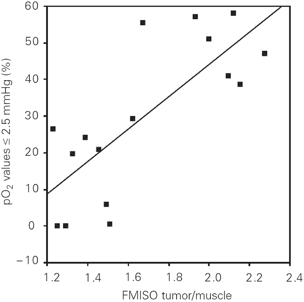
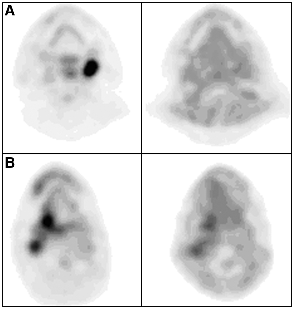
Preclinical in vivo studies revealed that 18F-FMISO uptake and pO2 values have an inverse linear relationship between the PET image intensity and oxygen levels (Pearson product coefficient R=−0.60 to −0.83) (36). In addition, autoradiographic comparisons between 18F-FMISO and 64Cu-ATSM in rodent models showed a strong correlation in the update of the two tracers 24 h after 64Cu-ATSM injection (R 2=0.86) (62). The uptake of 18F-FMISO linearly corresponds to pO2 levels H&NC patients (HP2.5, r=0.75–0.78; HP5.0, r=0.76–0.79) (96, 302), making it a relevant tracer for imaging tumor hypoxia (Fig. 8). Interestingly, no significant correlation exists between electrode measurements and 18F-FMISO T:M ratios in patients with either STS (3 benign, 11 malignant) or benign tumors (n=4) (14, 195).
Pretherapy 18F-FMISO uptake is an independent prognostic factor in H&NC. In a multivariate analysis performed on 73 patients, T:Bmax was highly predictive for OS over a 9 year period (P=0.006) (225). In a population of 25 H&NC patients, the pretreatment 18F-FMISO T:B ratio above 1.6 predicted 11 out of 13 recurrences of H&NC 1 year after radiotherapy (84). A second report also observed that DFS rate for H&NC patients was negatively correlated with both baseline 18F-FMISO scanning (P=0.04) and scans performed during radiotherapy (P=0.02) over a 5 year period (70). When used in combination with intensity-modulated radiation therapy (IMRT vide infra) and platinum-based chemotherapy, 18F-FMISO PET imaging could detect a reduction of uptake in 16 out of 18 oropharyngeal cancer patients (168). No patients experienced local failure, and the 3-year PFS rate was 100%. Although this study was a single-arm study, it suggests that reductions in hypoxia during treatment provide a strong indication that patients will likely have improved outcomes. A recent prognostic clinical study suggests that early 18F-FMISO imaging provides critical prognostic information related to tumor reoxygenation (303). While the baseline 18F-FMISO imaging data were moderately prognostic for PFS (P=0.139), 18F-FMISO hypoxia PET imaging data obtained 2 weeks after the initiation of chemoradiation therapy were a better predictor for local PFS (P=0.001). The researchers explained that the stronger relationship observed between early imaging time points and PFS can conceivably arise from an improvement of tracer kinetics resulting from treatment-related changes in tumor perfusion. These results support a possible patient selection strategy that identifies patients in need of an adaptive therapy planning.
However, there are limitations associated with 18F-FIMSO PET imaging. First, due to its relatively lipophilic nature and slow tissue washout, a suitable contrast between hypoxic and normal tissues is achieved no earlier than 2 h postinjection, and often requires approximately 4 h. This long wait time can be inconvenient to the patient. In addition, the relatively short half life of 18F-fluorine restricts the length of prescanning uptake times as the radioactive signal continuously weakens. Second, a preliminary study of the reproducibility of 18F-FMISO PET imaging revealed a considerable variability in scans performed 3 days apart in the same patient (199). This variable uptake would complicate radiation dose planning based on hypoxic areas within the tumor (i.e., IMRT and “dose painting” strategies, vide infra). However, a recently published report disclosed that reproducible imaging of 18F-FMISO is feasible (208) when using next-generation PET imaging technology with increased sensitivity.
Given the known imaging characteristics of 18F-FMISO, several other molecules utilizing the 2-nitroimidazole scaffold have been developed in an effort to optimize the in vivo imaging properties en route to developing new PET hypoxia imaging tracers.
b. 18F-fluoroazomycinarabinofuranoside
18F-fluoroazomycinarabinofuranoside (18F-FAZA) is a hydrophilic (partition coefficient=1.1) (153), ribose-containing PET hypoxia imaging agent with improved clearance and hypoxia targeting properties. 18F-FAZA diffuses into cells faster than 18F-FMISO (153) and clears from bodily organs more rapidly than 18F-FMISO in preclinical models (219). In humans, the tracer eliminates predominantly via hepatic metabolism and biliary excretion as well as urinary excretion. As a result, liver, gallbladder, colon, and kidneys typically exhibit moderate to high tracer uptake. Uptake of 18F-FAZA in the lungs, bone, fat tissue, and brain was reported to be relatively low (250). The accumulation of 18F-FAZA correlates well with PIMO uptake in murine models (r=0.41–0.73 (multiple tumor types); P<0.001) and tumor pO2 data (30, 194, 269). 18F-FAZA is reported to produce images of adequate quality with T:B ratios ranging from 1.05 to 15.6 among a mixed population of cancer patients (117, 222), comparing favorably against 18F-FMISO (228). 18F-FAZA PET imaging is feasible in H&NC patients (174), showing promise for selecting candidates for TPZ-augmented chemoradiation (228) and IMRT therapy planning (113). However, a recent report found that neither 18F-FAZA nor CA-IX IHC staining was suitable for detecting resectable, high-grade primary prostate tumors (99).
In a recently published report, H&NC patients enrolled into the Danish Head and Neck Cancer Group's (DAHANCA) 24 protocol were imaged with 18F-FAZA before and after treatment, which consisted of primary radiotherapy, nimorzaole, and concomitant cisplatin (196). Using a T:M cut-off of 1.4, 63% of the patients were identified as having hypoxic tumors, with the location of hypoxia remaining generally localized during treatment. 18F-FAZA was associated with poor LRC (93% vs. 66%, p=0.07) and DFS (93% vs. 60%, P=0.04), and all treatment failures were found in patients with hypoxic tumors. There was a positive correlation between the tracer's uptake in the primary tumor and the lymph node, although some discordance in uptake was reported. Although it is not possible to determine the extent at which nimorazole ameliorated the effects of tumor hypoxia, the results indicate how 18F-FAZA can enable the selection of patients at risk for treatment failure.
c. 18F-EF5 (pentafluorinated etanidazole)
It is an 18F-radiolabeled analog of the exogenous hypoxia marker EF-5, and it is the third iteration of the fluorinated “EF” etanidazole derivatives. It is characterized by a markedly high lipophilic character (partition coefficient=5.7) and multiple fluorine atoms (146). Unlike other hypoxia tracers that are designed to possess low lipophilic characteristics, 18F-EF5's high lipophilicity affects a rapid and homogenous distribution throughout all bodily organs, including the brain as confirmed from biodistribution studies in both rodents and humans (176, 299). The increased lipophilicity of 18F-EF5 extends the blood half life to a range of 7.5 to 10 h (176), which is longer than the blood half life of 18F-EF3. From a human patient radiation dosimetry study, 18F-EF5 was reported to be safe for PET imaging (176). The urinary bladder received the highest radiation-absorbed dose (0.12±0.034 mSv/MBq) with an average fractional urinary excretion of 25% over an average of 320 min postinjection (176). The tracer was found to be very stable with the intact tracer being virtually the only radioactive species present in the blood, with polar metabolites being excreted into the urine (146). In rodent tumor models, uptake of 18F-EF5 was low in 9L (oxic) tumors and elevated in large Q7 (hypoxic) tumors, suggesting that the tracer's side chain does not impede the localizing properties of the 2-nitroimidazole pharmacophore (299). 18F-EF5 has recently undergone clinical evaluations in humans showing significant accumulation on head and neck tumors (149) as well as brain malignancies (146). In brain gliomas, areas of increased 18F-EF5 uptake by PET imaging matched areas of increased EF5 binding by IHC staining (88).
d. 18F-flortanidazole
A recently developed tracer of 2-nitroimidazole family was designed to achieve better water solubility and faster background clearance, resulting in improved pharmacokinetic and clearance properties as compared with 18F-FMISO. 18F-flortanidazole (18F-HX4) is a hydrophilic molecule (partition coefficient=0.21, log P=−0.69) incorporating a polar 1,2,3-triazole moiety within its molecular scaffold. Accordingly, the dosimetry profile of 18F-HX4 is comparable to both 18F-fluorodeoxyglucose (18F-FDG) and 18F-FETNIM; its bio-distribution is characterized not only by reduced brain and heart uptake as compared with 18F-FMISO (74, 148) but also by diminished GI uptake, enabling imaging in the abdominal region. The clearance of 18F-HX4 from normoxic tissues is more rapid than 18F-FMISO, suggesting that PET imaging can be performed at an earlier time point. Its metabolic stability is comparable to 18F-FMISO, with 82% of the tracer intact in human plasma at 135 min postinjection. The in vivo uptake of 18F-HX4 has been correlated with hypoxic IHC staining using PIMO in rodents, and its tumor uptake is dependent on tissue oxygenation (75). In patients with lung, thymus, and colon tumors, the observed (T:M) ratios ranged between 0.63 and 1.98 two hours postinjection (272). The uptake of 18F-HX4 among various tumors is highly reproducible, with tumor-to-blood ratios taken from different days correlating well with each other (r=0.945, P<0.001, 90% CI 0.904–0.967). In H&NC patients, 18F-HX4 uptake correlated well with 18F-FMISO uptake, suggesting that both tracers target the same biology (41). In addition, 18F-HX4 demonstrated a better sensitivity and specificity for hypoxia than 18F-FMISO based on CA-IX IHC staining of resected tumors (41).
e. Copper (II) (diacetyl-bis (N4-methylthiosemicarbazone))
Copper (II) (diacetyl-bis (N4-methylthiosemicarbazone)) (Cu-ATSM) is a hypoxia tracer utilizing an oxidation/reduction of chelated copper ion for the selective accumulation into hypoxic tissue (20). Various positron-emitting isotopes of copper can be used: 60Cu (t1/2=0.39 h), 61Cu (t1/2=3.33 h), 62Cu (t1/2=0.16 h), and 64Cu (t1/2=12.70 h). The tracer has been validated in vivo (170) and is currently under clinical investigation. The two major advantages of using Cu-ATSM over 2-nitroimidazole derivatives are the shorter imaging times (as low as 30 min after administration (123)) and a higherT:B ratio despite its relatively lipophilic character [log P=2.2 (10)]. The tracer tends to accumulate in the liver with moderate accumulation observed in both the kidneys and spleens in humans (156). For the discrimination of hypoxic tumor tissue, CuATSM T:B ratios from 2.0 (38), 3.0 (61), and approximately 4 (123) have been used as hypoxia demarcation thresholds. 60Cu-ATSM has been shown to identify NSCLC patient responders from nonresponders undergoing radiation with or without chemotherapy (P=0.002) (61). The tracer uptake is inversely related to 3 year PFS and OS rates in cervical cancer patients (59, 60).
f. 18F-FDG imaging of hypoxia
As previously mentioned, malignant and hypoxic tumors have increased levels of glucose transporters and hexokinase overexpression, suggesting a link between hypoxia and glucose metabolism. However, 18F-FDG PET imaging, a measure of glucose metabolism, does not distinguish between hypoxic and normoxic tumors when compared against several hypoxia assessment modalities. According to three separate publications, the uptake of 18F-FDG (SUVmax or SUVmean) in a combined total of 47 H&NC patients was not correlative against common pO2 descriptors, including HP2.5 and HP5 as measured by oxygen-sensitive polarographic electrodes (95, 96, 302). While there was a reported trend of increasing 18F-FDG SUVmax values among hypoxic tumors, this relationship was observed only among a small number of patients. A similar discordance between 18F-FDG uptake and 18F-FMISO hypoxia imaging data has also been reported. PET imaging data collected from head and neck, sarcoma, breast cancer, and brain tumors revealed no clear correlation between 18F-FMISO uptake and 18F-FDG uptake (18F-FDG SUVmean in hypoxic tumors [6.2±3.0] versus normoxic tumors [6.9±3.2, P=0.478]) (95, 96, 302). While both 18F-FDG and 18F-FMISO are known to identify aggressive tumors, their mechanisms of tumor localization are not clearly linked. Further reports have cited a lack of correlation between 18F-FDG uptake and 60Cu-ATSM uptake in both rectal tumors (r=0.4; P=0.9) (68) and oropharynx carcinomas (R=0.50) (207). The observed lack of correlative uptake between 18F-FDG and 60/61Cu-ATSM mirrors the reported discordance in uptake between 18F-FMISO and 18F-FDG, despite mechanistic differences between the uptake of 60/61Cu-ATSM and 18F-FMISO in hypoxic tissue. While 18F-FDG does not appear to be a specific means for assessing tumor hypoxia, its intrinsic value may reside in an ability to help define specific tumor phenotypes that are overtly aggressive, providing mutually complementary information which can be used in conjunction with PET hypoxia data (Fig. 9) (95, 224).
IV. Modifying Hypoxia to Improve Therapeutic Outcomes
The poor prognosis associated with tumor hypoxia, especially in head and neck tumors, has led to the development of distinct hypoxia-targeted therapies for the improvement of treatment outcomes. While data from more than 30 distinct clinical trials demonstrates that hypoxic modification improves LRC and disease-free survival for H&NC patients, the reported improvements are generally modest in magnitude (Table 5, Studies I and J) (213, 214). However, despite the overall heterogeneous distribution of tumor hypoxia and the lack of hypoxia assessment in these studies, the improvement in patient outcomes supports the concept that hypoxia remains a relevant therapeutic target.
ARCON, accelerated radiotherapy with carbogen and nicotinamide; OR, odds ratio; LRF, locoregional failure; DSD, disease-specific death; DSS, disease-specific survival; OD, overall death; DM, distant metastasis; LRTF, locoregional tumor failure; DSM, disease-specific mortality; HPV, human papillomavirus; 18F-FMISO, 18F-fluoromisonidazole; FFS, failure-free survival; LRFF, locoregional failure free; RC, regional control; RR, relative risk; PL, placebo; AR, accelerated radiotherapy; R, radiation; R+C, radiation plus chemotherapy; NM, nimorazole; RT, radiotherapy; TPZ, tirapazamine; OPN, osteopontin; CA-IX, carbonic anhydrase IX.
In retrospect, the lack of hypoxia assessment in these trials (15) has made it difficult to extract the maximal benefit of hypoxia modification therapies for patients, consequently hindering the development of hypoxia modification therapies. The apparent lack of efficacy has also diminished the urgency for future work in this area and stalled efforts to improve outcomes in radiation therapy by overcoming the effects of hypoxia. The development of hypoxia amelioration strategies for patients with radioresistant tumors is an unmet medical need, especially considering the high percentage of cancer patients who undergo radiotherapy as a part of their treatment (211). In recognizing the potential increase in cure rates and OS by coupling radiation therapy with radiosensitizers, the NCI-RTOG recently published strategic guidelines for the early-stage development of radiosensitizers to accelerate drug development with radiation (159).
Recognizing the critical need for hypoxia assessment, recent clinical hypoxia-modification trials have begun to identify patient subgroups according to their hypoxia status. Such hypoxia stratification data now identify patients with hypoxic tumors at risk for treatment failure and reveal how hypoxia-modification therapy benefits patients with hypoxic tumors as evidenced by an overall increase in LRC, DSS, and local failure free rates. In addition, hypoxia stratification identifies patients with normoxic tumors who, as nonresponders to modification therapy, would forego unnecessary treatment and avoid the negative treatment side effects.
The clinical value in assessing tumor hypoxia now becomes compelling as researchers successfully identify high-risk “hypoxic” patient subgroups as candidates for alternate therapy management strategies, which may include hypoxia-modification therapy. The use of biomarkers and functional imaging to identify sensitive or resistive tumor types that link the biological target with a targeted therapy is consistent with a personalized medicine approach. Biomarkers may also aid in the monitoring of therapy to assess the response to therapy. The incorporation of hypoxia assessment for patient stratification can be valuable to radiation oncologists, surgeons, and biotechnology and pharmaceutical companies who are developing tumor hypoxia therapies or other new treatment strategies for treating hypoxic tumors (13, 17, 26, 130, 192, 211, 214, 230).
A. Use of hypoxia information in radiation therapy planning
IMRT is a recently developed radiotherapy technique that incorporates tomographic imaging data to deliver nonuniform radiation intensities to regions of interest within a tumor (38, 169, 221). The imaging information supplied for IMRT planning comes from anatomical tomographic imaging modalities such as MRI and CT, or functional imaging (i.e., metabolic), such as 18F-FDG-PET (46, 112). However, the reported failure mode of IMRT is locoregional recurrence within the clinical tumor volume (57) ascribable to the presence of undetectable radio-resistant hypoxic cells (39, 243). The incorporation of PET hypoxia imaging is uniquely suited for IMRT because of the full utilization of the spatial tomographic PET data for therapy planning. While there is currently no validated hypoxia imaging technique used for IMRT planning, several studies described next used PET hypoxia imaging data to model how dose escalation to hypoxic (radioresistant) regions within a tumor increased tumor control. The use of 18F-FMISO-PET imaging for IMRT planning was first explored by Lee and co-workers in H&NC patients (168). The hypoxic regions delineated by 18F-FMISO-PET received a 20% dose escalation (84 Gy), while nontarget tissue doses were maintained below normal tissue tolerances, making this a feasible strategy for IMRT therapy planning. In a separate study in 10 H&NC patients, IMRT planning and dose escalations using 18F-FMISO PET imaging data was shown to increase tumor control probability by an average of 17% as a result of the modeled dose increases (119). Dose painting by numbers (DPBN), which escalates radiation doses based on the intensity of the 18F-FMISO PET imaging data, enabled additional precision in therapy planning over uniform dose estimates (uniDE) derived from 18F-FDG (265, 266). In this model, the average increase in tumor-control probability (TCP) in 13 patients was ∼15% for 18F-FMISO DPBN, which was higher than the 2% increase in tumor control probability for the 18F-FDG uniDE protocol based on modeled dose increases to the hypoxic regions of the tumor. A recent biological modeling study comparing hypoxia dose-painted plans against both standard and uniform dose escalation plans confirms the increases in tumor control probability. A cohort of eight H&NC patients underwent 18F-FMISO PET imaging to define the regions of tumor hypoxia. From the biological modeling analyses, the hypoxia dose plan had higher TCP (93% [hypoxia plan] vs. 73% [standard plan]), equivalent normal tissue complication probability, and higher uncomplicated tumor control probability relative to the standard plan (66% [hypoxia plan] vs. 48% [standard plan]). Compared with the uniDE plan, the hypoxia plan was equivalent in TCP, but higher in the UTCP (66% [hypoxia plan] vs. 37% [uniDE plan]). Consequently, the researchers concluded that the hypoxia plan may have a beneficial impact on the therapeutic ratio. These techniques are still under clinical evaluation and are in need of a validated means for assessing hypoxia to further their development. Fortunately, these techniques are feasible, and demonstrations of increased tumor control probability are a necessary step toward the development and incorporation of these methods into clinical practice.
B. Use of hypoxia assessment for selection of patients responsive to nimorazole
Hypoxic cell sensitizers modify a hypoxic tissue's response to radiation by mimicking oxygen's ability to induce the formation and stabilization of toxic DNA radicals (235). The most common hypoxic sensitizers are based on the 2-nitroimidazole scaffold that can localize into hypoxic regions of tumors and tissues, especially at pO2 levels below 10 mmHg (111). A meta-analysis of more than 7000 patients treated with nitroimidazole-based radiosensitizers across various tumor types of unknown tumor hypoxia status demonstrated an overall improvement in patient outcomes, including both LRC and OS, with greatest treatment benefit occurring in head and neck tumors (Study I) (213). Subsequent clinical trials of various 2-nitroimidazole analogs performed among an undefined hypoxic population in H&NC has not demonstrated improvements in treatment outcomes, citing both minimal therapeutic efficacy and competing toxic side effects (165, 166, 285).
A notable exception is the DAHANCA study (Fig. 10), which incorporated a less toxic 2-nitroimidazole analog, called nimorazole, as a part of their clinical research radiation treatment protocol (Study K, Table 5). Radiotherapy augmented with nimorazole has been subsequently introduced into clinical practice for treating patients with head and neck tumors in Denmark (217).The incorporation of the nimorazole-based treatment for patients with head and neck tumors may not be beneficial to all H&NC patients, so the question of how nimorazole directly impacts the outcomes of patients with hypoxic tumors remains unanswered. Accordingly, three important retrospective studies using patient data from the nimorazole DAHANCA study have examined whether hypoxia assessment could identify patients with radioresistant tumors who would respond positively to nimorazole-augmented radiotherapy (Table 5, Study K). The first report disclosed by Overgaard and coworkers used plasma OPN levels, an endogenous marker linked to hypoxia, as a means for identifying patients with hypoxic tumors (Fig. 11) (215). Overall, patients with high OPN levels in the placebo group were associated with significantly poorer 5-year outcomes, increased locoregional failure (LRF), and higher disease-specific mortality than the moderate-to-low OPN groups (Fig. 11, column 3). Conversely, patients with high OPN levels receiving nimorazole augmentation had both a lower LRF and disease-specific mortality as compared with the high OPN placebo group. Based on the stratification information, nimorazole-augmentation offers a clinical means for modifying hypoxia-mediated radioresistance in head and neck tumors, but does not significantly improve treatment outcomes in patients with low to moderate OPN levels. The results from this study clearly indicate a potential therapy decision: H&NC patients with high plasma OPN levels are candidates for nimorazole-supplemented therapy.
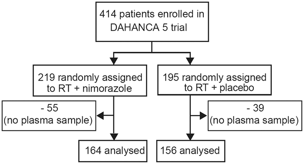

In a second analysis, CA-IX expression levels were evaluated in head and neck tumor tissues in an attempt to identify differences in treatment results between the control and nimorazole treated groups, consistent with the purported link between CA-IX expression and tissue hypoxia (82). No significant trend was observed when comparing either treatment group against the various expression levels of CA-IX, and neither a prognostic value nor a predictive value for patients receiving nimorazole was observed. This suggests that tissue CA-IX is not an optimal prognostic marker for H&NC, possibly because it has been found to not correlate well with the hypoxia status as determined by pO2 measurements (202).
In a third study analysis, patient samples from the DAHANCA study were stratified by using a panel of 15 hypoxia-related genes to determine whether radioresistant tumors could be identified and their hypoxic status could be altered using nimorazole (268). Patients classified as “more hypoxic” receiving nimorazole had a significant benefit in both 5 year LRC (Fig. 12, column 1) and 5 year DSS. The effects of nimorazole on the “less hypoxic” groups for 5 year LRC and 5 year DSS were not significant. The stratification of patients by their human papillomavirus (HPV) status revealed further outcome information: The effect of nimorazole treatment on the “more hypoxic” HPV-negative group showed a large difference in 5 year LRC, indicating that these patients are at high risk for treatment failures (Fig. 13). The remaining groups “less hypoxic” HPV-negative/positive and the “more hypoxic” HPV-positive did not achieve a statistical difference between the treatment arms. The authors noted that the distribution of tumors being “more” or “less” hypoxic occurred essentially equally between the HPV-positive and HPV-negative groups, which was consistent with earlier reports noting no statistical relationship between HPV status and intratumoral pO2 levels (151). This analysis supports the hypothesis that HPV-negative patients identified with hypoxic tumors are at high risk for treatment failure and, therefore, candidates for nimorazole supplementation; whereas the other treatment groups are unlikely to receive any additional benefit from nimorazole-supplemented therapy.
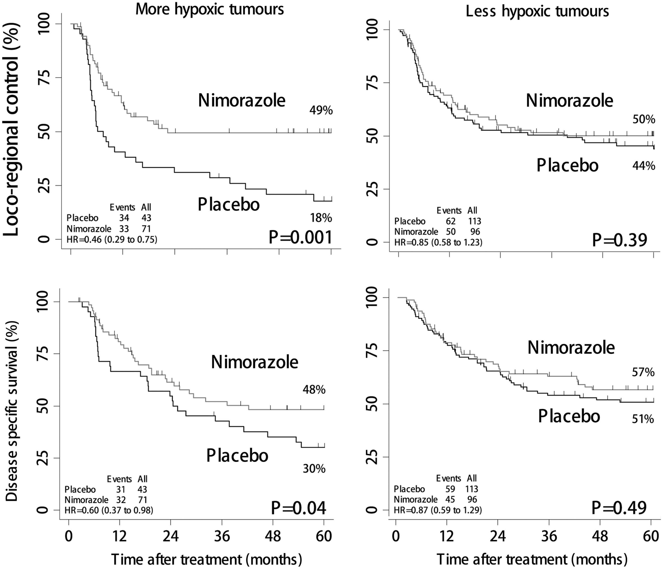

C. Use of hypoxia assessment for selection of patients responsive to tirapazamine
Another hypoxic modifier is tirapazamine (TPZ), a cytotoxin that synergistically enhances the cytotoxic effect of cisplatin in hypoxic cells by 1.2×to 3×over cisplatin alone (25). As opposed to nimorazole, TPZ does not enhance the effects of radiation, but confers its own toxic effects in hypoxic environments by inducing double-strand DNA breaks. Early phase clinical trials have reported mixed results on the effectiveness of tirapazamine-supplemented chemotherapy in cancer patients (163, 231, 282). Additional clinical studies have focused on using hypoxia detection strategies in an effort to identify patients responsive to tirapazamine-supplemented therapy.
Rischin and co-workers revealed how the detection of intratumoral hypoxia using 18F-FMISO PET imaging illuminated the relationship between tumor hypoxia treatment and local failure for patients treated with chemoradiation therapy with or without TPZ (Table 5, Study L) (229) (Fig. 14). As a sub-study of the promising Phase II TROG 98.02 study (230), 18F-FMISO imaging identified patients with high-risk hypoxic tumors who ultimately performed poorly in the absence of TPZ augmentation. Those patients with hypoxic tumors in the control group (i.e., chemoboost only, no TPZ radiosensitizer) had the lowest complete response rate and were at risk for increased LRF and shorter OS over 6 years (232). Conversely, the complete response rate for hypoxic tumors treated with TPZ was nearly as high as the complete response rate for the normoxic group receiving chemoboost only. Interestingly, patients with baseline hypoxic tumors were more likely to experience distant metastases (8 out of 32) compared with patients with normoxic tumors (1 out of 13) as the cause of first failure. This is consistent with the finding that hypoxia is linked to increased metastatic potential (255). Analysis of this data clearly indicates another potential therapy decision: H&NC patients with hypoxic tumors are candidates for TPZ-supplemented therapy, while normoxic patients would not benefit from such therapy.

The TROG 02.02 HeadSTART Phase 3 trial (Trans-Tasman Radiation Oncology Group) was designed to examined TPZ augmenting with cisplatin and radiation treatment H&NC patients of unknown hypoxia tumor status (Table 5, Study M). Despite the promising effects of TPZ reported in earlier trials, no evidence of patient benefit of TPZ augmentation was observed for patients in this trial as reported by the 2 year OS, failure-free survival or locoregional failure-free (LRFF) rates (232). The authors noted that it was not possible to determine the maximal effect of TPZ-augmentation on patients without a hypoxia-based diagnostic tool and suggested a PET-based hypoxia imaging approach (225, 229). The authors also suggested that future hypoxia-based treatment trials in H&NC would be worthwhile only if patients were identified as being at high risk for LRF as determined by their hypoxic status (218).
In an attempt to identify high-risk patients with hypoxic tumors, Rischin and co-workers examined the predictive potential of plasma OPN using samples from the previously mentioned TROG 02.02 Phase 3 study (175). Unlike the results from the DAHANCA study, there were no associated differences between the treatment groups (cisplatin vs. TPZ) as determined by the 2-year OS or LRFF rates. The discrepancy in these results as compared with the DAHANCA results (vide supra) were rationalized on the basis of the different treatment protocols, that is, chemoradiotherapy in the HeadSTART trial as opposed to radiation therapy alone used in the DAHANCA study. In addition, the authors cited the lack of a standardized procedure for assigning high, middle and low OPN thresholds for separating patients into the various hypoxia groups. The discordant results of this study highlight the complicated nature of defining hypoxia, especially when using endogenous biomarkers (168).
D. Use of hypoxia assessment for selection of patients responsive to oxygen delivery therapies
Oxygen delivery therapies are designed to increase local oxygen concentration in hypoxic tumors (116). One method for improving intra-tumor oxygenation is through the normalization of tumor vasculature, which transiently improves oxygenation and creates a therapeutic window of increased treatment sensitization (32). Historically, several therapeutics were developed to inactivate VEGF by halting angiogenesis and isolating the tumor from a recurring blood supply. Unfortunately, prolonged inhibition of vessel growth using such therapies triggered an adaptation response to treatment, causing poor treatment efficacy. However, there is an initial transient normalization of tumor vessels during anti-angiogenic therapy that is characterized by improved blood flow, increased local oxygenation concentration, and enhanced tumor treatment sensitivity. In addition, by repairing leaky vasculature, both tumor cell intravasation and extravasation are considerably reduced, lowering the propensity for metastatic formation. Selective targeting of critical proteins, including VEGF, VEGF receptor 2 (VEGFR2) (290), regulator of G-protein signaling 5 (RGS5) (115), angiopoietin-2 (ANG2) (35), placenta growth factor (PlGF) (234), and prolyl hydroxylase domain protein 2 (PHD2) (189), is known to lead to transient improvements in vascular function, including the stabilization or amelioration of tumor hypoxia. Up-regulation of proteins involved with endothelial cell function, including angiopoietin-1 (ANG1) (267), semaphorin 3A (Sema3A) (185), and PTEN (114), also lead to tumor vessel stabilization and decreased tumor hypoxia. Since the reduction of hypoxia is transient, it becomes critical to identify opportune states of oxygenation for maximizing treatment efficacy. Vessel normalization, rather than destruction, is a new therapeutic strategy for treating cancer (32), and its combination with hypoxia modification therapies may usher in a new paradigm for treating hypoxic tumors.
Several preclinical studies have investigated the transient improvement of tumor vessel normalization and comparing treatment regimens based on changes in vessel normalization and hypoxia in an effort to improve outcomes. Across several different human tumor cell lines, including gliomas, mammary ovary, and melanoma carcinomas, VEGF-targeting therapies, including antibodies (e.g., DC-101, bevacizumab) and kinase inhibitors (e.g., sunitinib and semaxanib), create a transient vessel normalization window, leading to improvements in vessel function and a concomitant decrease in hypoxia as assessed by PIMO, PET imaging, or pO2 electrode measurements (69, 78, 271, 290). In addition, supplemental radiation therapy administered during this normalization window decreases in tumor growth compared with radiation administered outside the normalization window (P<0.05), ascribable to a transient decrease of hypoxia. The treatment of hypoxic squamous cell carcinoma xenografts with the EGFR inhibitor erlotinib resulted in decreased tumor hypoxia with a concomitant increase in tumor vascular normalization and blood flow, which enhanced the effect of adjuvant treatment using either cisplatin or radiation therapy (34). Paclitaxel has also been reported to also induce vessel normalization in hypoxic liver tumors, leading to a decrease in hypoxia assessed by EPR (55). Mice treated with combined paclitaxel and radiation therapy outperformed mice receiving monotherapy (P<0.01), suggesting a synergistic treatment effect made possible through vessel normalization and tumor oxygenation.
Given the unique molecular signaling that affects vessel normalization and tumor oxygen, unique molecular targets may identify new therapeutic strategies that improve treatment outcomes. A recent report describes inducing tumor vessel normalization and reversing tumor hypoxia during treatment by activating endothelial PTEN, an enzyme that inhibits PI3K and leads to tumor vessel normalization (141). The administration of inositoltrispryrophosphate (ITPP) in combination with paclitaxel and cisplatine decreased tumor size (0.5 g [treated] vs. 2.5 g [un-treated]) with no visible signs of metastatic invasion in nearby organs. Tumor pO2 measurements, PIMO staining, and 18F-FMISO PET imaging confirmed an increase in pO2 levels after the administration of ITPP, suggesting a normalization of tumor function and creation of a normalization window for treatment. MRI imaging revealed vessel reorganization, and CD31 staining was better delineated in treated tumors compared with nontreated tumors, which was consistent with the effects of vessel normalization. PCR analysis showed a strong down-regulation of hypoxia-related mRNAs for HIF-1 and HIF-2 along with a reduction of PHD-2 protein. There was also an enhancement of CD31, VEGFR1, and VEGFR2 mRNAs, which was consistent with the notion of vessel maturation. Although still in preclinical development, emerging treatment strategies that improve vessel growth, reduce tumor hypoxia, and improve the effectiveness of chemo- and radiotherapy may lead to new treatment paradigms for improving outcomes in patients with hypoxic tumors. Identifying the normalization window in cancer patients using perfusion and hypoxia assessment techniques will aid in the transition of this therapeutic strategy into clinical practice.
Another method for improving intra-tumor oxygenation is through vasodilatation that is achievable by combining carbogen inhalation with the administration of nicotinamide. ARCON couples carbogen and nicotinamide treatments with accelerated radiotherapy (AR). ARCON therapy was studied in H&NC patients in a Phase 2 clinical trial in which high local and regional control rates (>80%) were achieved in patients with advanced stage squamous cell carcinomas of unknown hypoxic status in the larynx and oropharynx; however, other tumor types such as oral cavity (37%) were still poorly controlled (135).
Due to the high LRC rates (80%) observed for patients with larynx carcinomas receiving ARCON treatment, researchers proceeded with a multi-center Phase 3 trial comparing AR against ARCON (Table 5, Study N). Given the importance of hypoxia information in understanding the effects of hypoxia-modification therapy, a sub-study of this trial gathered tumor hypoxia data from tumor biopsies using PIMO staining. For the entire patient population (lacking hypoxia assessment), there were no significant differences for patients with regard to the 2- and 5-year LCRs or in the DFS or OS rates, suggesting that ARCON treatment did not provide a significant patient benefit relative to AR (Fig. 15, column 1) (131).

However, when the sub-group data were analyzed using tumor hypoxia information, an obvious difference in the response rates emerged between the AR and ARCON treatment arms. There was a significant improvement in regional control in the high hypoxia group treated with ARCON Figure 15, column 3). A similar trend was observed for the hypoxia group treated with ARCON, resulting in improved 5 year DFS as compared with the AR group. In addition, the patient group with hypoxic tumors was at high risk for treatment failures as evidenced by their depressed regional control and DFS percentages. ARCON treatment had little effect on patients with normoxic tumors, as there was no statistical difference in regional control or DFS in either treatment regimen (cf. Fig. 15, column 2). The authors noted that selecting patients based on the treatment mechanism is critical, as the hypoxia status determination revealed those patients more likely to experience improved outcomes when receiving ARCON treatment. In summary, the analysis of the ARCON data supports the application of hypoxia assessment for both patient selection and therapy decisions: Patients assessed early for hypoxic head and neck tumors are candidates for ARCON therapy, while normoxic patients would not experience a benefit from this therapy.
V. Concluding Remarks
Tumor hypoxia is a well-established biological phenomenon that is often present in malignant solid tumors, including tumors of head and neck, cervix, breast, prostate, and lung. Tumor hypoxia disperses heterogeneously, and is independent of tumor size, stage, grade, or histology. Hypoxic tumors employ several different survival mechanisms, which may result in a loss of apoptotic potential, increased proliferative potential, and formation of new blood vessels that encourages the evolutionary selection for a more malignant phenotype. As such, hypoxia affects the curability of solid tumors, regardless of treatment modality.
Given the compelling link between tumor hypoxia and negative treatment outcomes, clinical research has turned to studying the effectiveness of hypoxia-targeted therapies. However, several trials lacked an appropriate method for accurately identifying and selecting patient subgroups with hypoxic tumors. As a result, the indiscriminate use of hypoxia-modification therapy, as evidenced by several meta-analyses, showed marginal benefits of such therapy when given to a patient population having both normoxic and hypoxic tumors. Due to the invasive nature of the “gold standard” method for measuring tumor oxygenation (pO2 electrode measurements), hypoxia-based patient stratification has not been readily adopted into clinical practice. Sponsors currently face the choice of either enrolling both tumor types in their clinical trials, which will dilute their efficacy results and threaten the success of the trial, or utilizing an unapproved diagnostic tool (e.g., hypoxia PET imaging) for selecting patients with hypoxic tumors. This situation impedes the clinical development of hypoxia-based therapies. Several studies, including recent secondary analyses from larger trials, suggest that hypoxia assessment is predictive for outcomes and can identify high-risk patients in need of treatment modification.
Given the negative treatment and outcome problems associated with tumor hypoxia, a major goal for clinicians has been to identify hypoxic tumors through a number of different diagnostic approaches. The polarographic electrode has emerged as the “gold standard” for detecting and characterizing hypoxia, which enabled clinical studies to establish the significance of tumor hypoxia in patients. The presence of tumor hypoxia is prognostic for LRC and OS in cervix, breast, lung, prostate, and H&NC supported by data from more than 2000 patients. However, despite its ability to generate prognostic data, the use of polarographic electrodes is not viable for the widespread clinical assessment of tumor hypoxia given the technical limitations of this instrument. Several different methods for assessing tumor hypoxia have evolved and replaced polarographic pO2 measurements. These modalities, either individually or in combination with other modalities, provide functional and physiological information on tumor hypoxia that has prognostic and predictive value.
There is an acute need for an approved diagnostic technology for determining the hypoxia status of cancer lesions, as it would enable the clinical development of personalized, hypoxia-based therapies, which will, ultimately, improve outcomes. The assessment of tumor hypoxia will be valuable to radiation oncologists, surgeons, and biotechnology and pharmaceutical companies who are engaged in developing hypoxia-based therapies or treatment strategies.
Footnotes
Acknowledgments
The authors would like to thank Helen Su and Katrin Szardenings for both helpful discussions and proofreading this article.
Author Disclosure Statement
Joseph C. Walsh is an employee of Siemens MI, a company that developed PET biomarkers for imaging hypoxia. Artem Lebedev and Hartmuth C. Kolb are former employees of Siemens MI. Kathleen Madsen is a consultant statistician to Siemens Molecular Imaging, with compensation based on fair market value and not tied to outcomes. Edward Aten is acting as Interim Medical Director to Siemens Molecular Imaging, with compensation based on fair market value and not tied to outcomes. Liane Marciano is a consultant to Siemens Molecular Imaging, with compensation based on fair market value and not tied to outcomes.