
Abstract
Significance:
MicroRNAs (miRNAs) are important regulators of gene expression and define part of the epigenetic signature. Their influence on every realm of biomedicine is established and progressively increasing. The impact of environment on human health is enormous. Among environmental risk factors impinging on quality of life are those of chemical nature (toxic chemicals, heavy metals, pollutants, and pesticides) as well as those related to everyday life such as exposure to noise or mental and psychosocial stress.
Recent Advances:
This review elaborates on the relationship between miRNAs and these environmental risk factors.
Critical Issues:
The most relevant facts underlying the role of miRNAs in the response to these environmental stressors, including redox regulatory changes and oxidative stress, are highlighted and discussed. In the cases wherein miRNA mutations are relevant for this response, the pertinent literature is also reviewed.
Future Directions:
We conclude that, even though in some cases important advances have been made regarding close correlations between specific miRNAs and biological responses to environmental risk factors, a need for prospective large-cohort studies is likely necessary to establish causative roles. Antioxid. Redox Signal. 28, 773–796.
Introduction
S
The miRNAs are encoded in different locations in the genome, including intronic and intergenic regions. Interestingly, intronic miRNAs often control the expression of genes associated with the same cellular functions regulated by the host gene where they are encoded. This elegant mechanism of gene regulation is exemplified by the sterol response element binding protein (SREBPs)/miR-33ab gene loci. SREBP2 and SREBP1 regulate the synthesis and uptake of cholesterol and synthesis of fatty acid, respectively. Coinciding with the transcription of SREBP2 and SREBP1, miR-33a and miR-33b are cotranscribed and negatively regulate the expression of a number of genes involved in regulating cholesterol efflux and fatty acid oxidation. Both negative feedback loops cooperate to enhance intracellular cholesterol and fatty acid levels by simultaneously balancing transcriptional activation and post-transcriptional repression of lipid homeostasis genes (156). In addition to lipid metabolism, most of cellular processes have been shown to be regulated by miRNAs. Importantly, recent work has demonstrated that miRNAs are able to “fine-tune” the regulation of redox signaling, by direct interaction with nuclear factor erythroid 2 like 2 (NFE2L2; also known as Nrf2), the major transcriptional regulator of defense against reactive oxygen species (ROS) (63, 103). The miRNAs may also interact with its coregulators Kelch-like erytroid cell-derived protein with cap ‘h’ collar homology (ECH)-associated protein 1 (Keap1) and Broad-complex, Tramtrack and Bric a brac (BTB) domain and cap ‘h’ collar (CNC) homolog 1 (Bach1), or regulate the generation of ROS. These new subsets of miRNAs that either regulate redox pathways or are themselves regulated by the cellular redox state have been termed “redoximiRs” (23). Redox regulation affects gene expression as well as translational processes at multiple levels, including the classical pathways (activity of transcription factors, mRNA stability) but also epigenetic processes (miRNA signaling, DNA methylation, histone modifications) and DNA damage/repair, all of which contribute significantly to overall genome stability (Fig. 1) (125).

Environmental Risk Factors and Scope of the Review
It has become increasingly recognized that chronic human diseases are primarily associated with environmental factors as opposed to genetic factors (155). Environmental risk factors comprise a large number of determinants related to populations sharing common living or working spaces. They can be of physicochemical or social nature and it is now quite clear that their influence on individual and social health differences is a concern of increasing magnitude (1). Nearly all human diseases result from a complex interaction between an individual's genetic profile and his/her exposure to environmental factors. Since adverse regulation of miRNA has a substantial impact on the development and progression of cardiovascular disease, environmental changes of miRNA expression and activity likely affect cardiovascular health (31). The concept of the “exposome” has been recently established to study gene–environment interactions, identify novel biomarkers, and discover key regulators of adaptive response after exposure to environmental risk factors (126). Importantly, environmental exposure may lead to epigenetic reprogramming (including changes in miRNA signatures), which can be a contributor to disease development later in life (Fig. 2). Specific miRNA expression profiles have been linked to several toxic environmental risk elements, including radiation, air pollution, and cigarette smoke (128). Efforts have been made to model the complexity of the networks and regulatory mechanisms of miRNAs involved in environmental gene regulation using computational tools (104, 176). In this review, we delineate three specific settings of environmental risk factors and the reciprocal influence of miRNAs as well as their connection to oxidative stress: (i) namely environmental chemicals, (ii) noise-induced hearing loss (NIHL), and (iii) mental stress.

Environmental chemicals
In this section, we discuss the critical involvement of miRNAs in xenobiotic-induced disease pathogenesis (Fig. 3). The important environmental risk determinants to be covered in this section include persistent environmental chemicals (polychlorinated biphenyls [PCBs], polybrominated diphenyl ethers [PBDEs], perfluorocarboxylic acids [PFCAs], and perfluorooctanesulfonate [PFOS]), benzene, bisphenol A (BPA), heavy metals, air pollution, and pesticides.
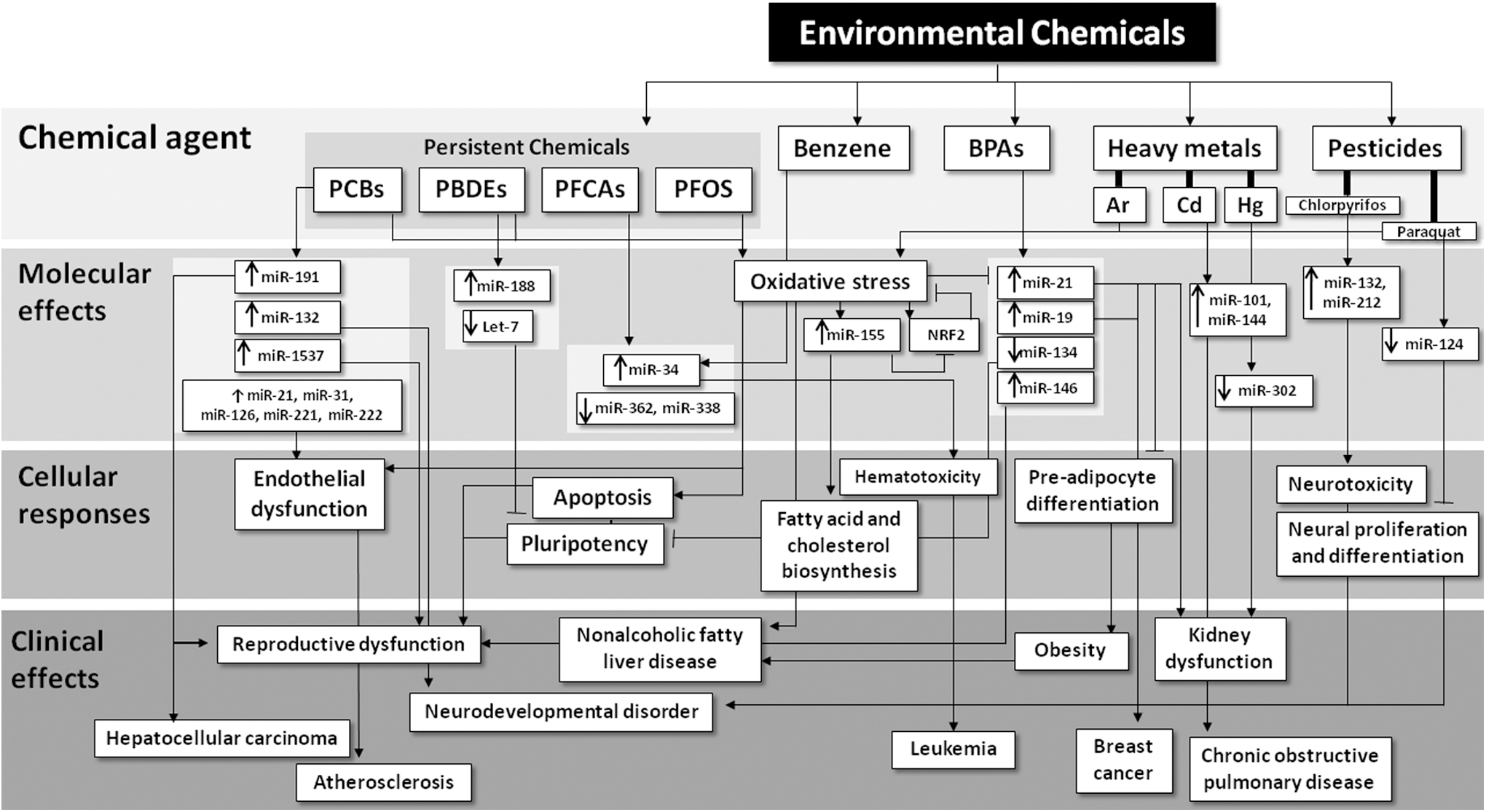
PCBs are environmental toxicants that produce a wide spectrum of toxicities in humans such as reproductive dysfunction (183), neurodevelopmental disorders (166, 167, 211), and obesity (53). Although the production of PCBs was banned in the United States in 1979, these chemicals are still widespread in food, drinking water, and soil because of their highly persistent nature, raising great safety concerns (54, 66). PCBs are activators for xenobiotic-sensing transcription factors, which, in turn, upregulate their target genes such as cytochrome P450s involved in xenobiotic biotransformation. Whereas the coplanar (also known as “dioxin-like”) PCBs are activators for the aryl hydrocarbon receptor (AhR), the noncoplanar PCBs are activators for the pregnane X receptor and the constitutive androstane receptor (4, 55).
PBDEs were used as flame retardants incorporated into plastics, rubbers, and textiles, and were recently banned because of their wide spectrum of toxicities such as thyroid hormone disorders (50, 58, 186, 232, 233), neurotoxicity (95), oxidative stress in liver (2, 227), and carcinogenesis (132). However, owing to their highly persistent and bioaccumulative nature, PBDEs still raise growing safety concerns, as certain PBDE congeners are enriched in seafood, breast milk, household dust, and in electric waste dismantling sites (110, 116, 169, 212).
The persistent perfluorinated compounds, especially PFCAs and PFOS, have been detected in humans and wildlife, raising health concerns (18, 35, 45, 170, 174). PFCAs have been extensively used in Scotchgard products and in making Teflon brand products. PFOS is used in industrial and consumer applications as surfactants and building material components (136).
Endocrine disruptors such as BPA, dichlorodiphenyltrichloroethane (DDT), and phthalates interfere with the body's endocrine system through modulating the activities of hormonal receptors such as the estrogen receptor (ER), producing a wide spectrum of developmental, reproductive, neurological, and immune effects in both humans and wildlife (150). BPA is widely used as a plasticizer in epoxy resins and in thermal papers. Thus, humans are regularly exposed to BPA, and this may lead to chronic diseases such as hormone-dependent cancers. Of particular concern are exposures to BPA that occur early in life, a period of susceptibility that can have a life-long impact on disease risk (149, 150).
Heavy metals and metalloids such as lead, cadmium, mercury, selenium, chromium, and arsenic toxicity are highly diverse, and are dependent upon many factors including the organ targeted, exposure route, time of development, gender, and dietary factors (20). The body has mechanisms to deal with heavy metal exposures including their excretion through the upregulation of cationic transporters, sulfhydryl-based scavengers (e.g., glutathione and metallothionein) (94), reduction of metabolism and methylation (87) at the transcriptional, translational, and post-translational levels to deal with metal exposures (87). Ambient air pollution is a term used to describe outdoor air pollution, which most often is a mixture of particulate matter (PM), volatile organic compounds, ozone, oxides of nitrogen and sulfur, and in some cases industrial emissions rich in heavy metals and toxic organics. Exposure to ambient air pollution has been associated with adverse outcomes in many organ systems, including the lung, the cardiovascular system, the liver, and the central nervous system (CNS) (20). As with other complex mixtures, the effects are variable depending on the sources, distances from those sources, and variations in climate, sunlight, or traffic loads. Therefore, there has been a need to identify biomarkers of exposure that are convenient, robust, reproducible, and ideally specific of the exposure.
miRNAs and persistent environmental chemicals (PCBs, PBDEs, and PFCAs/PFOS)
Polychlorinated biphenyls
For the dioxin-like PCBs, in human peripheral blood mononuclear cells, miR-191 expression correlates with total blood concentrations of PCBs, and in particular with the dioxin-like congener PCB169 (57). The blood levels of PCB169 significantly correlate with miR-191 in pregnant women living in a PCB-contaminated area who have undergone therapeutic abortion because of fetal malformations. Of note, miR-191 is also known to be upregulated by dioxin in hepatocellular carcinoma cells in vitro (Table 1) (57).
BPA, bisphenol A; HESC, human embryonic stem cells; miRNAs, microRNAs; N/A, not available; PBDEs, polybrominated diphenyl ethers; PCBs, polychlorinated biphenyls; PFCAs, perfluorocarboxylic acids; PFOS, perfluorooctanesulfonate; MAPK, mitogen-activated protein kinase.
The nondioxin-like PCBs have been linked to neuropsychological dysfunction in children. Specifically, they increase spontaneous Ca2+ oscillations in neurons by stabilizing ryanodine receptor (RyR) calcium release channels in the open configuration, leading to cAMP response element-binding (CREB)-dependent dendritic outgrowth (122). The nondioxin-like congener PCB95 at nanomolar concentrations promotes synaptogenesis via RyR-dependent upregulation of miR-132 and inhibition of RyR, CREB, or miR-132 block PCB95-mediated effects. Interestingly, miR-132 is also dysregulated in Rett syndrome and schizophrenia. Therefore, miR-132 is an important risk factor for PCB-mediated neurodevelopmental disorders (102).
PCBs have also been correlated with multiple vascular complications such as endothelial cell dysfunction and atherosclerosis, through producing oxidative stress and induction of proinflammatory cytokines and cell adhesion proteins (65). In primary human endothelial cells, the commercial PCB mixture Aroclor 1260 alters the expression of 557 out of 6658 miRNAs, and 21 of them have been associated with vascular diseases according to the MetaCore database (191). Specifically, Aroclor 1260 increases the expression of miR-21, miR-31, miR-126, miR-221, and miR-222. Whereas miR-21 has been implicated in cardiac injury, miR-126 and miR-31 have been shown to modulate inflammation (191). Last but not least, the National Children's Study (NCS) has established the associations between miRNA expression profiles and various environmental pollutants including PCBs, and specifically, PCBs positively associate with miR-1537 expression in term placentas (106). These studies have suggested that miRNAs may serve as potential biomarkers to stratify distinct mechanisms of various diseases associated with PCB exposure.
Polybrominated diphenyl ethers
In the NCS, it was reported that BDE-209 positively correlates with miR-188-5p, whereas PBDE-99 inversely correlates with the miRNA let-7c in term placentas (106). In human embryonic stem cell (ESC) lines (FY-hES-10 and FY-hES-26), BDE-209 at nanomolar concentrations reduces expression of pluripotent genes such as OCT4, SOX2, and NANOG and induces apoptosis (39). In addition, the downregulation of OCT4 is accompanied by hypermethylation of the OCT4 promoter and increased expression of miR-145 and miR-335, which inhibit OCT4 expression (106). BDE-209 also produces ROS and decreases superoxide dismutase (SOD)2 expression. Therefore, the authors have concluded that BDE-209 decreases pluripotent gene expression via epigenetic regulation (e.g., miRNAs) and induces apoptosis through ROS generation (39).
Perfluorocarboxylic acids
Among various types of PFCAs, perfluorononanoic acid (PFNA), which is a PFCA with a nine-carbon backbone, produces hepatomegaly, increases hepatic triglycerides and total cholesterol, and increases serum transaminases (154). Many miRNAs were differentially regulated by PFNA in a dose-dependent manner, including an upregulation of miR-34a and a downregulation of miR-362-3p and miR-338-3p at both doses tested, whereas miR-34a regulates fucosyltransferase 8 and lactate dehydrogenase expression. Therefore, the authors have concluded that PFNA exerts its hepatic effect at least partially through miRNA-mediated post-translational downregulation (196).
Perfluorooctanesulfonate
PFOS has been shown to induce adipogenesis and glucose uptake in preadipocytes and this was associated with activation of the oxidative stress responsive transcription factor Nrf2, which is important for upregulating antioxidant genes and metabolic reprogramming (219). In addition, it has been shown in rats that PFOS tends to accumulate in the liver, resulting in hepatomegaly, actin filament remodeling, endothelial permeability changes, and ROS production. This coincides with PFOS-mediated upregulation of miR-155, which appears to suppress Nrf2 signaling, because pretreatment of HepG2 cells with catalase (CAT) decreases miR-155 expression, increases Nrf2 expression and activation, and reduces PFOS-induced cytotoxicity and oxidative stress (192). Therefore, PFOS-induced oxidative stress is at least partially dependent on miRNA-mediated downregulation of Nrf2 signaling.
miRNAs and benzene
Exposure to benzene such as that of paint sprayers may result in hematological disorders of significant severity. The aberrant expression of miRNAs in workers exposed to benzene has been analyzed (7). It was found that miRs 34a, 205, 10b, let-7d, 185, and 423 5p-2 were upregulated and 133a, 543, has-130a, 27b, 223, 142-5p, and 320b were downregulated. Several pathways involved in cell proliferation and differentiation including vascular endothelial growth factor (VEGF), transforming growth factor beta (TGF-β), and Wnt signaling were shown to be affected, thus providing the opportunity to explore them as causative links or to exploit selected miRNAs as potential biomarkers.
miRNAs and BPA
In human placental cells, miRNA microarrays were used to identify several miRNAs that were significantly altered in response to BPA treatment, exemplified by a marked upregulation of miR-146 that leads to slower proliferation and higher sensitivity to the DNA damaging drug bleomycin (6, 171). Its expression correlated significantly with BPA accumulation in the placentas from pregnant women living in a polluted area and undergoing therapeutic abortion because of fetal malformations (34). Therefore, miR-146 expression may serve as a biomarker for developmental exposure to BPA.
In both ESCs and embryoid bodies (EBs) of mouse origin, BPA decreases the expression of miR-134, which is a suppressor of the pluripotency markers Oct4, Sox2, and Nanog, suggesting that miR-134 may play a role in BPA-mediated disturbances in pluripotency in ESCs and EBs (22).
In sheep, gestational BPA exposure at environmentally relevant doses altered the expression of steroidogenic enzymes Cyp19 and 5α-reductase in the ovaries at gestational day 65, and the expression of fetal ovarian miRNAs (45 downregulated at gestational day 65 and 11 downregulated at gestational day 90). Importantly, miRNAs that target Sry-related high-mobility-group box (SOX) family genes, kit ligand, and insulin-related genes were downregulated by BPA, suggesting that miRNAs may play a role in BPA-mediated disturbances in gonadal differentiation, folliculogenesis, and insulin homeostasis (190).
Exposure to BPA is also implicated in breast cancer. In the ER-positive and hormone-sensitive human MCF-7 breast cancer cell line, BPA potentiates ER transcriptional activity and this coincides with alterations in the expression profiles of certain miRNAs including miR-21, which is an onco-miR that is frequently upregulated in solid tumors (182). BPA also increases the expression of the onco-miRs miR-19a and miR-19b and dysregulates the expression of the miR-19-related downstream proteins such as PTEN, p-AKT, p-MDM2, and p53 in MCF-7 cells. Interestingly, the chemopreventive drug curcumin reverses these effects, suggesting that curcumin may suppress BPA-induced breast cancer through modulating the miR-19/PTEN/AKT/p53 axis (109). Regarding the effect of BPA on intermediary metabolism, in epidemiological and animal studies, BPA exposure has been associated with type-2 diabetes and especially gestational diabetes mellitus (GDM), and there is emerging evidence showing that placental-derived exosome miRNAs may serve as predictors for GDM (43). In rats, miR-21a-5p overexpression attenuated BPA-induced obesity in vivo (214). In preadipocytes, BPA-induced cell differentiation is suppressed by miR-21a-5p by targeting map2k3 in the MKK3/p38/mitogen-activated protein kinase (MAPK) pathway, suggesting that miR-21a-5p mimics may serve as a potential therapy for BPA-induced obesity (214).
miRNAs and heavy metals
Arsenic
Earlier work published by Marsit et al. (119) investigated the relationship between arsenic exposure and miRNA expression in TK6 cells (an immortalized human lymphoblast cell line). Treatment with sodium arsenite led to decreased miR-210 expression and increases in miR-22, miR-34a, miR-221, and miR-222, similar to those that would be observed with cellular nutritional stress such as folate deficiency. These effects were then confirmed in human peripheral blood samples similarly exposed to arsenite.
Kong et al. (97) assessed microalbuminuria in adolescents, and its relationship among urinary metals (Hg, Pb, As, and Cd) and the levels of miR-21, miR-126, miR-155, and miR-221. They found no relationship between metal levels and microalbuminuria, but miR-21 and miR-221 were negatively associated with this arsenic and lead levels, and miR-21 was associated with microalbuminuria. Thus, miRNA levels were proposed as biomarkers of kidney function in the context of heavy metal exposure.
In a study of arsenic exposure to pregnant mothers and the consequences for their infants, Rager et al. (153) examined the relationship between arsenic in drinking water and maternal urine, and the expression of miRNAs in cord blood. There were significant associations between a number of cord blood miRNAs (let-7a, miR-107, miR-126, miR-16, miR-17, miR-195, miR-20a, miR-20b, miR-454, miR-96, and miR-98) and urinary arsenic. These miRNAs have been linked to cancer and diabetes. Furthermore, there was a depression in the expression of a number of immune response-related mRNAs that were predicted to be partially caused by changes in these miRNAs.
Bollati et al. (16) studied the expression of miR-21, miR-146a, and miR-222 in peripheral blood leukocytes (PBLs) obtained from steel workers occupationally exposed to PM containing arsenic, iron, nickel, lead, cadmium, chromium, and manganese. They also examined the impact of exposure on the oxidative stress biomarker 8-hydroxyguanine, and the effects the individual metals have on miRNA expression in isolated PBLs. Both miR-21 and miR-222 were increased when comparing baseline expression (start of work week) to 3 days of exposure, and miR-222 levels were correlated with lead exposure. Conversely, miR-146a was inversely correlated with exposure to lead and cadmium. Furthermore, miR-21 was associated with 8-hydroxyguanine levels. The authors thus suggested that the expression of these miRNAs could represent novel mechanisms of response to PM and its associated metals. Exposure to lead associated with atmospheric PM is also related to the expression of miRNAs that are involved in oxidative stress and inflammation (16), all of which contribute to cardiovascular risk and mortality (29).
Cadmium
Cadmium (Cd) is an especially toxic and persistent heavy metal. Although many studies have examined the effects of Cd on mRNA transcript levels, few have examined the miRNAs-mediated Cd effects. Fabbri et al. (44) investigated the effects of Cd exposure in HepG2 human hepatoma cells on global gene expression and miRNA levels. Transcriptional changes at higher Cd exposure included those related to cancer and depressed hepatic function, and a number of let-7 miRNA family members were differentially expressed by Cd, suggesting a connection between their tumor suppressor roles and cadmium carcinogenesis.
Cd is also a major component of cigarette smoke. In a study designed to assess the effects of cigarette smoke and Cd on cystic fibrosis transmembrane regulator (CFTR) function, Hassan et al. (62) showed that cigarette smoke and cadmium increased miR-101 and miR-144 expression in human airway epithelial cells, which suppressed the expression of CFTR protein. They also showed that cigarette smoke exposure caused similar changes in miR-101 in the lungs of mice. Moreover, chronic obstructive pulmonary disease (COPD) patients had increased pulmonary expression of miR-101 than patient controls, suggesting a link between cigarette smoking, Cd exposure, and suppression of CFTR in COPD.
Mercury
Mercury in its inorganic form (Hg++ ion) has deleterious effects on the kidney, whereas the methylated form of mercury (MeHg) targets the CNS, especially during embryonic and fetal development (20, 163). In an in vitro model of CNS differentiation, Pallocca et al. (141) treated mixed neuronal/glial cell cultures derived from NT2 cell ESC precursors with MeHg chloride during differentiation. As the cells differentiated, there was a decrease in the stem cell miRNA expression signature (downregulation of miR-302 cluster that reflects stem cell character) and an upregulation of miRNAs emblematic of neuronal differentiation (let-7, miR-125b, and miR-132). When exposed to MeHg, these cultures showed differential regulation of several miRNAs (miR-141, miR-196b, miR-302b, miR-367, and miR-372) whose targets were mapped to pathways important for axonal guidance, learning, and memory.
Changes in the expression of circulating miRNAs with occupational exposure to mercury were recently examined in a pilot case–control study by Ding et al. (37). High-level Hg exposure was associated with increases in the expression of miR-92a and miR-486, suggesting that these two miRNAs may be suitable biomarkers in larger cohorts occupationally exposed to Hg at lower levels.
miRNAs and air pollution
Changes in the expression of miRNAs have also been investigated as biomarkers of air pollution exposure (70, 83, 113, 146, 204) (Table 2). Susceptibility to particle health effects, microRNAs and exosomes (SPHERE) study (15) is focused on evaluating the adverse health effects of air pollution on obese subjects. This same group has shown that exposure to PM is associated with changes in the exosomal miRNA profile in humans, with similar changes in A549 human lung epithelial cells (14). In addition, Rodosthenous et al. (157) found an association between long-term exposure to ambient PM of <2.5 μm in diameter (PM2.5) and increased levels of extracellular vesicle miRNA circulating in the serum of subjects in the aging cohort study. These included miR-126-3p, miR-19b-3p, miR-93-5p, miR-223-3p, and miR-142-3p with 6 months of exposure, and miR-23a-3p, miR-150-5p, miR-15a-5p, miR-191-5p, and let-7a-5p with 1 year of PM2.5 exposure. Pathway analysis revealed gene targets of these miRNAs that were associated with cardiovascular disease, including oxidative stress, inflammation, and atherosclerosis.
BP, blood pressure; PM, particulate matter.
Although human epidemiology studies are clearly important for evaluating such risks, mechanistic information has often come from cell culture and animal studies. Several in vitro studies have examined the effects of various air pollutants on miRNA expression. Bleck et al. (11) found that diesel exhaust and ambient particulate exposures were associated with miR-375-mediated regulation of thymic stromal lymphopoietin (TSLP; a potent proinflammatory chemokine important in both innate immunity and tail homology 2 (TH2)-adaptive immunity) in human bronchial epithelial cells. This effect was likely mediated via miR-375 targeting the AhR, thus relieving AhR suppression of TSLP. Zhou et al. (229) examined the effects of diesel exhaust particles on miRNA-21 in human bronchial epithelial cells and the potential carcinogenic mechanisms associated with such exposures. Diesel particle exposure caused an increase in the expression of miR-21, which, in turn, upregulated PTEN/PI3K/AKT signaling, a pathway that is often activated in cancer cells.
In recent studies, Li et al. (108) found that miR-1228(*) was able to inhibit apoptosis in A549 human alveolar epithelial cells exposed to fine PM. Using the same cell line, Jeong et al. (84) conducted an integrative analysis of mRNA and miRNA expression of these cells exposed to aqueous and organic-soluble extracts from PM2.5. This comprehensive analysis found that a large number of miRNAs were altered (37 and 62 miRNAs for aqueous and organic extracts, respectively), which mapped to a number of pathways important for nutrient sensing, nucleic acid synthesis, DNA synthesis, and cell cycle regulation.
Animal studies have also been conducted to investigate the roles of miRNAs in the adverse effects of PM exposure (213). Farraj et al. (47) exposed rats to ambient PM and noted changes in cardiac functional parameters (ST-segment depression in the electrocardiogram, arrhythmia, and vagal dominance), which was associated with a general decrease in the expression of miRNAs in cardiac tissue.
An earlier human study by Wilker et al. (209) noted associations between exposure to ambient black carbon (BC; a marker of traffic-related air pollution), blood pressure (BP) changes, and single nucleotide polymorphisms (SNPs) in several miRNA processing genes (DICER, GEMIN4, and DGCR8). This study did not evaluate the potential mechanisms by which these SNPs might be causative for the association between BC exposure and increased BP. In the normative aging study, Wilker et al. (208) investigated the relationships between ambient air pollutants, polymorphisms associated with miRNA processing, and the concentrations of circulating soluble cellular adhesion molecules, which are correlates of atherosclerosis and cardiovascular disease. Seven-day moving averages of PM2.5 exposure were associated with higher soluble intercellular adhesion molecule-1 (sICAM-1) and soluble vascular adhesion molecule-1 (sVCAM-1) levels. Sulfates and sulfur dioxide 7-day moving averages were associated with higher sICAM-1 and a suggestive association was observed with sVCAM-1 in aging men (117). They also noted that SNPs in miRNA-processing genes may modify associations between ambient air pollutants and sICAM-1 and sVCAM-1. Of note, miRNA polymorphisms have also been encountered in patients with esophageal squamous cell carcinoma heavily exposed to environmental smoke (202).
In follow-on studies, Fossati et al. (49) investigated the relationships between miRNAs in PBLs and PM exposure. The mRNAs miR-1, miR-126, miR-135a, miR-146a, miR-155, miR-21, miR-222, and miR-9 were all associated with PM exposure. When mapped to targeting pathways, miR-126, miR-146a, miR-155, miR-21, and miR-222 were strongly associated with changes in the high-mobility group chromatin proteins. The relationships between the expression of these mRNAs and PM exposures were also influenced by polymorphisms in the RNA processing genes GEMIN4 and DGCR8. More recently, Motta et al. (129) found that miRNAs are a likely molecular mechanism underlying the BP-related effects of air pollution exposure, and indicated that changes in miR-101 expression are important as an epigenetic mechanism for this relationship.
Although less commonly investigated than the effects on cardiopulmonary function, a recent study found an association between miRNA expression, air pollution exposure, and lung cancer. Pan et al. (142) found that miR-144 was downregulated in air pollution-related lung cancer, and this could be related to the fact that it targets the oncogene Zeb1.
PM is also known to adversely impact vascular function, which has been elucidated, by measures of BP and flow-mediated vessel dilation (99). Imaging of the retina provides another measure of vascular function. Louwies et al. (115) measured microvascular responses to PM with retinography and investigated the roles that oxidative stress-associated miR-21 and miR-222 might have on PM-induced changes in these microvessels. Both miR-21 and miR-222 were found to correlate with PM-induced abnormalities in retinal microvessel diameter, suggesting a role for oxidative stress and inflammation in these effects.
In a study that investigated the relationship between air pollutants and potential adverse perinatal effects, Tsamou et al. (185) measured the expression of six candidate miRNAs in placental tissue from 210 mother–newborn pairs. Of the six miRNAs examined, miR-22, miR-146a, and miR-222 expression were negatively associated with PM2.5 exposure, and the tumor suppressor PTEN was identified as a common target of these miRNAs. Importantly, its expression was increased with exposure to PM2.5 in the third trimester, suggesting a mechanistic link between PM2.5, miRNAs, and PTEN expression.
Regarding the potential role of miRNAs in the effects of PM on CNS function, long-term exposure to BC was shown to be associated with cognitive impairment in older men, which was also associated with SNPs in miRNA processing genes (27).
Finally, there is recently published evidence that PM is associated with suppression of innate immunity and decreased clearance of viruses. Hou et al. (73) investigated associations of short-term PM2.5, EC, and PM10 with miRNA expression. They found correlations between EC exposure and viral miRNA expression, suggesting that latent viral miRNAs are potential mediators of air pollution-associated health effects.
miRNAs and pesticides
Pesticides are substances intended for preventing, destroying, repelling, or mitigating pests such as insects, rodents, weeds, and many other unwanted organisms (20) and adversely impact human health ranging from skin irritation to more severe effects such as neurological disorders, reproductive problems, and cancer. Many pesticides can modulate the expression of miRNAs associated with certain diseases (28).
For organophosphates, which are a classic group of insecticides that inhibit acetylcholinesterase, urinary miRNAs have been suggested to be biomarkers for human exposure. Significant differences in miRNA profiles have been observed between farmworkers and nonfarmworkers, as well as between farmworkers during thinning and during postharvest agricultural seasons. Importantly, there is a positive dose–response relationship between certain miRNAs and organophosphate insecticide metabolites in farmworkers (206).
Subchronic exposure to the organophosphate insecticide chlorpyrifos is implicated in cognitive dysfunctions such as learning and memory deficits. In chlorpyrifos-exposed rats, miR-132 and miR-212 are elevated in the hippocampus CA1 region, and this has been suggested to play a role in the disruption of neurotrophin-mediated cognitive processes after chlorpyrifos exposure (101).
Another organophosphate, namely dichlorvos, can produce both neurotoxicity and non-neuronal toxicity. In porcine kidney epithelial cells, dichlorvos produces aberrant expression of miRNAs, and this coincides with inhibition in the cell proliferation in a dose- and time-dependent manner, which has been suggested to be a result of dichlorvos-induced apoptosis (107).
The phenylpyrazole insecticide fipronil and the broad-spectrum insecticide/miticide triazophos have been shown to alter miRNA expression in zebrafish and have been suggested to serve as biomarkers for toxicity (198).
The conazole fungicides triadimefon and propiconazole are mouse liver carcinogens, whereas another conazole fungicide myclobutanil is not. There are upregulated miRNAs in livers of mice that are treated with carcinogenic conazoles as compared with mice treated with noncarcinogeneic conazole, suggesting the important roles of miRNAs in certain conazole-mediated formation of liver cancer (158).
Paraquat is another extensively studied environmental chemical that is used as a herbicide. Paraquat produces toxicity in the lung through redox cycling and formation of superoxide anion and eventually hydroxyl radicals leading to lipid peroxidation (20). In human neural progenitor cells, 66 miRNAs have been found to be differentially regulated in proliferating cells upon paraquat treatment, and in silico analysis has shown that the targets of these miRNAs include genes involved in neural proliferation and differentiation, as well as cell cycle and apoptosis (75).
miRNAs regulation of NIHL
The mammalian inner ear comprises two main organs, the cochlea, responsible for hearing, which contains the organ of Corti, an extremely sensitive sensory epithelium, comprising specialized sensor hair cells, and the vestibule that is responsible for the perception of balance (51).
Since 2006 when miRNAs appeared in the mammalian inner ear field, they have exploded as an additional layer of gene regulation in both inner ear development and disease (172). miRNAs show particular expression patterns in the inner ear. Thus, 74 miRNAs have been differentially expressed in the auditory and vestibular portions, whereas the conserved miRNA cluster, which includes miR-96, miR-182, and miR-183, presents a well-defined pattern of expression along inner ear development (162, 207). In addition, mutations in the seed region of miR-96 are associated with hearing loss in humans and mice (123). Thus, miRNAs have also been described as effective elements in ear-related diseases and hearing loss (118).
The early tissue-specific deletion of the mouse Dicer1 gene, involved in the processing of mature miRNAs, results in gross inner ear malformations, suggesting that miRNAs are crucial for inner ear development (178). Inner ear-specific Dicer1 deletion decreases the expression of fibroblast growth factor (FGF) ligand FGF10, a critical signaling molecule of inner ear morphogenesis (144). These mice also have defects in prosensory cell proliferation and hair cell fate specification, which are likely because of derepression of Wnt signaling frizzled-related proteins Sfrp4 and Sfrp5. The expression of these genes in the developing cochlea is likely repressed by miR-124, which is selectively present in the differentiating auditory sensory epithelium (30, 78). In addition, Dicer1-deficient auditory prosensory cells do not properly exit the cell cycle, partially because of the loss of let-7 miRNA and the increased expression of its target genes N-Myc and cyclin D1(17). Furthermore, miR-200b, which is selectively expressed in cochlear and vestibular epithelial cells, regulates the critical processes for inner ear morphogenesis, epithelial-to-mesenchymal transition, and its reversal (mesenchymal-to-epithelial transition), underlying the negative feedback loop between members of miR-200 family and the transcription factors zinc finger E-box binding homeobox 1 (ZEB1) and ZEB2. Twirler mice, which have a noncoding point mutation in the first intron of the Zeb1 gene, have severe vestibular and auditory defects (67). Finally, the identification of specific miRNAs in age-related hearing loss (AHL) suggests that proapoptotic miRNAs and those promoting proliferation and differentiation are both involved in age-related degeneration of the organ of Corti (226).
Noise toxicity key factors on hearing loss
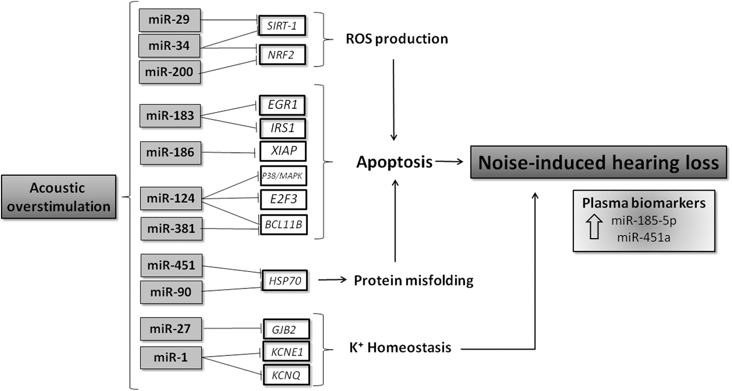
NIHL is a complex disease that results from the interaction of genetic and environmental factors with a susceptibility that differs remarkably among individuals. Long or repeated exposure to sounds at or >85 dB can cause hearing loss. Shearing forces from an excessive sound can cause cell death of the hair cells of the basilar membrane in the cochlea when they impact on the stereocilia. Hair cells are completely differentiated, and after becoming apoptotic, it is impossible to regenerate them. For that reason, an excessive noise exposure causes an irreversible degeneration in the basilar membrane of the cochlea (151). Different association studies have identified a cohort of genes involved in NIHL. These genes can be classified into different pathways: oxidative stress, potassium recycling, monogenic deafness, and heat shock protein (HSP)-related genes. In addition, post-translational regulation through miRNAs has also emerged as an additional layer of noise-induced gene regulation (173).
Acoustic contamination leads to sensory cell degeneration through well-differentiated pathways, resulting in severe apoptotic and/or necrotic phenotypes (13). For example, Taok1, involved in the stress response MAPK pathway, may be responsible of cochlear acoustic trauma via apoptosis modulation (195). Among the targets of miR-183 are early growth response 1 (Egr1) and insulin receptor substrate 1 (Irs1). Acoustic overstimulation in the rat cochlea increases the expression of Egr1 (24). However, even though Irs2-deficient mice develop sensorineural hearing loss (130), Irs1 has not been characterized in the cochlea.
Surprisingly, many predicted and validated target genes of NHIL-dependent miRNAs have been previously associated with sensorineural hearing loss. Xiap, a miR-186 target gene, has been involved in the protection against NHIL when it was overexpressed in a transgenic mouse model (194). The stress response P38α/MAPK is a predicted target of miR-124, and it has been linked to stress-related pathways in the cochlea and in apoptosis responses induced after acoustic damage (180). miR-124 target E2F3, this transcription factor, was upregulated 2 h postnoise exposure in the Chinchilla lanigera cochlea. This E2f3 increase was also involved with the p38/MAPK signaling pathway (82). Inhibition of Bcl11b, an antiapoptotic target of miR-124 and miR-381, both in vitro and in vivo, induces hair cell apoptosis (89).
Oxidative stress regulation
Recent publications have shown that increased oxidative stress in the inner ear is closely related to NIHL and AHL. These studies suggest that prevention of oxidative damage might be a solution to prevent development and progression of the majority of hearing loss cases (161).
During noise exposure, vasoconstriction increases after 8-isoprostane-F2a release (112). Then cochlear blood flow recovers to normal levels inducing ischemia reperfusion. This ischemia-reperfusion injury induces mitochondrial dysfunction and release of ROS in the cochlea. Antioxidant treatments have been shown to protect and decrease the progress of hearing loss in laboratory animals (181). Nrf2 is one of the master regulators of antioxidant genes. Although Nrf2 is not essential for normal development and cochlea function, Nrf2-/- mice seem to be more susceptible to noise exposure and have an impaired recovery and increased oxidative stress accumulation. Two groups of antioxidant enzymes, both tightly regulated by Nrf2, are active in the cochlea: glutathione metabolism enzymes and peroxide/superoxide scavengers, including CAT and SOD1 and SOD2 (72). Rabinowitz et al. have shown that noise-exposed workers that carry glutathione S transferase M1 (GSTM1) were protected from NIHL (152). Moreover, the SNP V16A in SOD2 in Chinese workers was associated with an enhanced sensitivity to NIHL. Some miRNAs expressed in the inner ear have been previously associated with impairment of Nrf2 function, as miR-34a and miR-200, but further investigation about their role in the Nrf2 pathway needs to be done.
Activation of NADPH oxidase enzymes, in particular NOX3, has been described in drug, noise, and AHL (160). Du et al. suggested that NOX3-associated oxidative stress may contribute to the accumulation of mtDNA mutations and activate a caspase 3-dependent apoptotic signalling pathway in the cochlea (40). However, the relationship between NOX3 and miRNAs has to be further investigated. Another interesting protein is pejvakin (PJVK). PJVK function is still unknown but PJVK-deficient mice are exceptionally vulnerable to sound. Noise damage in the cochlea induces upregulation of PJVK transcription and triggers peroxisome proliferation, resulting in an enhanced antioxidant effect in the auditory system (36).
AHL, also known as presbycusis, is also caused by cochlear hair cell degeneration and it is the most common form of hearing loss. Oxidative stress is also enhanced with aging, and has a fundamental role in inducing hair cell apoptosis in AHL. However, the mechanisms that mediate this effect need further investigation (92). One of the proteins previously related to hearing loss and aging is Sirtuin-1 (SIRT-1). SIRT-1 is a highly conserved NAD-(+)-dependent protein deacetylase, which has protective effects against age-related diseases. This protein acts like a sensor to regulate the internal oxidative stress by deacetylation of its substrates, like proliferator-activated receptor gamma coactivator 1α (PGC-1α) or the tumor suppressor protein p53 (147). SIRT-1 expression in cochlea has been well described and its reduction is associated with elevated hearing threshold and hair cell loss during aging, becoming a protective molecule (216). Several publications have shown differences in the expression of miR-29 and miR-34a families in the inner ear during aging (188). mir-34 has previously described to provide robustness to stress response programs by controlling noise in the DAF-16/FOXO-regulated gene network in Caenorhabditis elegans (79). Pang et al. described an increase in miR-34a in the cochlea, auditory cortex, and plasma from C57BL/6 mice during aging experiments and they corroborated these pieces of evidence in human patients with AHL. This was accompanied by a decrease in SIRT-1 and other miR-34a targets (143). Xiong et al. studied this mechanism by analyzing SIRT-1-dependent p53 acetylation. Their results showed that an increase in miR-34a and the consequent decrease in SIRT-1 lead to an increase in p53 acetylation and apoptosis (217). SIRT-1 and miR-34a have also been involved in other hearing pathologies as cisplatin-mediated hearing impairment, especially when together with SIRT-1 decreased expression; its function is also compromised by the reduction of intracellular NAD(+) (93).
miR-29b has been shown to be involved in aging, cellular senescence, and apoptosis, and one of its confirmed targets is also SIRT-1 (231). In this scenario, Xue et al. studied the correlation between miR-29 and SIRT-1 that also leads to hair cell apoptosis. They demonstrated that not only SIRT-1 but also PGC-1α, which plays an essential role in apoptosis and mitochondrial metabolism, are affected. Inhibition of miR-29b in in vitro studies with HE1-OCI cells, a hair cell line, increased the expression of these targets while decreasing apoptosis (220). However, the role of miR-34a and miR-29 in redox-dependent NIHL as well their utility as biomarkers in early detection of NIHL awaits further confirmation.
Finally, miR-451, a DICER-independent miRNA (222), previously described as protective against erythroid oxidative stress (224), was reported to be upregulated in a HEI-OC1 cell model of oxidative stress after treatment with tert-butyl-hydroperoxide (t-BHP) (203). This miRNA is one of the most upregulated in NIHL patients versus individuals exposed to noise, but it is significantly downregulated compared with nonexposure controls. These findings underscore its relevance in oxidative stress modulation in hearing loss pathology.
Potassium recycling pathway genes
The sensory cells of the inner ear are bathed in the endolymph, an extracellular fluid that is rich in potassium ions. Potassium is mainly responsible for the sensory transduction. Its proper recycling is necessary for the hearing mechanism (189). Multiple mutations in these potassium ion transporter genes (gap junction beta-2 protein [GJB2], gap junction beta-3 protein [GJB3], gap junction beta-6 protein [GJB6], potassium voltage-gated channel subfamily E member 1 [KCNE1], potassium voltage-gated channel subfamily Q member 1 [KCNQ1], and potassium voltage-gated channel subfamily Q member 4 [KCNQ4]) lead to both syndromic and nonsyndromic forms of hearing loss (189). Different susceptibility to noise is also associated with SNPs in these genes (145). A deletion in GJB2, also known as Connexin 26, in the cochlear sensory epithelium leads to increased apoptosis. In this loss-of-function model, miR-27 showed an increased expression. Wang et al. observed that the use of miR-27 shRNA inhibited GJB2 knockout-induced apoptosis (201). Moreover, Zhu et al. found that deletion of GJB2 reduced miR-96 expression in the cochlea during postnatal development. This reduction is associated with a cochlear tunnel developmental disorder in these knockout mice (234). In the case of KCNE1 and KCNQ, no evidence for miRNA modulation has been found. However, in the electrical remodeling of atrial fibrillation, miR-1 is responsible for the decreased expression of these genes (85). Thus, the link between miR-1 and KCNE1 and KCNQ in the cochlea needs further investigation.
Monogenic deafness genes
Most of the cases of genetic deafness recognized today are monogenic disorders. The failure of the activity in these genes has also been associated with sensitivity to NIHL. One of the most relevant is related to mutations in cadherin 23 (Cdh23) and protocadherin 15, molecules that link the sensory hair cells in the cochlea. These genes are fundamental for a right mechanoelectrical transduction (42). Mutations in Cdh23 disrupted the stereocilia organization on hair cells, inducing deafness and vestibular dysfunction in the model of the Ames waltzer mouse; the 753A variant of the Cdh23 gene was correlated with increased sensitivity to NIHL. In humans, PCDH15 and CDH23 gene mutations have been described in syndromic and nonsyndromic hearing loss (134).
MYH14 encodes one of the proteins of the myosin superfamily. They are actin-dependent motor proteins regulating cochlear hair cells motility and polarity. Mutation in MYH14 results in autosomal dominant hearing impairment in humans (DFNA4) (98). Nevertheless, no relationship between these genes and miRNAs in the inner ear or other tissues was found. miRNA mutations related to human deafness have been found in three families (123, 175). Mutations in the seed region of miR-96, specifically expressed in the inner and outer hair cells, are associated with hearing loss in human and mice. Studies addressing whether or not a larger number of miRNAs contribute to human deafness have been done, even though some authors propose this is unlikely to be the case (68).
HSP genes
HSPs form a group of conserved proteins assisting in synthesis, folding, assembly, and intracellular transport of many other proteins, whose expression increases under stressful conditions, including noise exposure. Variations in HSP70-1, HSP70-2, and HSP70-hom genes were shown to be associated with susceptibility to NIHL (200). Several publications have shown a correlation between HSP70 and some resident miRNAs in the inner ear such as miR-451 and miR-29 (25, 48, 184). Overexpression of miR-34 and miR-451 in cortical neurons increases HSP70 and vulnerability to apoptosis in the transfected cells (184). This might also be the case in miR-34-induced apoptosis in the cochlea.
Biomarkers
The epidemiological studies directed toward elucidating the potential effects of noise exposure or chronic NIHL on miRNA expression also questioned whether miRNAs can represent biomarkers as indicators of responses to noise exposure or occurrence of NIHL. Extracellular miRNAs in plasma have the potential to serve as stable noninvasive biomarkers in physiological and pathological conditions (165). However, few studies have specifically addressed the problem of miRNAs as biomarkers in NIHL. In a recent publication, Ding et al. studied differences in plasma miRNAs in male textile workers diagnosed with NIHL. They compared a population of 10 noise-exposed individuals and 10 NIHL patients and then attempted to further validate their results in a population of 46 noise-exposed textile workers, which included 23 NIHL patients. More than 73 miRNAs demonstrated significant differential expression in NIHL patients. The sequential validation restricted this group to four significantly upregulated miRNAs (miR-16-5p, miR-24-3p, miR-185-5p, and miR-451). After exclusion analysis, just two of these miRNAs, miR-185-5p and miR-451a, have been identified and validated as biomarkers. The plasma levels of both miRNAs were significantly downregulated in the noise-exposed individuals than in the nonexposed individuals, whereas they were slightly elevated in the NIHL patients than in the noise-exposed individuals (38). miR-451, as mentioned before, has been related to the regulation of oxidative stress in the inner ear. In the case of miR-185, its presence in the cochlea has not been established, but its role in DNA damage responses has been well described (193). In the case of other hearing loss pathologies, miR-34a has been correlated with AHL in mice and humans and its upregulation in plasma has been proposed as a possible biomarker (143). In idiopathic sudden sensorineural hearing loss (SSNHL), the protein Argonaute-2 (AGO2), an essential component of the RISC, was upregulated in SSNHL patients versus healthy control patients (59). However, further studies are necessary to prove the causal association between changes in the expression of miRNAs and noise exposure.
NIHL is a complex disease that results from the interaction of environmental and genetic factors. Recently, miRNAs have emerged as an additional mechanism of noise-induced gene regulation (Fig. 4). Some miRNAs such as miR-96, miR-182, and miR-183 family are highly specific in the inner ear in development and in hearing loss pathology (32). Some other miRNAs such as miR-34a and miR-29, previously involved in apoptosis and oxidative stress in other pathologies, also modulate key targets as SIRT-1 and Nrf2 in the cochlea (221). Other miRNAs have been weakly related to essential proteins in the cochlea function as is the case for miR-27 and miR-96 with the potassium ion transporter GJB2 (234). In addition, some miRNAs such as miR-185-5p and miR-451a have emerged as potential biomarkers in this pathology, leading to an easier and earlier diagnosis. Further studies are needed to complete this puzzle and connect highly regulated miRNAs in cochlea after damage exposure with hearing loss-relevant enzymes. Besides, a comparative analysis in all the different hearing loss pathologies is required to identify potential common causative factors, including hair cell apoptosis (38).
The role of miRNAs in mental stress and neuropsychiatric disorders
Studies from the past decade have highlighted the role of miRNAs in mental stress, psychiatric disorders, and the response to antipsychotic and antidepressant drugs. miRNAs are involved in a variety of psychiatric disorders such as schizophrenia, anxiety, major depression disorder (MDD), bipolar disorder, or post-traumatic stress disorder (PTSD). Psychiatric disorders are often accompanied by alterations in neuron architecture, function and survival, and synaptic plasticity and are the product of a combination of genetic and environmental factors (140). The molecular mechanisms by which environmental factors contribute to the onset and progression of psychiatric disorders are currently unclear. In this regard, it has been shown that the modulation of epigenetic mechanisms by environmental factors can have an effect on brain plasticity and behavior (159) as well as in the development of mental illness (133).
miRNAs are an important subset of these epigenetic regulators. Recent studies have shown that they can be mediators of the onset and progression of psychiatric disorders as well as modulate the response to treatment with antipsychotic and antidepressant drugs (69). Increasing evidence suggests that psychoactive agents including antidepressants and mood stabilisers utilize miRNAs as downstream effectors. Altering miRNA levels has been shown to alter behavior in a therapeutically desirable manner in preclinical models (135). For instance, miRNAs dysregulation may underlie many of the molecular changes observed in PTSD pathogenesis (56). Thus, Zhou et al. analyzed the role of miRNAs on the immunological dysfunction associated with PTSD. They found 7 upregulated and 64 downregulated miRNAs in combat veterans with PTSD. Specifically, miR-125a downregulation was suggested to be responsible for the increase in interferon gamma (IFNγ) observed in PTSD patients (230). Another study showed that DICER1 levels were reduced in the blood of PTSD patients with comorbid depression, suggesting that this decrease could be responsible for the general downregulation of miRNA levels observed in PTSD patients (210). However, this decrease in blood DICER1 levels was not found in a study of major depression (10). Thus, further human studies must be conducted to discern whether DICER1 downregulation is a specific characteristic of PTSD but is not present in other psychiatric disorders. MDD is the most common psychiatric illness. Recent studies have shown that MDD is associated with alterations in the levels of several miRNAs in whole blood, plasma, or serum. Expression levels of miR-34b-5p and miR-34c-5p are higher in leukocytes of MDD patients than in controls, and the levels of these two miRNAs correlated with the severity of the depression status (179). The miR-34 family is involved in hypothalamic-pituitary-adrenal (HPA) axis that has a prominent role in stress response (69). In fact, it has been reported that miR-34c regulates corticotrophin releasing hormone receptor 1 (CRHR1) mRNA, which modulates anxiety-like behavior and affects regulation (60, 64). According to the role of miR-34 family on depression, it has been shown that miR-34a is downregulated by a combination of lithium and valproic acid, two well-known mood stabilizers, in cultured rat neuronal cells (77).
Brain-derived neurotrophic factor (BDNF) is a neurotrophin that plays essential roles in neuronal development and plasticity. Serum BDNF levels have been proposed to be a good biomarker of depression and it is known that BDNF is associated with the risk of schizophrenia and with aggressiveness and anxiety (120). Serum BDNF levels are lower in patients with depression and, concomitantly, serum levels of the BDNF-targeting miR-132 are higher. Serum miR-132 levels were also positively associated with depression severity (111). Interestingly, BDNF increases the expression of miR-132 in cultured cortical neurons. In contrast, glucocorticoid receptor (GR) activation has been demonstrated to reduce miR-132 expression (90). These authors also showed that miR-132 partly contributes to the BDNF-mediated increase of postsynaptic proteins (90).
Glucocorticoids are important mediators of neuronal function and are associated with behavior, cognition, memory, and emotions (76). Uchida et al. demonstrated that GR protein was lower in the paraventricular nucleus of restrain-stressed F344 rats, although they failed to observe a similar decrease in the hippocampus and prefrontal cortex. They found that miR-18a inhibited translation of the GR mRNA, although this miRNA was not modulated by restrain stress in this model (187). In another study, Fan et al. found that miRNA-26b, miRNA-1972, miRNA-4485, miRNA-4498, and miRNA-4743 were significantly upregulated in the blood of MDD patients. A functional analysis of targets of these miRNAs showed that most of them were involved in axon guidance, glutamatergic synapsis, and long-term potentiation, but they were also involved in mTOR signaling and carbohydrate metabolism (46). Blood miR-26b levels have also been found to be increased after chronic academic stress and decreased once this stress disappears (71). This study also showed academic stress-mediated upregulation in miR-16, miR-20b, miR-126, miR-144, and miR-144*. Moreover, miR-16 levels were positively correlated with anxiety levels. MiR-451a, miR-17-5p, and miR-223-3p have also been found to be upregulated in MDD patients in a cohort from Turkey and miR-451a levels seemed to be correlated with the duration of the depressive episode (19).
Selective serotonin reuptake inhibitors (SSRIs) are the most common antidepressant drugs currently in use. It has been shown that some miRNAs are modified by SSRI treatment and may be used as potential biomarkers of treatment response (41). For instance, escitalopram increased blood levels of 28 miRNAs including those of the let-7 family and miR-29b and decreased levels of miR-34c and miR-770. Escitalopram-modulated miRNAs are involved in fundamental neurological functions, including neuroactive ligand–receptor interaction, axon guidance, long-term potentiation, and depression. However, it is noteworthy that these miRNAs are also involved in metabolism, nutrient sensing (MAPK, insulin, and mTOR signaling), and vascular smooth muscle cell contraction (12). Belzeaux et al. found that miR-589, miR-579, miR-941, miR-133a, miR-494, miR-107, miR-148a, miR-652, and miR-425-3p were upregulated, whereas miR-517b, miR-636, miR-1243, miR-381, and miR-200c were downregulated in patients suffering from a major depressive episode compared with those of normal controls. Furthermore, miR-20b-3p, miR-433, miR-409-3p, miR-410, miR-485-3p, miR-133a, and miR-145 were modified by antidepressant treatment in patients with clinical improvement, suggesting that these miRNAs are potentially accessible biomarkers of treatment response (10). Plasma levels of miR-144-5p and miR-30c-5p are significantly increased after 8 weeks of treatment and SSRIs and plasma levels of miR-144-5p are also inversely correlated with a depressive score (197).
Another study carried out by Lopez et al. measured levels of miRNAs in the ventrolateral prefrontal cortex of depressed individuals compared with those of psychiatrically healthy controls and found that miR-1202 was significantly downregulated. miR-1202 is a primate-specific miRNA that targets metabotropic glutamate receptor 4 (GRM4). Interestingly, brain miR-1202 levels were higher in depressive patients who had been subjected to antidepressant treatment than in those without a previous history of antidepressant treatment. Blood levels of miR-1202 also increased in patients who responded to citalopram treatment, but not in nonresponding patients (114). One of the described mechanisms by which SSRIs reduce depression is the inhibition of serotonin transporter (SERT). A recent study showed that SSRIs increased the expression of miR-135a in the raphe nuclei of a mouse model of depression (81). miR-135 directly targets SERT and, therefore, the authors suggested that the increase in miR-135a levels is a potential mechanism that could explain the antidepressant action of SSRIs. Moreover, authors showed that overexpression of miR-135 resulted in increased resilience in mouse anxiety and depression models. On the contrary, miR-135a knockdown resulted in decreased SSRI treatment efficacy (81). This study also showed that miR-135a levels were lower in the raphe nuclei of depressed patients who had committed suicide. Circulating levels of this miRNA were also found to be lower in depressed patients (81). miR-16 also targets SERT and is increased by fluoxetine, an SSRI, in mouse serotoninergic raphe nuclei (9).
To sum up, these studies suggest that miRNAs play essential roles in the onset and progression of psychiatric disorders. In addition, they show that miRNAs are associated with treatment response and blood and circulating miRNAs are potential biomarkers of both disease progression and treatment response. However, it should be noted that few studies consistently found similar dysregulated miRNAs in brain and blood. This could be because of the small sample size of many of them as well as other factors such as ethnicity and sociodemographic factors.
Environmental factors that modulate neuropsychiatric miRNAs
All these miRNAs are associated with psychiatric disorders and treatment response. Also recent studies have revealed that patients with psychiatric disorders have altered miRNA expression profiles in the circulation and brain and have shown that manipulating the levels of particular miRNAs in the brain can alter behavior (80). Environmental factors can modify miRNA expression, leading to both short- and long-term effects on mood and behavior through the modification of neuronal plasticity or inducing neuronal structural changes (69). The effect of environmental factors on psychiatric miRNAs has been studied mainly in animal models exposed to different types of stressors. It should be borne in mind that depression and other mood disorders are difficult to study in murine models because they are complex disorders with highly heterogeneous symptoms. Moreover, to determine the severity of those disorders, subjective tests are often used that cannot be applied to animal models. Despite these limitations, some animal studies have demonstrated the potential of some environmental factors to contribute to psychiatric disorders through the modulation of related miRNAs. For instance, adult male rats subjected to immobilization stress showed changes in amygdala and hippocampus miRNA profiles. Among the dysregulated miRNAs, miR-134 and miR-183 were both upregulated by acute stress in the central amygdala (121). One of the targets of both miRNAs is Sc35, whose protein accumulates in the prefrontal cortex after acute stress and regulates the stress-induced alternative splicing of acetylcholinesterase mRNA in brain neurons (124). miR-134 has also been involved in synaptic plasticity and long-term memory formation (52).
Environmental pathogens, environmental chemicals, dietary stress, and drugs/alcohol abuse are other environmental factors that can potentially alter psychiatric-related miRNAs. For instance, dietary stress induced by caloric restriction (CR) or high-fat diet (HFD) modifies hypothalamic miRNA profiles in rats. It has been shown that miR-30, miR-200b/c, and let-7 miRNA families were deregulated after persistent nutritional challenge. Specifically, miR-30 and let-7 miRNAs increased by either HFD or CR or both, whereas miR-200 miRNAs were downregulated after CR (164). Let-7 and miR-200 miRNA families were similarly modified in the brain of mice exposed to hexahydro-1,3,5-trinitro-1,3,5-triaxine (the explosive known as RDX), which is a common environmental contaminant. Interestingly, let-7 family was downregulated in the liver of mice exposed to this pollutant (225). Cocaine increased the expression of miR-34b, miR-34c, miR-134, and miR-181, among others, in the hippocampus of addicted rats. However, miR-34b and miR-34c levels decreased after addiction extinction (21). miR-190 levels were higher in hippocampi of rats after cocaine addiction extinction than in control nonaddicted rats (21). miR-190 was also downregulated by fentanyl in mice hippocampi (228).
Exposure to environmental stress during embryonic development can result in altered neurodevelopmental processes that lead to impaired hippocampal development, impaired HPA axis activity and responsiveness, and impaired synaptic plasticity. There is increasing evidence that maternal mental stress induced by anxiety or depression results in neurodevelopmental disorders and higher risk of psychiatric illnesses in the offspring (69). Recently, Zucchi et al. showed that gestational stress induced by restrain of the body or forced swimming in pregnant rats leads to altered miRNA profiles in the hippocampus of newborn offspring. Gestational stress induced upregulation of miR-98, miR-103, and miR-323 and downregulation of miR-145, miR-151, and miR-425 (235). The same authors showed that gestational stress can even have transgenerational effects because they showed that growth retardation and behavioral disorders, while evident in the F1 generation, were even stronger in subsequent F2 and F3 generations. These alterations were accompanied by changes in the level of brain miRNAs in F2, including increases in miR-23b and miR-200c and decreases in miR-200a, miR-200b, miR-96, miR-182, miR-183, miR-141, miR-429, and miR-451 (223).
miRNAs link oxidative stress and mental stress
Proper mitochondrial function is fundamental for neuronal survival. Oxidative stress, especially in astrocytes and microglia, deeply impacts mitochondrial function (105). Mitochondrial dysfunction caused by oxidative stress is associated with psychiatric and mood disorders like bipolar disorder or MDD (26, 100). Recent emerging pieces of evidence suggest that miRNAs play a role in the regulation of oxidative stress-mediated neuronal mitochondrial disease. Six miRNA families have been consistently associated with neuropsychiatric disorders and mental stress and have been shown to be modulated by antipsychotic and antidepressant treatments: miR-29, miR-30, miR-200, miR-34, miR-181, and let-7. These miRNA families also play a role in the cellular response to oxidative stress (Fig. 5).
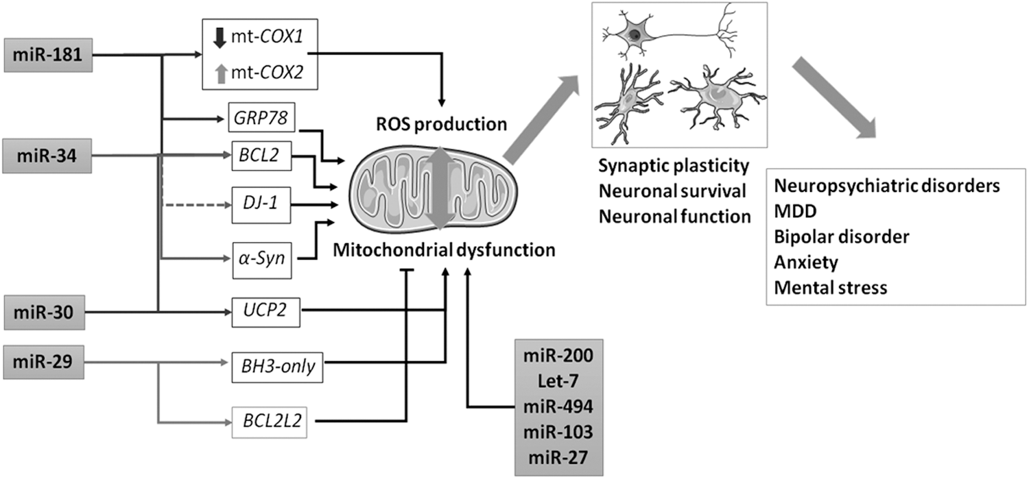
miR-181c plays a role in mitochondrial function regulating cytochrome c oxidase subunit 1 (mt-COX1) mRNA by producing electron transport chain complex IV remodeling through the increase of mt-COX2 production. This effect of miR-181c on the mitochondria results in higher oxygen consumption and ROS production (33). miR-30e targets uncoupling protein 2 (UCP2) (86). UCP proteins are mitochondrial proton transporters that play key roles in reducing ROS production and have been shown to promote neuroprotection. In particular, UCP2 protects astrocytes and microglia from oxidative stress, contributing to neuronal survival (61). miR-30e, miR-34c (91), and miR-181d (199) target BCL2, which controls mitochondrial outer membrane permeability and Ca++ homeostasis in brain stress conditions (105). In fact, Ouyang et al. showed that all members of the miR-181 family target other members of the BCL2 family. They also showed that miR-181 overexpression induces higher ROS production and lower astrocyte survival after glucose deprivation (137). In another study, these authors showed that, in addition to BCL2, miR-181 also targets GRP78 (138), a fundamental regulator of endoplasmic reticulum stress, which is also involved in ROS production and mitochondrial function (105). The miR-29 family is enriched in astrocytes and its expression increases during neuron maturation. It also enhances neuron resistance to ER stress and death by targeting BH3-only genes that inhibit BCL2 and favor cytochrome c release during apoptosis (96). The role of miR-29 as a promoter of neuron survival was confirmed by Ouyang et al. who showed that miR-29a targets BCL2 binding component 3 (BBC3), a member of the BH3-only-family. They additionally showed that miR-29a overexpression reduces ROS production and increases mitochondrial membrane permeability after glucose deprivation (139). However, it has also been reported that miR-29b promotes neuronal cell death by targeting BCL2L2, a member of the BCL2 family of antiapoptotic mediators (168). Further research is needed to clarify the role of miR-29 in neuronal survival. miR-34b and miR-34c levels are also increased in differentiated SH-SY5Y cells compared with those in undifferentiated cells, suggesting that this miRNA family is also important in neuronal maturation. miR-34b/c downregulation has been shown to be associated with higher ROS production and mitochondrial dysfunction. This effect could be partly because of an indirect effect on DJ-1 (127), which is involved in the maintenance of mitochondrial complex I integrity, and increases neurodegeneration in mice (218). miR-34b/c downregulation also increases alpha-synuclein (α-SYN) expression at both mRNA and protein levels (88). α-SYN is the main component of the neuronal cytoplasmic intrusions called Lewy bodies, whose accumulation is associated with neurodegeneration, partly caused by complex I impairment and ROS production (215). miR-34b/c directly targets α-SYN 3′-UTR (88). In hippocampal HT-22 cells subjected to a cycle of hypoxia/reoxygenation, an increase in ROS production occurs that could be mediated by an increase in miR-200a-3p and miR-200b-3p expression, although the mechanism by which miR-200 miRNAs affect ROS production in neuronal cells is still unclear (205). In B2B human bronchial epithelial cells, it has been shown that members of the let-7 family are downregulated by oxidative stress and, in turn, let-7a overexpression protects cells from the oxidative damage-mediated death by reducing oxidative stress through the direct inhibition of arginase 2 (177). In human Huh-7 hepatocytes, let-7 miRNAs also protect cells from oxidative injury through the direct inhibition of BACH1, a key repressor of the cytoprotective enzyme heme oxygenase 1 (74).
Other miRNAs associated with neuropsychiatric disorders and mental stress are also involved in mitochondrial function and oxidative stress. For instance, miR-494 increases ROS production in neuro2a cells through the direct targeting of DJ-1 gene (218). miR-103 and miR-27a/b become upregulated in SH-SY5Y cells treated with tumor necrosis factor α (TNF-α), whereas miR-23a/b shows downregulation. TNF-α induces mitochondrial oxidative stress in these cells. Among the predicted targets of these miRNAs, there are several mitochondrial complex I subunits, a complex that is compromised by TNF-α treatment (148). In SH-SY5Y cells, it has been demonstrated that miR-27a also directly targets NRF2 (131), a key component of the defensive cellular mechanism against oxidative stress (215).
Taken together, these studies highlight the role of miRNAs in mental, mood, and behavioral disorders and show that many of these neuronal-related miRNAs are involved in the regulation of ROS production, mitochondrial function, neuronal survival, and response to oxidative injury.
Summary and Conclusions
It is evident that the three types of environmental risk factors covered in this review, physicochemical, noise, and mental stress, promote responses involving specific sets of miRNAs. These, in turn, have important regulatory roles upon cellular processes such as apoptosis, signal transduction cascades, and DNA repair mechanisms. Nevertheless, in the overwhelming majority of cases, the available data in the literature have not established definitive or causative roles for miRNAs in the pathological phenotypes associated with the noxious effects of the environmental risk factors covered. Thus, the design of large cohort studies in selected populations exposed to these environmental risk factors is a major unmet need.
Footnotes
Acknowledgments
This work was supported by grants from the Ministerio de Economía, Industria y Competitividad (MINECO) SAF 2015-66107-R; Consolredox Network SAF 2015-71521-REDC; Instituto de Salud Carlos III network REDinREN RD16/0009/0016; and Fundación Renal “Iñigo Alvarez de Toledo,” all from Spain. It also has been supported by European Cooperation in Science and Technology/COST action BM-1203 (EU-ROS). The Centro de Biologia Molecular “Severo Ochoa” (CBMSO) receives institutional support from Fundación “Ramón Areces.” V.M. is supported by the MINECO program of Formación de Personal Investigador (FPI BES-2013-065986). L.D.'s research is supported by a grant from Instituto de Salud Carlos III, Fondo de Investigaciones Sanitarias (PI14/01374). IMDEA Food belongs to Madrid Institute for Advanced Studies Network promoted by the Madrid Regional Government (Comunidad de Madrid) and is funded by European Regional Development Funds from the European Union. C.F.-H. is supported, in part, by NIH Grant R35HL135820, Foundation Leducq Transatlantic Network of Excellence in Cardiovascular Research, and AHA Established Investigator Award (16EIA27550004).