
Abstract
Significance:
Efficient targeted therapy with minimal side-effects is the need of the hour. Locally altered redox state is observed in several human ailments, such as inflammation, sepsis, and cancer. This has been taken advantage of in designing redox-responsive nanodrug carriers. Redox-responsive nanosystems open a door to a multitude of possibilities for the control of diseases over other drug delivery systems.
Recent Advances:
The first-generation nanotherapy relies on novel properties of nanomaterials to shield the drug and deliver it to the diseased tissue or organ. The second generation is based on targeting the drug or diagnostic material to the diseased cell-specific receptors, or to a particular organ to improve the efficacy of the drug. The third and the latest generation of nanocarriers, the stimuli-responsive nanocarriers exploit the disease condition or environment to specifically deliver the drug or diagnostic probe for the best diagnosis and treatment. Several different kinds of stimuli such as temperature, magnetic field, pH, and altered redox state-responsive nanosystems have educed immense promise in the field of nanomedicine and therapy.
Critical Issues:
We describe the evolution of nanomaterial since its inception with an emphasis on stimuli-responsive nanocarriers, especially redox-sensitive nanocarriers. Importantly, we discuss the future perspectives of redox-responsive nanocarriers and their implications.
Future Directions:
Redox-responsive nanocarriers achieve a near-to-zero premature release of the drug, thus avoiding off-site toxicity associated with the free drug. This bears great potential for the development of more effective drug delivery with better pharmacokinetics and pharmacodynamics.
Introduction
E
The factors that dictate the NP's targeting ability are particle size, hydrophobicity, surface charge, and modification. The particle size determines its interaction with biological membranes and its ability to penetrate physiological barriers (endothelium, cell membrane, blood–brain barrier, blood–cerebro spinal fluid barrier, etc.). It is desirable that NPs have increased circulation time and do not aggregate in body fluids. Conventional first-generation nanosystems are hydrophobic in nature and are rapidly opsonized and cleared by the resident macrophages of the mononuclear phagocytic system. To prevent this clearance, NP surfaces are modified by the addition of certain hydrophilic molecules. Surface charge is important in that it determines whether NPs get attracted to each other or the oppositely charged cell membrane (11). Cationic charge promotes internalization since cell membranes are negatively charged. Anti-adhesive properties, imparted to the surface of NPs by extended configurations that act as steric barriers, minimize the chance of clearance by hepatic macrophages and increase the intracellular permeation (111). Drug release is modulated by the molecular weight of the polymer used and, as a rule of thumb, the higher the molecular weight of the polymer, the slower is the in vitro release (132). Even though these drug delivery systems are in the order of nanometers and can circulate in the blood for several hours, they hardly make it into the tissue interstitium owing to the high cell density and interstitial fluid pressure (101).
Further advances in nanotechnology led to the development of next-generation nanosystems for targeted drug delivery, which incorporates ligands into the nanostructure that is specific to the target of interest. These ligand-conjugated nanostructures outperformed the first-generation nanosystems, demonstrating several therapeutic benefits involving thernostic as well as tissue engineering applications (16, 85, 88, 110). These second-generation nanostructures are especially helpful in cancer treatments where the tumor cells need to be specifically targeted compared with their normal counterparts (85). This is also useful for the transport of therapeutic drugs across the blood–brain barrier to treat brain diseases (33).
Polymeric NPs can also be activated by local environmental changes such as pH, redox state, temperature, the presence of a particular molecule, and external stimuli. These “smart” third-generation nanosystems take advantage of disease conditions, such as imbalanced redox state or decrease in pH. Altered redox state of the disease site has been employed for the on-demand release of the cargo from redox-sensitive nanocarriers (RSNs). Different combinations such as RSNs coupled with a diagnostic probe or conventional drug or siRNA have been exploited for the diagnosis and treatment of human diseases (7, 62).
Of particular interest in recent years are the redox-sensitive nanostructures that have been exploited to control the release of the drugs for various therapeutic applications (7, 44, 46). Redox potential is one of the major physiological differences between tumor and normal tissues that is advantageous for scientists to construct redox-responsive nanostructures to deliver the chemotherapeutic drugs (85). Polymers that can self-assemble into nanostructures on oxidation–reduction conditions provide great advantage toward site-specific controlled delivery of drugs (131, 133). Recent studies with inorganic nanocarriers have also been found to provide a stable structure to entrap the drug, allowing surface modification with necessary anchoring groups coupled to a variety of responsive building blocks (120). The unique physiochemical characteristics of inorganic nanocarriers and their facile synthesis make them advantageous. A prime example is mesoporous silica nanocarriers (MSNs), which have been chemically altered and combined with different inorganic nanocarriers such as quantum dots and magnetic nuclei or macromolecules (27, 44, 61, 71). These inorganic MSNs were tethered with organic macromolecules by either a redox-sensitive or pH-sensitive bond to form so-called “gatekeepers” to achieve the exclusively redox-dependent release of the cargo (73). Redox-responsive nanocarriers have gained substantial attention due to their exceptional ability to release the cargo exclusively at the diseased site. Different material configurations have been experimented with to enhance drug release stimulated by internal cues as well as external physical inputs. In this review, we summarize the evolution of nanomedicine and describe stimuli-responsive nanosystems with special emphasis on different platforms of redox-responsive nanocarrier systems and their implications in the cure of human diseases.
First-Generation Nanosystems
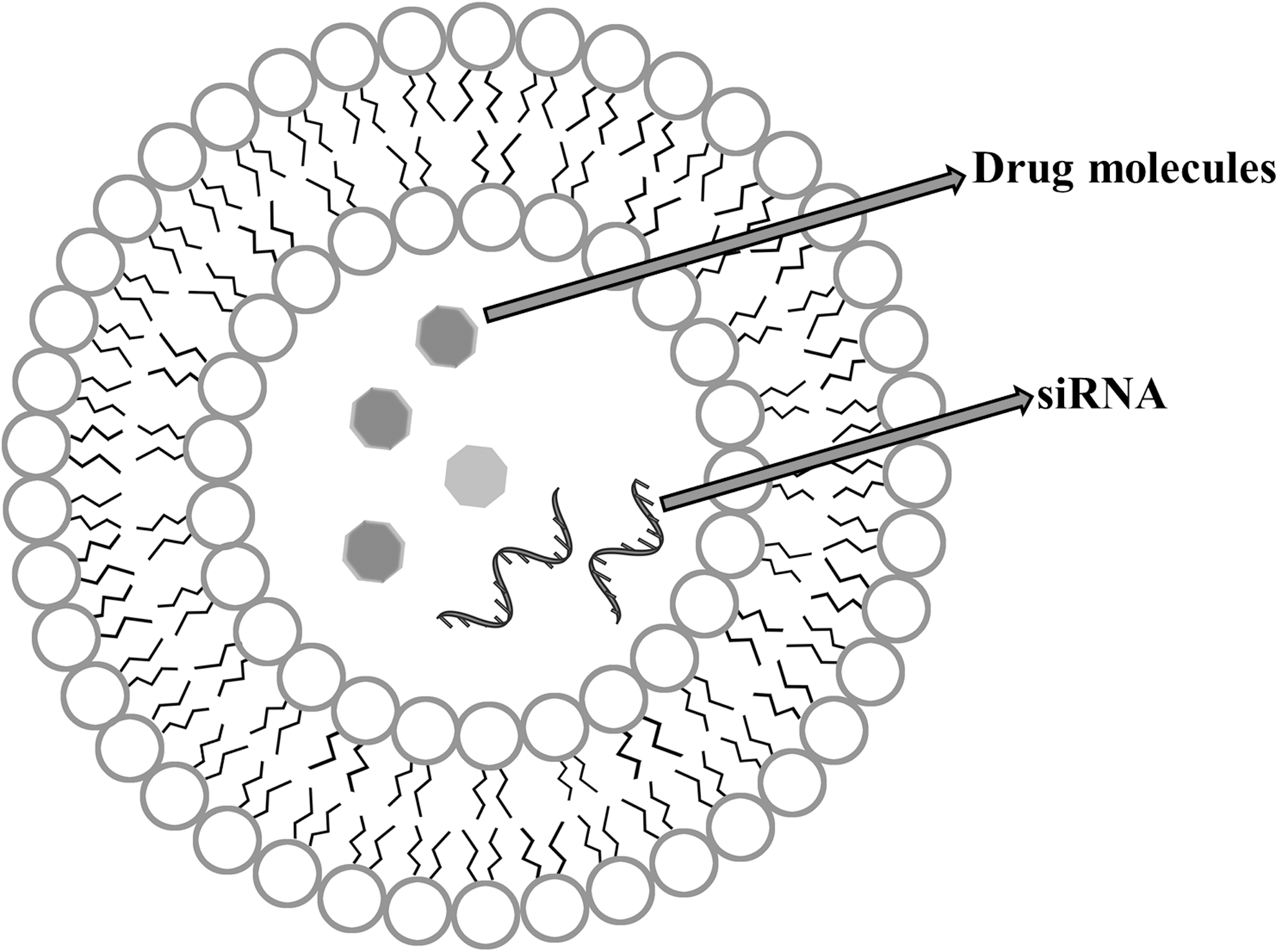
To increase a drug's bioavailability, a drug carrier ideally should spend maximal time in the blood avoiding high non-specific accumulation in tissues. The nanoscale of the carrier and surface modifications such as the addition of polyethylene glycol (PEG) allow the carrier to avoid renal clearance and uptake by cells of the reticulo endothelial system (RES) (39). Leaky vasculature and poor lymphatic drainage are hallmarks of cancerous tissue, sites of inflammation, and bacterial infection. Certain nanoscale drug carriers exploit this and can extravasate and later accumulate in these tissues. This is known as enhanced permeation and retention (EPR) effect (83, 84), which results in enhanced theranostic potential of the drugs with subsequent reduction of side-effects. Liposomes and polymeric NPs employ this EPR effect to deliver the cargo at the diseased site. Liposomes have the unique ability to protect drugs from degradation and to reduce the toxicity of the drugs by allowing lower dosages to be used. Hence, they are used as potential drug carriers instead of conventional drug therapy (79) (Fig. 1). Biodegradable polymeric NPs are promising drug delivery systems because they provide controlled/sustained release, are of a sub-cellular size, and are biocompatible (94). In addition, they are stable in body fluids, non-toxic, non-immunogenic, non-inflammatory, non-thrombogenic, and avoid the RES (26). Biodegradable polymers such as poly(lactic-co-glycolic)acid (PLGA) have had a high success rate in nanomedicine therapy primarily because they are biodegradable and undergo hydrolysis in acidic medium to produce glycolic acid and lactic acid, both of which are effectively dealt with in the body through the tricarboxylic acid cycle. In our lab, we have employed polysorbate-80 as a stabilizer to synthesize disulfiram-loaded PLGA NPs for the treatment of liver cancer (50, 51).
Second-Generation Nanosystems
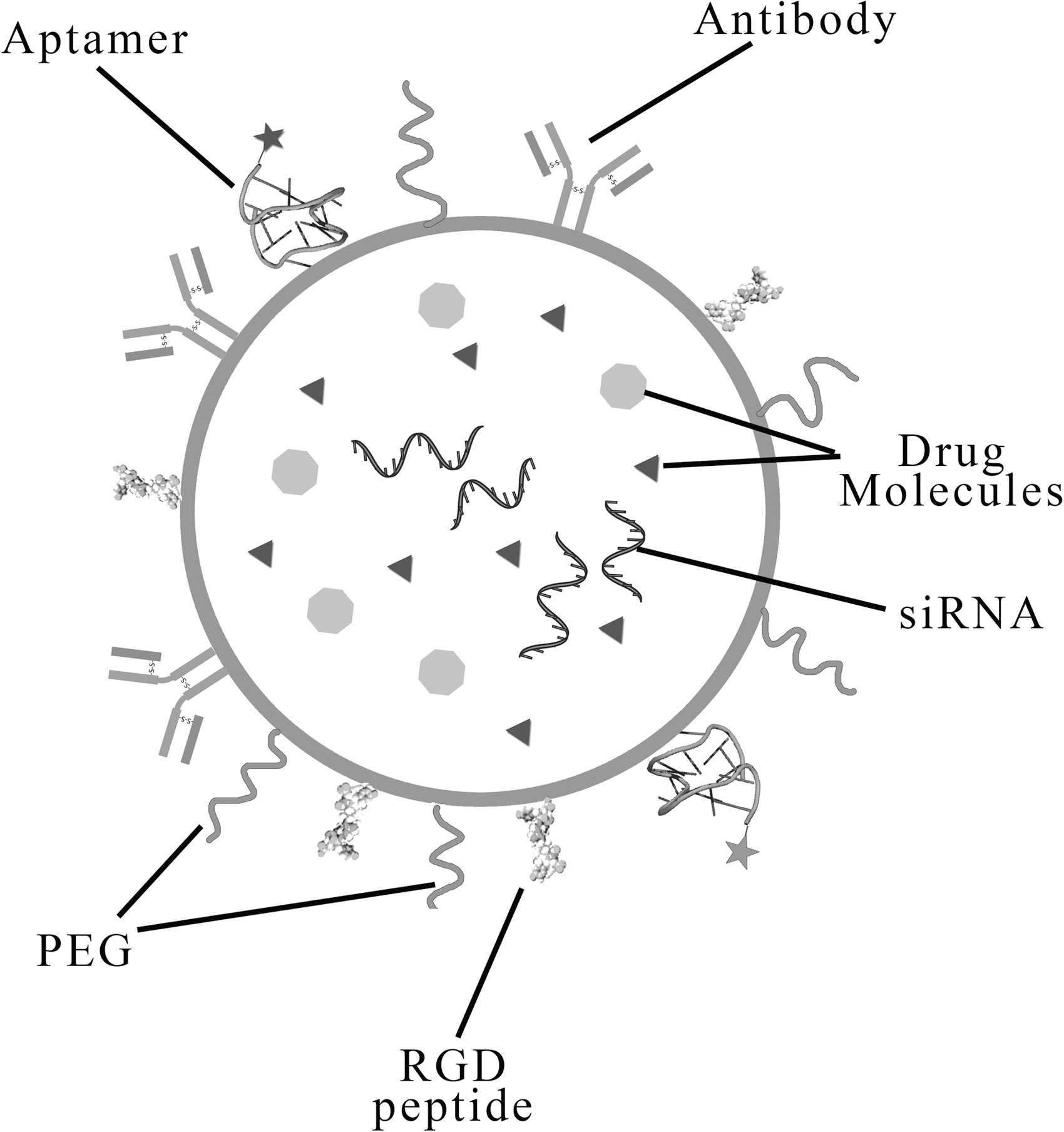
Several cancer cells overexpress particular cell surface receptors. NPs can be customized by functionalizing with molecules such as folic acid, Arg–Gly–Asp (RGD) peptides, and other molecules or antibodies (8, 109) to recognize these overexpressed receptors to improve cancer targeting and therapeutic efficacy (Fig. 2). In vitro, these have been validated and in the broad sense shown to significantly increase the uptake of drug delivery systems in cancer cells in vivo (139). These nanostructures could attain the advantage of both sustained release and targeted delivery, thus behaving as a double-edged sword in controlling the disease. In this section, we will briefly discuss some of the major types of second-generation nanosystems (Table 1).
Various Generations of Nanomedicine
Each generation of nanosystem along with their representative formulation and scopes are summarized.
DOX, doxorubicin; EGFR, epidermal growth factor receptor; HER2, human epidermal growth factor receptor 2; P-gp, P-glycoprotein; PLGA, poly(lactic-co-glycolic)acid.
Cellular receptors such as epidermal growth factor receptor (EGFR) and human epidermal growth factor receptor 2 (HER2), which play key roles in cancer progression, are found to be overexpressed in certain solid tumors (65). Qian et al. conjugated cetuximab (a commercial monoclonal antibody [mAb] against EGFR) on the surface of gold NPs and found that it was significantly selective toward cells displaying a high level of EGFR (106). In another report, Long et al. synthesized doxorubicin (DOX)-loaded anti-EGFR mAb conjugated gelatin nanoparticles and tested its targetability in the A549 lung cancer cell line (78). This formulation also induced a reduction in tumor growth by 90% with a survival rate of 100%, in contrast to the non-targeted formulation and free drug.
Integrins are transmembrane proteins that are expressed exclusively in human endothelial cells that act to harbor extracellular matrix proteins containing RGD sequence (25). Integrins were shown to be upregulated in endothelial malignancy and many other cancer cells (98). Cisplatin-loaded polymeric NPs decorated by cyclic penta peptide (RGDfK) are shown to exert improved in vitro cytotoxicity in breast and prostate cancer cell lines (38). This formulation showed promising results in vivo, in contrast to the free drug in a xenograft model of human breast cancer (Fig. 2).
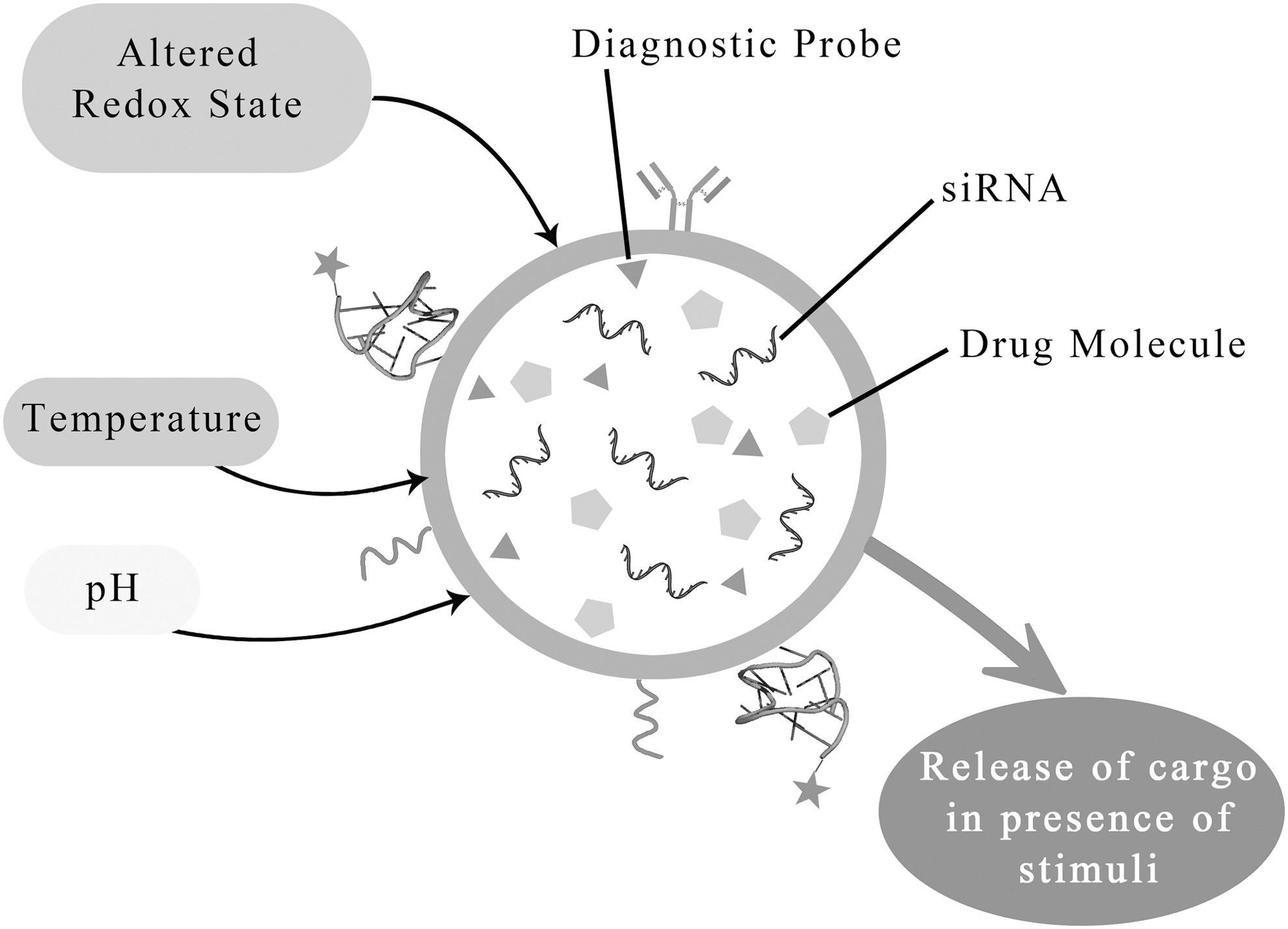
Third-Generation Nanosystems: Stimuli-Responsive Nanocarriers
Rationale of developing stimuli-responsive nanocarriers
Chemotherapeutics, especially anti-neoplastic drugs over the ages, have faced a common persistent problem of limited water solubility and non-specific cytotoxicity, giving rise to a myriad of adverse side-effects (21). A majority of anticancer drugs are mitotic inhibitors that target actively dividing cells (47). Due to the lack of specificity of conventional drugs, non-cancerous actively dividing cells share the same fate like cancerous cells and are also condemned to death. This seriously limits the dosage and treatment period of the drug. Stimuli-responsive drug delivery systems that have the ability to trigger drug release on demand could be an alternative to site- and time-controlled drug release. Such systems encapsulate the drug such that premature release at undesirable places in the body does not occur. Drug release using stimuli-responsive nanosystems can be triggered by external stimuli (heat, ultrasound [US], light, magnetic field, etc.) or physiological cues (pH, redox potential, metabolite or enzyme concentration, etc.) at the target site (Fig. 3). An example of this concept is ThermoDox, which is a heat-activated liposomal formulation of DOX (91). DOX is released from the aqueous core into the outer medium by enhancing the permeability of the liposome membrane. This is achieved by heating to temperatures above the used phospholipid's transition temperature (41°C). ThermoDox along with radio frequency ablation have undergone phase III clinical trials in hepatocellular carcinoma patients (90). The mechanism of action of these systems relies on abrupt changes in volume in response to temperature, pH, electric field, or protein concentration (40, 112). In the next section, we discuss some of the types of stimuli-responsive nanosystems that exist till date (Fig. 3).
Exogenous stimuli-controlled nanosystems
External stimuli such as light, temperature, magnetic field, and US can be employed to trigger the release of the drug or cargo from the nanoformulations at the destined site (66). Photothermal therapy (PTT) or photodynamic therapy (PDT) works by introducing light or altering the temperature locally to initiate the release of the drug or diagnostic material from the nanoformulation at the diseased site.
Light- and temperature-controlled nanostructures
Light-responsive nanocarriers have been designed based on the fact that several chemical reactions could be controlled by light. For example, Sakthi Kumar and colleagues reported the synthesis of PEGylated copper sulfide nanocrystal (PEG-Cu2S NC), which releases the encapsulated drug DOX in a light-responsive manner (102). Moreover, this formulation was shown to be multimodal, being responsive to pH and N light, and it also acts as an x-ray contrasting agent. In addition, the PEG-Cu2S NC was surface decorated with folate for the active targeting. It was observed that photo excitation leads to a four-fold increase in drug release and DOX was found in the nucleus of the photoexcited cells treated with PEG-Cu2S NC within 10 min, which takes place only after 1 h in the non-exposed cells. Subsequently, Wang et al. reported the synthesis of chlorine e6 (Ce6)-based photosensitive micelles loaded with DOX for achieving the benefit of PDT and chemotherapy (123). Near-infrared (NIR) illumination of the treated cells/tumor cells resulted in reactive oxygen species (ROS) generation and helped chemotherapeutics to penetrate through solid tumors. In addition, the temperature of the NIR-exposed tissue becomes higher, which helps in photoaquostic imaging and photothermal therapy (Table 1).
Magnetic field and US controlled
The use of a magnetic field in the routine diagnosis is very common; for example, magnetic resonance imaging (MRI) provides fine details of the organ being investigated. Magnetic field-responsive nanomaterials can be used to accumulate the particles near the magnetic field and deliver the drug. Lin et al. developed surface-functionalized and DOX-loaded superparamagnetic iron oxide nanoparticles (SPIONs) for MRI contrast enhancement and chemotherapy (75). They found that exposure of the magnetic field to the tumor greatly enhanced the accumulation of DOX-loaded SPIONs at the tumor site, which resulted in a reduced tumor size than the group that was not exposed to the magnetic field. Also, the MRI of the magnetic field-exposed tumor yielded higher T2-weighted signals than the non-exposed tumor, signifying the potential of MRI-monitored magnetic field-induced chemotherapy.
US can be used for the local release of the drug/cargo from US-sensitive nanostructures. US produces an acoustic field in a solution that induces pressure variation, resulting in the formation of bubbles or cavities. When these bubbles collapse, they generate solvodynamic shear. Any polymer segment surrounding this shearing force travels toward the cavities. When a polymer chain approaches these bubbles, the solvodynamic shear elongates the polymer chain and finally breaks/cuts the polymer chain. This phenomenon has been employed in the design of a supramolecular cap such as 2-Tetrahydropyranyl methacrylate (THPMA), which bears a US labile acetal group. Exposure of THPMA to US irradiation leads to the formation of methacrylic acid (MAA). US irradiation also yields a phase transition: hydrophobic THPMA to hydrophilic MAA.
Paris et al. fabricated a MSN-based nanosystem for the US-triggered release of an anti-cancer drug DOX (96). They grafted the surface of the MSNs with a thermo-sensitive and US-sensitive copolymer that has a lower critical solution temperature (LCST). The thermo-responsive polymer they used was poly (2-(2-methoxyethoxy)ethylmethacrylate), in short p(MEO2MA). It is soluble in water at a low temperature such as 4°C, but it becomes hydrophobic as the temperature increases. At physiological temperature, it becomes completely hydrophobic, thus sealing the ends/pores of MSNs. This ensures no leaking of the cargo before it reaches the desired site. US irradiation causes the formation of hydrophilic MAA and the increase of LCST to more than 37°C, leading to the phase transition of the gatekeeper copolymer, which leads to the eventual release of the drug. It was shown that this hybrid system is not cytotoxic until it is exposed to US. DOX-loaded MSNs capped with this US and thermosensitive copolymer have also been shown to induce cell death when they were exposed to US.
Endogenous stimuli-controlled nanosystems
Endogenous cues, such as locally altered metabolite concentration, pH, or redox state, provide an exciting tool for the efficient design of stimuli-responsive nanosystems. Some of the nanoformulations have been designed such that they respond to external as well as internal stimuli to precisely tune the release of cargo. In this section, we discuss various types of endogenous stimuli-responsive nanocarriers used for the detection, diagnosis, and treatment.
Altered metabolite concentration-responsive nanostructures
Altered metabolite concentration-responsive regulated smart nanosystems have drawn attention due to their success in insulin delivery for controlling blood glucose level. Patients who suffer from diabetes mellitus experience high blood glucose level due to a lowered production of insulin or resistance to insulin. The current therapy relies on oral or subcutaneous insulin administration, which leads to great fluctuations in blood glucose levels owing to the time lag between delivery of insulin and glucose response. This problem was addressed by Gu et al., who devised a pH-responsive material loaded with insulin (41). The enzyme glucose oxidase was tagged with this system, which serves as a sensor to detect and convert glucose to gluconic acid, thus altering the local pH and leading to the release of insulin from the pH-sensitive material. The decrease in blood glucose level restores the blood pH to a physiological condition, resulting in the restrained release of insulin. This system, compared with a non-responsive one, yielded a clinically safe blood glucose level in a mouse diabetic model. Moreover, physiologically controlled nanosystems have been studied for their potential application in blood clotting diseases. Bhatia and colleagues developed a nano-device for the selective release of heparin that inhibits thrombin to prevent blood coagulation in a pulmonary embolism model (74).
pH-responsive nanosystems
A local decrease in pH can occur in some disease conditions such as cancer. Inside a normal cell, endocytic vesicles are trafficked to endosomes where they are directed either toward the endolysosomal pathway or the salvage pathway. The human influenza virus exploits the low pH in the endosome to protonate a few important residues that trigger a conformational change in the virus coat protein, enabling the viruses to come out to the cytosol (49). This has been a source of inspiration for studying the intracellular fate of the nanoscale materials. Saltzman and colleagues showed that polymeric biodegradable NPs find their way to the early endosomes, late endosomes, lysosomes, endoplasmic reticulum, and Golgi apparatus in three different human epithelial cell lines (13). In solid tumors, insufficient access to the vasculature leads to the accumulation of acidic metabolites, making the tumor microenvironment hypoxic and marginally acidic in nature than the physiological pH. Poon et al. employed this strategy to design polymeric NPs that undergo a transition between pH 6.5 and 7.4, releasing the cargo when pH decreases (100). This formulation could deliver the anti-cancer drug paclitaxel to the cytosol of the cells and tumors more efficiently than the non-pH-responsive counterpart, making it an appealing model for cancer therapy. The low pH in endosomes and lysosomes can augment solubility, binding affinity, and conformational changes in the NPs, leading to the release of cargo. In a study, Procko et al. developed pH-responsive targeted micelles for the delivery of B cell lymphoma 2 protein inhibitor for treating a lymphoma model (104). Almutairi and colleagues also reported a drug delivery system that exploits the pH and oxidative environment in the diseased tissue (57).
Redox-responsive nanostructures
Control over the time and the target site for the release of the drug is of most importance to avoid undesirable side-effects. Ideally, the retention of the drug within the NP should be strong enough to prevent premature leakage before reaching the site, but on the other hand, when reaching the site it should unload the bound drug cargo. A majority of the drug delivery systems fail because even though the drug-loaded NP targets the cancer tissue, the insufficient release of the drug results in lowered therapeutic potential (56, 59). This is why most of the currently approved drugs, even after formulating them in NP, have not shown increased clinical performance (121). Redox-responsive nanocarriers, which fall under the stimuli-responsive category, can overcome these problems due to their inherent property of releasing the cargo only in the presence of the altered redox state.
The inspiration
Some of the disease microenvironment exists in an altered redox state resulting from the buildup of free radicals [ROS, hydrogen peroxide (H2O2)] or reducing equivalents (glutathione, NAD+). The subcellular compartments such as the cytosol of human cells may attain a reducing environment in certain conditions. Imbalances in redox potential occur locally in some diseases such as inflammation, sepsis, hypoxia, and cancer. An observation that the human immunodeficiency virus-1 takes advantage of the reducing environment inside the host to cleave a disulfide bond to facilitate its interaction with the host cells has inspired scientists to develop smart redox-responsive nanocarriers (63). A major way by which the altered redox potential has been used is by fabricating the nanosystem such that the targeting moiety is tethered to the nanosystem by a disulfide bond that can be cleaved in a reducing environment. For example, siRNA against tumor necrosis factor-α was delivered in a high ROS environment inside the body in an intestinal inflammation model (128). This oral delivery system contained a thioketal linkage between siRNA and the NP, which is labile to ROS, leading to a 100-fold accumulation of siRNA in the inflamed tissue. In another siRNA delivery study, Bhatia and colleagues showed that gene silencing is achieved only when the siRNA is attached to the quantum dots by disulfide bonds (113).
Redox-responsive nanodrugs
The difference in the glutathione (reduced glutathione [GSH]) concentration in the cytosol and the extracellular matrices can be used as a trigger for the drug content release from the nanosystems (87). Paclitaxel-loaded hyaluronic acid (HA)-deoxycholic acid micelles released a higher percentage (about 55%) of the drug in the vicinity of the tumor cells compared with 14% release in the plasma (70). This is due to the fact that high concentrations of GSH are present in the tumor cells compared with the plasma. These micelles are particularly sensitive to GSH due to the cystamine reducible linker that links HA to deoxycholic acid, the hydrophobic core, which is destabilized by the reducing capacity of GSH. When further tested in breast cancer models, it was observed that there was a significant effect on tumor cells with the redox-sensitive micelles compared with the non-sensitive ones. Redox-responsive nanomicelles targeting hepatocellular carcinoma were also evaluated (43). These nanomicelles were made of PEG-pLys-pPhe polymers, with pLys forming redox-sensitive cross-links with disulfide-containing agents. High intracellular levels of GSH in tumor cells lead to increased drug release with enhanced efficacy.
Similarly, curcumin-loaded redox-responsive micelles were prepared from monomethoxy-poly (ethylene glycol)-chitosan-S-S-hexadecyl (C16-SS-CS-mPEG) (136). The inclusion of disulfide bonds was achieved through amidation reactions. Curcumin, an anticancer drug, was successfully delivered into the tumor cells through this nanostructure. Intracellular accumulation of the drug was effectively achieved through the redox reactions of the nanostructure with the intracellular redox environment.
Another redox-responsive nanostructure involves folic acid conjugated MSNs for the delivery of cisplatin to tumors (4). The MSNs were prepared through a surfactant-template approach, and the cisplatin (IV) prodrug was prepared through a procedure that involves oxidation through H2O2. The reducing agent found in the intracellular environment of cancer cells reduces cisplatin (IV) complexes to the active form of the drug cisplatin. The anticancer drug cisplatin has a tendency to bind guanosine to form Pt[(NH3)2Cl(guanosine)]. This was employed to study the reduction of cisplatin (IV) prodrug to cisplatin. In an electrospray ionization-mass spectrum experiment, with the absence of reducing agents such as GSH and ascorbic acid, no cisplatin-guanosine adducts were observed, indicating the successful release of cisplatin from the nanosystem under reductive conditions. The released cisplatin is shown to reach the nuclear DNA, eventually forming adducts and inducing apoptosis.
A redox-sensitive pluronic nanocarrier conjugated with disulfide-linked poly(ethylenimine) (PEI-SS) was evaluated for enhanced DNA delivery and transfection efficiency both in vitro and in vivo (36). The preparation of this nanosystem involves the synthesis of PEI-SS through a conventional Schotten-Baumann reaction and grafting Pluronic P123 to the amino groups of PEI-SS and PEI to form pluronic PEI-SS conjugate copolymer and Pluronic PEI, respectively. The study showed that the copolymer has a high affinity for DNA and also protects DNA from DNase I digestion, with high potential as a gene delivery system.
Hu et al. reported a redox-sensitive nanostructure that is a hydroxyethyl starch-Dox, HES-S-S-DOX. HES is a hydrophilic carrier, and disulfide bonds act as a redox linkage between HES and Dox (53). The nanostructure fabrication involved the synthesis of 3′-dithio dipropionyl ester of HES (HES-DTDPA) and a semi-octanedionyl ester of HES (HES-ODA), which were further processed into HES-S-S-DOX and HES-DOX, respectively, by conjugating with Dox through the disulfide bond. HES-S-S-DOX was shown to remain stable in the blood during in vivo circulation, and to release the drug in the reductive environment within the tumor. The study showed that this nanostructure is an effective and safe pro-drug of Dox for cancer chemotherapy. Ci and colleagues showed reduction-responsive HA–cholesteryl (Chol) based NP coupled with GE11 peptide for targeted drug therapy with controlled release (52). In this, cystamine was used as the detachable linker between HA and Chol, which acts as the redox-responsive unit. This nanosystem was used to test the delivery efficacy of Dox and was made target specific by attaching a peptide GE11, which specifically binds to EGFR, which is highly expressed in certain types of cancer. In vitro and in vivo results of this study confirmed the effects of this nanosystem in enhancing drug efficacy.
Hybrid organic–inorganic redox nanosystems
Recently, a novel redox-sensitive lipid-polymer hybrid-based nanostructure was synthesized by Wu et al. (129). The nanostructure comprised monomethoxy-poly(ethylene glycol)-S-S-hexadecyl (mPEG-S-S-C16), soybean lecithin, and PLGA. This was used for the co-delivery of Dox and triptolid, a diterpene triepoxide from a Chinese herb extract, for cancer treatment. Lipid-polymer hybrids comprise the advantages of both liposomes and polymer NPs and are, therefore, highly efficient in encapsulating drugs for therapeutic applications. The polymer mPEG-S-S-C16 contains a disulfide bond that acts as the redox-responsive structure and acts as a trigger switch to release the encapsulated drugs. In vitro and in vivo experiments of the study confirmed the increased cellular uptake of Dox using these nanostructures and the synergistic roles played by Dox and triptolid to suppress cancer.
Smart dual stimuli-responsive nanocarriers
Further investigations into the advanced type of nanosystems led to the development of dual stimuli-responsive systems. For example, ROS is present in high concentrations in tumor cells compared with normal cells due to the unregulated mitochondrial production, oncogene stimuli, and inflammatory responses associated with the tumor tissue (97). A nanosystem that could respond to both ROS concentrations and GSH concentrations could be much more tumor specific, leading to controlled drug release to these tumor sites. Smart nanosystems have also been engineered such that they can respond to multiple stimuli. For instance, Lo and colleagues developed dual (ROS and GSH-redox) responsive micelles loaded with anticancer drug camptothecin (CPT) for the treatment of several cancers, including lung, gastric, and colon cancer (18). This formulation is stable at low or balanced ROS or GSH levels found in normal cells, such as fibroblast cells, but sensitive enough to release CPT in cancer cells/tissues with redox imbalance and a high GSH level. These dual-responsive CPT micelles showed exceptional anti-tumor activity in vivo. These dual redox-responsive micelles comprised the ROS-sensitive diethyl sulfate component, which gets oxidized in the presence of high ROS concentrations, leading to micelle swelling, and the GSH-sensitive cystamine component, which in the presence of a high GSH concentration leads to micelle dissociation and release of the drug content (18).
Triblock copolymer nanomicelles were synthesized for a combinatorial cancer therapy for the co-delivery of supercoiled mini-circle DNA along with the anti-cancer drug. These nanomicelles comprised poly (2-ethyl-2-oxazoline)-poly(
In a recent study, a redox/pH dual-responsive hybrid polypeptide nano-vector for the delivery of tumor-associated macrophage-targeted microRNA was synthesized by Liu et al. (76). The nanostructure is based on galactose-functionalized n-butylamine-poly (
Redox-responsive cerium oxide nanostructures
Improved understandings of the diseased condition lead to the better development of nano-architectures that are pointed toward a specific set of diseased cells, leaving the healthy cells/tissue/organ unaffected. Enormous research on cancer in the past decade has shown that tumor progression vastly relies on the interaction between the tumor cells and the cells residing in their close vicinity, such as myofibroblast cells. Myofibroblasts help to remodel the tumor surrounding tissue and, thus, play a pivotal role in tumor invasion. The transforming growth factor β1 (TGF β1) was shown to promote the formation of myofibroblast cells. In addition, TGF β1 exhibited an increase in the cellular level of superoxide radicals and ROS (119). Antioxidant-mediated tumor therapy is found to be successful in inhibiting the formation of myofibroblast cells, thereby inhibiting angiogenesis. However, the tumor cells also benefit from the antioxidants and become more aggressive at the same time. Cerium oxide-based nanostructures (nanoceria or CNPs) were shown to possess both pro- and antioxidant activity due to their unique ability to switch between the oxidation states Ce3+ and Ce4+. Alili et al. showed that CNPs could prevent the TGF β1 and ROS, which trigger the formation of myofibroblast cells, thus inhibiting the invasiveness of the tumor by its anti-oxidant activity (3). Depending on the pH of the surrounding environment, the non-toxic concentration of CNPs (on normal cells) could induce an increase in intracellular ROS level in the tumor cells, resulting in cytotoxicity and decreased invasiveness. At the same time, Ce3+ can dismutate superoxide radicals to hydrogen peroxide (superoxide dismutase [SOD] activity), resulting in the formation of Ce4+. Once formed, Ce4+ exerts a catalase-like activity to convert hydrogen peroxide to water and oxygen (catalase activity) under physiological pH (99). The catalase mimetic activity of CNPs heavily relies on the pH of the surrounding environment. The catalase activity of CNPs (containing a mixture of Ce3+ and Ce4+) is reduced drastically from physiological pH to acidic. However, the SOD activity of CNPs remains unaltered with a change in pH. In a tumor microenvironment, a lowered pH hinders the catalase activity of the CNPs, but the SOD activity of CNPs continues to produce hydrogen peroxide from superoxide radicals, causing a buildup of hydrogen peroxide in the tumor tissue, showing a pro-oxidant effect, which, in turn, causes injury and damage to the tumor cells (Fig 4). Further, the nontoxic dosage of nanoceria (on normal cells such as fibroblast cells) displayed cytotoxic and anti-invasive properties on squamous tumor cells. These studies illustrate that understanding the disease condition may help to employ nanomaterials or develop nano-architecture in a more precise and intelligent way to combat human diseases in the near future (Figs. 4 and 5).


Ceria nanomaterials was extensively studied by Tana et al., who profiled the redox features of different platforms of ceria nanomaterials such as NPs, nanowires, and nanorods (118). Their results revealed that ceria rods yield more oxygen storage capacity and higher catalytic activity for CO oxidation. However, the cerium oxide (CeO2) nanowires produced more active planes on the surface, improving the activity of CO oxidation, and both the nanorods and nanowires were more effective in CO oxidation than the CNPs.
Ornatska et al.employed ceria NPs for a different purpose: as a colorimetric probe in a paper-based bioassay (93). They immobilized the enzyme glucose oxidase and CNPs on filter paper, which in the presence of glucose turns dark orange from pale yellow color. The intensity of the orange color is dependent on glucose concentration, and it shows linearity in detecting a wide range of glucose concentration (0.5–100 mM). In addition, the assay was reversible and the strips could be reused for at least ten times, making them a sustainable material for bioassay. Moreover, the strips could be stored at room temperature for at least 79 days without losing their analytical performance. This platform was also shown to detect the blood glucose in humans, making the cerium NPs a potential tool for developing colorimetric bioassays.
Redox-active MSN
MSN comprises a two-dimensional hexagonal structure bearing cylindrical pores of diameter in the range of 2–50 nm (mesopore) running through them, which makes them appear as a honeycomb. The cylindrical chamber can hold a significant amount of cargo while the release of the cargo can be precisely tuned and regulated by modifying the openings of the pores with several different materials. Each cylindrical pore exists in complete isolation from its adjacent one, making them an individual cargo carrier. The particle size and the pore size of the MSNs can be tuned from 50 to 300 nm and from 2 to 6 nm, respectively (115). This particle size range allows them to be endocytosed by the living cells and to deliver the cargo inside the cells. The pore distribution is dense and narrow, whereas the tunable size range of the pore diameter gives the provision to load different amounts of drug inside the cylindrical tube. MSNs also possess a high surface area and a large pore volume, allowing high loading capacity. It is also notable that MSNs are heat, pH, and mechanical stress resistant, which makes them an ideal choice for their use in drug delivery purposes. Several reports have shown that MSNs are endocytosed through clathrin-mediated endocytosis in a variety of mammalian cell lines such as HeLa, CHO, liver, endothelial, and macrophages (114). MSNs were further studied for their biocompatibility, and a preliminary in vitro report shows that they are non-toxic (55).
As mentioned earlier, an ideal drug delivery system should not release/leak the cargo until it reaches the destined site. To achieve this, MSNs were modified by another magnitude; the openings of the pores of MSNs were regulated in many ways, such as stimuli-sensitive NPs, organic molecules or supramolecular structures, popularly known as gatekeepers. Lai et al. exploited disulfide linkage to attach cadmium sulfide NPs onto the pores of MSN (64). To prove their hypothesis, they loaded ATP and Vancomycin inside the MSNs gated with CdS NP and kept the formulation in phosphate buffer saline to check for any leakage of either ATP or Vancomycin. As expected, no leaching was observed from the CdS-capped MSNs in aqueous solution till 12 h of incubation. However, ATP and Vancomycin release was observed only in the presence of dithiothreitol (DTT), a reducing agent that cleaves the disulfide bond between the CdS NPs and the MSNs, thereby opening the pores. The efficacy of this CdS-MSN system was studied in astrocyte cells. The fact that ATP induces an increase in calcium ion concentration in astrocyte cells was employed in this case. When ATP-loaded CdS-MSNs were added to the astrocyte cells followed by the addition of mercaptoethanol (reducing agent), the release of ATP from the redox-sensitive CdS-MSNs resulted in a marked increase in intracellular Ca2+ concentration, which was monitored by the fluorescence of a Ca2+-sensitive dye. This was one of the first reports of the use of stimuli-responsive redox-controlled MSNs (Fig. 6).

In a subsequent study, Radu et al. developed second-generation polyamidoamine (PAMAM) dendrimer capped MSNs for gene delivery, where the dendrimer functions as a supramolecular cap (108).The supramolecular caps open at the reducing environment, thus releasing the cargo in the cytosol of the cells (Fig 6). They rendered the PAMAM tethered MSNs to be positively charged followed by electrostatic attachment with a plasmid DNA vector that encodes enhanced green fluorescent protein (EGFP). The authors observed that PAMAM-capped MSNs could efficiently protect the plasmid from restriction digestion by BamHI. It was shown that this system could work significantly better than the commercial transfection reagents in expressing the EGFP in the plasmid vector. Cellular uptake studies and controlled release properties of these capped MSNs suggest that these nanostructures hold huge promise in the gene delivery mediated therapy.
In another study, Luo et al. reported the fabrication of collagen-capped MSNs for the redox-responsive controlled release of drug/cargo (81). Collagen was attached on the exterior surface of fluorescent probe (fluorescein isothiocyanate [FITC]) loaded MSNs by a disulfide linkage. In addition, lactobionic acid (LA) was embedded on the collagen-capped MSNs as targeting moiety. This system was tested in the HepG2 cell line for its targeted and controlled release of the guest (FITC). DTT was added as external stimuli to study the release pattern of FITC from Collagen-MSN-LA nanostructure. Cellular uptake studies and redox-response studies suggest that this system could serve as a potential targeted and redox-responsive controlled drug delivery device in future.
The combinations of these stimuli-responsive materials with MSNs have uncovered interesting applications. MSNs have found applications in gene delivery owing to their biodegradability, high stability, and less toxicity. Li et al. grafted a short-chain ammonium group modified with disulfide bonds and amide bonds onto MSNs (72). With about 80–110 nm in size, these NPs behave as efficient dual-responsive gene delivery vectors through the redox-sensitive disulfide linkages and pH-sensitive amide bond.
Redox-responsive nanocarriers for the delivery of siRNA
An interesting redox-responsive nanostructure was developed by Han et al., in which multi-layered nano-complexes were designed to co-deliver DOX and vascular endothelial growth factor (VEGF)-siRNA (44). These structures comprised the cationic core for Dox loading constructed through TAT peptide-modified MSNs and poly(allylamine hydrochloride)-citraconic anhydride as the anionic inner layer and galactose-modified trimethyl chitosan-cysteine conjugate as the outer layer to encapsulate siRNA. The strong pH stability of these nano-complexes protected them in the blood and tumor microenvironment. High levels of GSH in the cytosol favored the cleavage of the disulfide bonds in the galactose-modified trimethyl chitosan-cysteine conjugate layers and accelerated the VEGF-siRNA release. Dox-loaded cores were transported to the nuclei by the TAT peptide, which helped in sustained release. These multi-layered nano-complexes exhibited anti-tumor efficacies and maximized synergistic effects for combinatorial therapy with drugs and genes.
A redox-responsive nanocarrier for siRNA delivery was prepared with the controlled integration of hyperbranched PEI tethered with redox cleavable linkers (103). These nanosystems were effectively taken up by the MDA-MB-231 cancer cells and could release siRNA in a controllable manner though the intracellular redox conditions. The cleavable redox linkers were prepared through succinylation of the surface amines. This was followed by a conjugation step where cystamines were covalently coupled. Here, the MSN carriers were fluorescently labeled green and the siRNA were labeled with Alexa-555, which fluoresces red. Live cell imaging of this nanosystem in the MDA-MB-231 cells showed no premature release of the siRNA. The long-term sustained release of siRNA was achieved by these redox-responsive MSNs. A promising long-term gene knockdown efficiency was demonstrated by this organic-inorganic hybrid nanocarrier system.
Redox nanostructures targeting solid cancers
Han et al. demonstrated a novel self-carried redox-sensitive nanodrug (SAHA-S-S-VE) for solid tumor therapy (45). A novel nanodrug based on vorinostat (SAHA), a histone deacetylase inhibitor, was prepared in this study with disulfide linkages that could self-assemble into 148 nm NPs through redox mechanisms. This nanodrug was functionalized with
Redox-responsive nanocarriers, employed by Nagasaki and colleagues for the treatment of Crohn's disease-induced colon cancer, is an orally administrable redox nanoparticle (RNP) containing a nitroxide radical that operates by scavenging ROS (122). This design significantly enhanced the accumulation of the RNPs in cancer tissues than the normal tissues. Long-term treatment with RNPs was found to be non-harmful to the major organs in mice. This preparation, when combined with conventional chemotherapeutic agent irinotecan, exhibited a significant increase in the efficacy of the drug and reduced the adverse effects on the gastrointestinal tract.
Redox-responsive formulations for other diseases
The redox-sensitive NP has been employed for the treatment of other diseases such as intracerebral hemorrhage (ICH). In a model of focused US-induced intracerebral hemorrhage, Nagasaki and colleagues showed that the redox-responsive polymer NP could improve ICH-induced oxidative damage and brain edema (19).
One of the major reasons of liver damage is oxidative stress (OS), which leads to fibrosis and inflammation. Feldstein and colleagues developed self-assembled redox-responsive NPs bearing nitroxide radicals for the treatment of non-alcoholic steatohepatitis (NASH) (31). This preparation, when given orally to a mice model of NASH, resulted in a decrease in OS and inflammation in the liver while reducing the fibrosis (Table 2).
Stimuli-Responsive Nanomaterials Along with Their Applications and Mechanism of Actions Are Depicted
ALP, alkaline phosphatase; Ce6, chlorine e6; EPR, enhanced permeation and retention; GSH, reduced glutathione; MDR, multi-drug resistance; MPS, mononuclear phagocytic system; MRI, magnetic resonance imaging; MSN, mesoporous silica nanocarriers; NIR, near-infrared; PAH, poly(allylamine hydrochloride); PDPA, poly(diisopropanol amino ethyl methacrylate co-hydroxyl methacrylate); PEG, polyethylene glycol; PTT, photothermal therapy; PVP, poly(2-vinylpyridine); RGD, Arg–Gly–Asp; ROS, reactive oxygen species; TPE, thermoplastic elastomer.
Redox nanostructures and image-guided therapy
A redox-based nanomicelle was developed for image-guided cancer therapy and real-time pharmacokinetic monitoring (77). This nanomicelle consists of a boron dipyrromethene-based fluorescent probe as the hydrophobic core and a redox-triggered detachable PEG shell. The destabilization occurring through the PEG detachment on the redox reactions inside the cancer environment leads to fluorescence, due to the disaggregation of the boron dipyrromethene fluorochrome.The release of the contained drug also occurs simultaneously. These nanomicelles have a potential for image-guided drug delivery and pharmacokinetic monitoring in vivo. The nanomicelles were prepared with intermediate boron dipyrromethene dyes prepared with Knoevenagel-type condensation reaction and further hydrolyzed and reacted with PEI through amidation reaction. This was further reacted with disulfide-bearing PEG by N,N′-carbonyl diimidazole. These structures assemble into a spherical micellar structure that collapses on incubation with GSH (10 mM), indicating the redox-responsive nature of the nanomicelles.
A multipurpose redox-responsive NP for NIR/magnetic resonance imaging and for magnetically targeted PDT was synthesized by Ding et al. (28). PDT is used in cancer treatment that is site specific and poses minimal damage to normal tissues. This nanosystem was synthesized for improving the efficiency of PDT-contained Ce6 conjugated dextran NPs loaded with Fe3O4 NPs for dual-modality imaging and targeting. The preparation involved conjugating Ce6 to dextran through a disulfide linkage that formed self-assembled NPs in an aqueous solution. This was further modified to encapsulate the Fe3O4 NPs. The singlet oxygen generation by Ce6 remains unaltered by the loading of Fe3O4 NPs, which is very crucial for PDT. The nanostructures exhibited spherical morphology with Fe3O4 clusters. The high intracellular redox environment induces Ce6 from a self-quenching state to exhibit a fluorescence signal. The study also showed that these nanostructures exhibited properties relative to contrasting agents in MRI. Importantly, these nanostructures exhibited high accumulation in the tumors under the influence of an external magnetic field and also resulted in improved PDT efficiency.
Redox nanostructures and multi-drug resistance
A tri-responsive nanosystem, for the co-release of anticancer drug DOX and the chemo-sensitizer pyronaridine for multi-drug resistance (MDR) cancer, was synthesized by Wang et al. (126). This nanosystem comprises redox-responsive moiety for intracellular GSH content, pH-responsive moiety for acidic environments, and photothermal-responsive moiety such as NIR laser irradiation materials. The nanosystem is specialized for MDR through the incorporation of pyronaridine, a synthetic quinoline derivative, having high selectivity and affinity to P-glycoprotein of MDR cancer cells. The preparation involved the co-encapsulation of Dox and pyronaridine to dsDNA/PEI-functionalized gold nanorods. This was achieved through an electrostatic interaction by complexing drug-loaded dsDNA and biotinylated disulfide-linked PEI-modified gold nanorods. The study showed that this nanosystem could potentially reverse MDR and further kill cells through NIR laser irradiation.
Redox-active nanocarriers as theranostics
A high spatiotemporal redox environment inside the body is currently being monitored for diagnosing the disease site. A recent study employed MRI and fluorescence of the nanomaterial to demonstrate the redox state of laboratory animals. In this study, a paramagnetic nitroxide probe as MRI contrast agent and a near-infrared-sensitive fluorochrome were conjugated proximally to each other on a brush polymer (117). In normal condition, the nitroxides quench excited singlets of the fluorochrome, thus reducing the background. However, in reducing the environment, the paramagnetic nitroxides become diamagnetic, causing an enhancement in fluorescence signal under NIR. In oxidizing condition, the MRI probe on the nanosystem ensures high MRI contrast due to its inherent property. Together, this device provides a provision to pinpoint the redox state in a live animal.
An elevated level of hydrogen peroxide is detected in the tissues/organs in human diseases such as diabetes, cardiovascular diseases, and cancer. Tsien and colleagues investigated the role of a nano-device composed of a cell-penetrating cationic peptide linked with a polyanionic peptide by a hydrogen peroxide cleavable boronic acid bond to locate high H2O2 levels in the body (127). In addition, the two peptides contained a fluoresence resonance energy transfer acceptor or donor in either of them to assess the cleavage. In the H2O2-rich environment, the cleavage of the boronic acid bond results in separation of the peptides and fluorescence signal enhancement from the donor fluorochrome. This nano-device was shown to be efficient in detecting the H2O2 levels in a model of lung inflammation.
Intriguingly, the ever-expanding field of nanomedicine is such that it can be tuned and manipulated in many different ways to meet the purpose. Nano-architectures that provide provision for multiple controlling stimuli are always found to be more effective (Table 2). In a recent report, Xu and colleagues fabricated DOX-loaded gold/MSNs (Go/MSN) to achieve a synergistic effect of chemotherapy and photothermal therapy in a redox-triggerable drug release platform to kill cancer cells (17). This platform, coupled with 64Cu, was successfully used to detect the presence of clinically significant lung tumors in a mouse lung cancer model by positron emission tomography imaging, thus serving as both a therapeutic and a diagnostic (theranostic) agent.
In another attempt, Zhang and colleagues developed a dual-redox- and pH-responsive cisplatin-absorbed manganese dioxide (MnO2) nanosheet conjugated with HA for the guided release of cisplatin in tumor tissue (48). At the same time, a lower pH and higher GSH level in tumor tissue results in the formation of Mn2+, which, in turn, enables to detect the tumor tissue by MRI. Altogether, these multifunctional nanosystems show immense promise for better use in the diagnosis and treatment of human ailments.
Future Prospects
In recent years, NPs have demonstrated increasing applications and medical benefits. Through innovations in design and engineering of NPs, it is possible to incorporate functions that alter their shape and structure, specifically in response to the target biological environment, thereby increasing the potential for medical benefits several folds. Several polymers exist such as phenylboronic acid-containing polymers, poly (
Although there has been considerable progress in the design and synthesis of redox nanostructures, clinical understanding of these materials is still limited, preventing immediate use of these nanomedicines. Also, self-assembled nanostructures are not fully understood in terms of their mechanisms of formation. A detailed understanding of these concepts based on more mechanistic studies is required in nanotechnological processes. On the other hand, it is very crucial to evaluate resulting toxicity, not only for the assembled nanostructure as a whole but also for the individual structures after the disassembly through the redox reactions. This could potentially improve the safe use of redox-sensitive nanostructures in clinical settings. Rational design of novel redox nanostructures with the detailed understanding of the biological processes will ensure a rapid advancement in their medical applications. The combination of several advancements in nanomedicine, such as targeted drug delivery based on ligands and immobilized antibodies, coupled with redox-responsive nanostructures for controlled and targeted release, could provide the next big leap in the quest for precision in nanomedicine.
Footnotes
Acknowledgments
The authors thank all the authors who over the years have worked hard to bring the information that they summarized here. They also acknowledge those authors whose work has enriched their knowledge but who could not be cited. S.A.S. and V.M. thank DBT-IPLS, Govt. of India for fellowship. S.P. thanks DST-SERB, GOI for postdoctoral fellowship.