




Abstract
Significance:
The imbalance in redox homeostasis is known as oxidative stress, which is relevant to many diseases such as cancer, arteriosclerosis, and neurodegenerative disorders. Overproduction of reactive oxygen species (ROS) is one of the factors that trigger the redox state imbalance in vivo. The ROS have high reactivity and impair biomolecules, whereas antioxidants and antioxidant enzymes, such as ascorbate and glutathione, reduce the overproduction of ROS to rectify the redox imbalance. Owing to this, redox monitoring tools have been developed to understand the redox fluctuations in oxidative stress-related diseases.
Recent Advances:
In an attempt to monitor redox substances, including ROS and radical species, versatile modalities have been developed, such as electron spin resonance, chemiluminescence, and fluorescence. In particular, many fluorescent probes have been developed that are selective for ROS. This has significantly contributed to understanding the relevance of ROS in disease onset and progression.
Critical Issues:
To date, the dynamics of ROS and radical fluctuation in in vivo redox states remain unclear, and there are a few methods for the in vivo detection of redox fluctuations.
Future Directions:
In this review, we summarize the development of radiolabeled probes for monitoring redox-relevant species by nuclear medical imaging that is applicable in vivo. In the future, translational research is likely to be advanced through the development of highly sensitive and in vivo applicable detection methods, such as nuclear medical imaging, to clarify the underlying dynamics of ROS, radicals, and redox substances in many diseases. Antioxid. Redox Signal. 36, 797–810.
Introduction
The balance between oxidation and reduction (redox balance) is generally maintained in living organisms; however, when the redox balance is disrupted and oxidation is accelerated, irreversible oxidative degeneration occurs, leading to functional abnormalities. This imbalance in the redox state is called oxidative stress. Reactive oxygen species (ROS) are major contributors of oxidative stress. ROS, such as superoxide anion radical, hydroxyl radical, and hydrogen peroxide (H2O2), are highly reactive chemical species that oxidize biomolecules, including lipids, proteins, and DNA, causing them to lose their original functionality (39).
On the other hand, antioxidants and antioxidant enzymes, such as ascorbate, glutathione, and superoxide dismutase, are ROS scavengers and thus protect biomolecules from oxidative stress (5). Various studies have shown that ROS are involved in the onset and progression of many diseases such as cancer (40), stroke (19), arteriosclerosis (7), neurodegenerative disorders (38), inflammatory conditions (23), and aging (12). Therefore, the detection of ROS and other molecules that are relevant to redox imbalances is postulated to contribute to the early detection of oxidative stress-related diseases, elucidation of their mechanisms, and development of therapeutic methods.
To date, various detection methods have been developed, such as chemiluminescence, fluorescence, and electron spin resonance (ESR) (55). Owing to these methods, the involvement of oxidative stress in the pathophysiology of various diseases has been clarified by using cultured cells and animal models. To apply the results of the aforementioned laboratory-based research to clinical practice, it is necessary to understand the dynamics of ROS and radicals in vivo. Therefore, the development of detection techniques applicable to clinical use is necessary.
Nuclear Medical Imaging
In nuclear medical imaging, extremely small amounts of imaging probes labeled with radioisotopes are administered into the body, and the biodistribution and kinetics of the radioactivity are noninvasively measured by single-photon emission computed tomography (SPECT) and positron emission tomography (PET). Because of its high sensitivity (10−10 to 10−12 mol/L) and quantitative performance, the nuclear medical imaging technique involving the use of molecular-targeted imaging probes enables spatiotemporal visualization of biological processes at the cellular and molecular levels in vivo for biochemical, biological, or diagnostic applications (37, 43).
These characteristics make it possible to understand the mechanisms underlying disease initiation and progression, detect disease at an early treatable phase, and identify individuals at risk for disease. The spatial resolution achieved by SPECT and PET techniques is relatively low in sub-millimeter, though the optical imaging with the fluorescence and chemiluminescence has quite high resolution; however, the amount of probe required for SPECT and PET is quite small, 10−10 to 10−12 mol/L or even less, as almost same as optical imaging offering high sensitivity, whereas ESR imaging needs the probe concentration as high as 10−3 to 10−5 mol/L.
The quantitativity of SPECT and PET is superior to other imaging modalities, because the emitted radiation shows quite low absorption and scattering by biological tissues. In addition, high depth penetration of gamma rays is advantageous whereas the skin depth of a visible light for optical imaging is less than 1 cm, which is hard to measure in a deeper region of the body.
With the development of radiolabeled imaging probes for noninvasive diagnosis, gamma-ray emitters [e.g., iodine-123 (123I) and technetium-99m (99mTc)] and positron emitters [e.g., carbon-11 (11C), fluoride-18 (18F), copper-64 (64Cu), and gallium-68 (68Ga)] have been commonly utilized for SPECT and PET, respectively, taking into account their decay type, photon energy, and half-lives.
11C and 18F are positron-emitting radionuclides (annihilation photon energy: 511 keV), and their half-lives are ∼20 and 110 min, respectively. Various PET probes labeled with 11C and 18F are available for clinical use. Since fluorine, unlike carbon, is a nonconstituent element of biologically active substances, careful drug design is needed for the development of 18F-labeled probes. In contrast to 11C, 18F, and 64Cu (half-life: 13 h) produced by cyclotron, a 68Ga radiometal has recently received a lot of attention as a positron emitter because it shows suitable radiophysical properties (half-life: 68 min, photon energy: 511 keV) for PET and can easily be eluted onsite from germanium-68 (68Ge)/68Ga generators.
123I is one of the most widely used radioisotopes suitable for SPECT because of its superior radiation properties (photon energy: 159 keV, half-life: 13 h). 99mTc is also commonly employed for SPECT (photon energy: 141 keV, half-life: 6.01 h), because it is produced from molybdenum-99 (99Mo, half-life: 65.9 h) by commercially available generator systems.
During small molecule-labeling applications, 123I can form a covalent bond with carbon; however, the van der Waal's radius of iodine is relatively larger, making it is possible to induce problematic steric perturbations. On the other hand, 99mTc-labeling requires a chelating agent to produce a metal complex; therefore, it is possible to compromise the binding affinity of probes for target molecules by introducing chelating agents to key compounds.
The expression levels of target molecules related to redox (e.g., ROS, free radicals, and some redox substances) are considered extremely low; thus, redox imaging using highly sensitive nuclear medical imaging techniques has been broadly investigated. In the past two decades, several nuclear medical imaging probes have been reported (Table 1). The strategy of the probe design is very different from that of fluorescent or ESR probes, in which the signal is switched off and on depending on the reaction.
Radiolabeled Probes for the Detection and Monitoring of Reactive Oxygen Species, Radicals, and Redox Status
DFA, 6-deoxy-6-fluoro-L-ascorbic acid; DHA, dehydroascorbic acid; DHMT, 6-(4-((1-(2-fluoroethyl)-1H-1,2,3-triazol-4-yl)methoxy)phenyl)-5-methyl-5,6-dihydrophenanthridine-3,8-diamine; DOX, doxorubicin; FASu, 5-fluoroaminosuberic acid; FDHM, 6-(4-fluorophenyl)-5-methyl-5,6-dihydrophenanthridine-3,8-diamine; FDMT, 6-(4-((1-(2-fluoroethyl)-1H-1,2,3-triazol-4-yl)methoxy)phenyl)-5-methyl-5,6-dihydrophenanthridine-3,8-diamine; LPS, lipopolysaccharide; ROS, reactive oxygen species; SNP, sodium nitroprusside.
On the other hand, a precise molecular design for radiolabeled probes is required because the radiation from these probes is emitted at all times, affording that the signal is always on. Therefore, in nuclear medical imaging, a sufficient amount of time is required for the clearance of the unreacted probe before attempting to obtain images to achieve a good signal-to-noise ratio. In this review, we outline the detection probes of redox imbalances, including ROS and other chemical species, utilizing nuclear medical imaging techniques.
Superoxide and Hydroxyl Radicals
Dihydroethidium analogues
Dihydroethidium (DHE) is the reduced form of ethidium bromide (EtBr) (Fig. 1A) used for the detection of nucleic acids. In contrast to EtBr, DHE is a neutral molecule with a non-planar phenanthridine moiety. When DHE is oxidized, it becomes positively charged and planar in orientation, allowing it to intercalate into DNA and remain in the cell. From the mechanism cited earlier, DHE has been well used as a “turn-on” type fluorescent probe by the reaction with superoxide, but it has been pointed out that its fluorescence spectrum was slightly differed from that of EtBr (56). In 2006, Kalyanaraman and colleagues reported the formation of 2-OH ethidium form (2-OH-E+) after the reaction of DHE with superoxide (Fig. 1A) (57).

Radiolabeled dihydroethidium analogues for superoxide detection.
In the presence of other one-electron oxidants such as ferricytochrome c (even in the absence of superoxide), DHE has been reported to produce ethidium without a hydroxyl group (E+), or a dimer (E+−E+) (Fig. 1A) (58). These products have similar structural characteristics with 2-OH-E+ and EtBr and would show the accumulation in the cell.
On the basis of the reaction of DHE with superoxide and other one-electron oxidants including hydroxyl radical mentioned earlier, the radiolabeled DHE derivatives
In 2016, Zhang et al. reported an automated synthetic system for
Probe
The radiolabeled probe
Further, in 2020, Egami et al. synthesized another 18F-labeled dihydromethidine analogue
In the same year that
In addition, the same group reported the accumulation of radioactivity derived from
The accumulation of radioactivity was decreased after treatment with dimethylthiourea (DMTU), a hydroxyl radical scavenger, indicating that the ROS produced by ischemia-reperfusion could be detected. Moreover, the same group of researchers investigated the accumulation of
The 11C-labeled analogue
With regard to synthesis and probe preparation, the authors mentioned some difficulties, namely direct methylation of phenanthridine (oxidized form of 5,6-dihydrophenanthridine) by nucleophilic substitution was not feasible due to the low nucleophilicity of the attached nitrogen atom with a preference for methylating the amine groups at the 3,8-positions. Boc protection of amine groups is necessary for direct methylation; however, the authors opted out of this method, citing stability concerns of the reduced form and thus making it unsuitable as a precursor in radiosynthesis. Moreover, because of the high susceptibility of
In addition to the concerns mentioned earlier, Kalyanaraman and colleagues have highlighted other shortcomings of DHE derivatives, including instability, complexity of reaction products, and potential interference with heme enzymes (58). The compounds are light sensitive and are prone to autoxidation. When DHE derivatives react with superoxide, they only give off 2-OH-E+ (Fig. 1A). In other words, to prove the presence of superoxide, 2-OH-E+ or 2-OH-methidium (2-OH-M+) must be detected.
However, in the presence of other one-electron oxidants (even in the absence of superoxide), these derivatives produce E+, methidium without a hydroxyl group (M+), or a dimer (E+−E+ or M+−M+) (Fig. 1A), which have similar fluorescence characteristics to 2-OH-E+ and 2-OH-M+. This also applies to radiolabeled DHE derivatives
Recently, the reaction mechanism and kinetics of DHE derivatives with superoxide and other biological radicals were reported (22). Radical cations were reported to be initially formed by one-electron oxidation, and the spin density distribution was localized at the 2-position of 5,6-dihydrophenanthridine based on density functional theory calculations. Superoxide then reacted with atoms at the aforementioned position to form a 2-OH form or intramolecular dimerization.
In the same study, methylation of the amine moiety was found to prevent the formation of dimers, but not the prevention of the reaction between the radical cation and the superoxide. This chemical modification may be useful as a new scaffold for the sensitive detection of superoxide.
Hydrocyanine derivative
Hydrocyanine is a reduced form of cyanine dye that typically has a polymethine structure and nitrogen-containing heterocyclic rings at both ends. Cyanine dyes have a positive charge on nitrogen atoms and emit strong fluorescence, whereas hydrocyanine is a neutral molecule with less fluorescent emission properties. Hydrocyanine is a “turn-on” type probe for oxygen radicals such as superoxide and hydroxyl radical, which attract hydrogen atoms from hydrocyanine, triggering oxidation to form cyanine dye.
Next, electronically neutral hydrocyanine is converted to cationic cyanine, which is expected to be trapped in the cell after the reaction with ROS. The 18F-labeled hydrocyanine probe
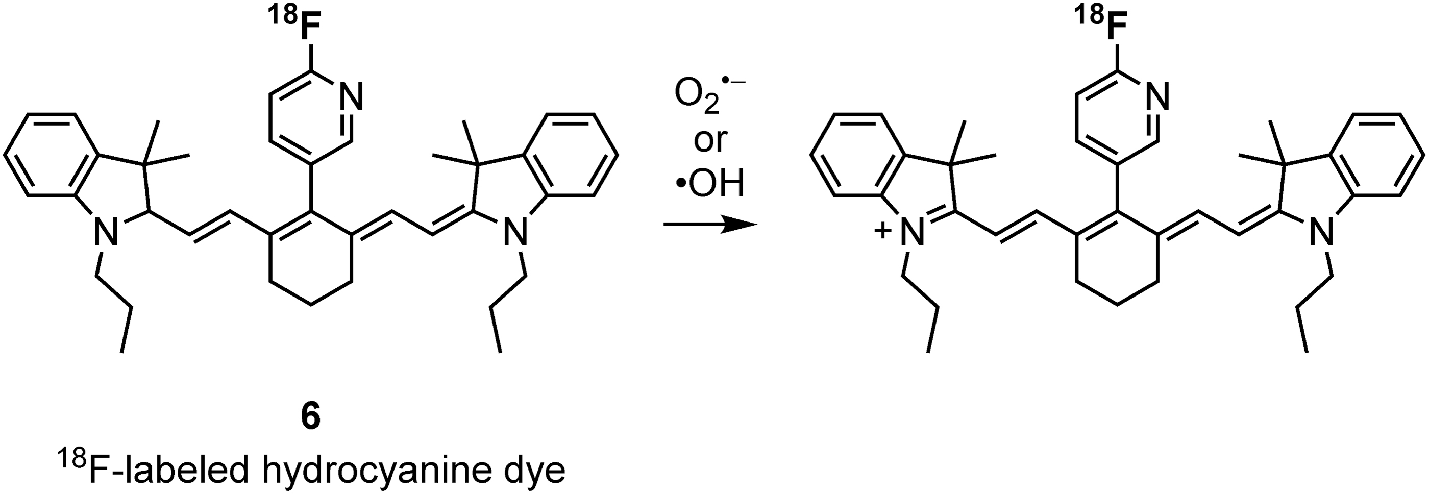
Radiolabeled hydrocyanine derivative for superoxide and hydroxyl radical detection.
During stability studies of
Fluorescein derivative
As a superoxide reacting probe, fluorescein-based radiolabeled probe

Radiolabeled fluorescein derivative for superoxide detection.
When this protection is offered by a chemical moiety that is selectively cleaved by target molecules, fluorescein works as a sensor by reacting with targets, thus offering high fluorescence capabilities. Huang et al. used trifluoromethanesulfonyl as a superoxide reactive group and the protective group of the hydroxyl group in the xanthene backbone and labeled the benzene moiety with radioiodine.
The log p value of the resultant compound was −1.62 (log p = 2.46 before the reaction), and it was thus postulated to accumulate intracellularly after reacting with intracellular superoxide. In an LPS-induced inflammation mouse model, SPECT/CT imaging showed that the accumulation of
Hydrogen peroxide
Thymidine analogue
H2O2 is an ROS and is considered to be an important messenger molecule because of its relatively long half-life and extensive diffusion distance compared with other ROS. PC-[18F]FLT-1 (8, Fig. 4) was developed by Carroll et al. (8) as a PET probe for H2O2. [18F]FLT (Fig. 4) was used as a PET probe in clinical settings for cancer diagnosis. It is a thymidine analogue that is phosphorylated by thymidine kinase and incorporated into DNA when taken up by cells, mimicking endogenous thymidine. Owing to this, 3′-deoxy-3′-fluorothymidine (FLT) accumulates in cancer cells because of their high rate of proliferation.
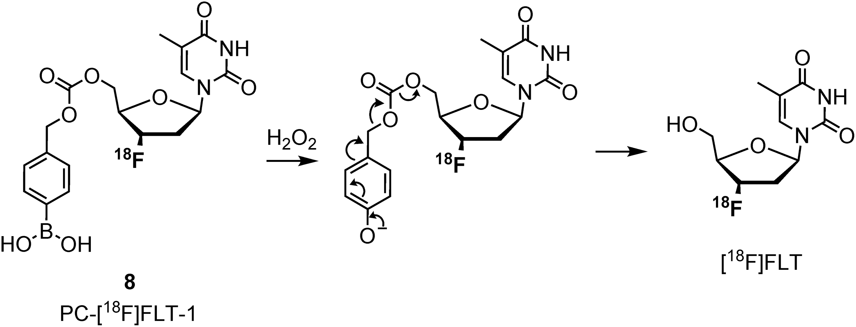
Radiolabeled thymidine analogue for H2O2 detection. H2O2, hydrogen peroxide.
The authors designed and synthesized a molecule consisting of boronic acid, which is often used as an H2O2 reactive substituent, attached to the 5′-OH of FLT by a linker and labeled with 18F at the 3′ position. Before uncaging by H2O2, compound
In other words, it accumulated in cells where both H2O2 and thymidine kinase activity were elevated. The accumulation of radioactivity in UOK262 renal carcinoma cells was increased in a concentration-dependent manner after the addition of H2O2, and a significant increase in cellular uptake was observed in UOK262 cells treated with paraquat, an ROS inducer, from 4 h after treatment compared with the untreated group. There are potential limitations to the uncaging of 18F by ROS and cleaving of the linker because of its short half-life.
Boronic acids or boronate esters are the only caging moieties for H2O2; however, the reaction rate between boronic acid and H2O2 is relatively slow (∼1 M−1 s−1) (31). Therefore, it is desirable to develop a more reactive caging moiety for H2O2 detection.
Total ROS and other radicals
Luminol derivative
In 2020, 68Ga-labeled probe

Radiolabeled luminol derivative for ROS detection. ROS, reactive oxygen species.
To assess the potential ROS imaging ability of
Then, this probe has a potential to monitor the ROS activity in acute lung injury. On the other hand, the biodistribution data in control mice at 60 min showed that most of the probe were accumulated in the bladder, next in the lung, and then in the kidney. This supposed that the probe was readily reacted in the blood, as luminol is a famous reagent for blood detection and excreted immediately into the urine. Then, the intravenous administration may lead to the loss of probe in the blood before reaching the target site in the unreacted form. Therefore, the result of accumulation needs to be precisely considered whether the probe reacted with the target molecule onsite, or the product reacted elsewhere was just distributed to the target site.
Edaravone derivative
There has been a report on the use of 3-methyl-1-phenyl-2-pyrazolin-5-one (edaravone, Fig. 6A), which is a potent free radical scavenger, for detecting free radicals such as hydroxyl radical (28). Edaravone has been developed and used as a medicine to protect the brain from damage after acute cerebral infarction. When edaravone, a lipophilic molecule, scavenges free radicals in the brain, it converts to a carboxylic acid [2-oxo-3-(2-phenylhydrazono)butanoic acid (OPB)] (Fig. 6A), which is a hydrophilic compound, leading to retention in the brain.
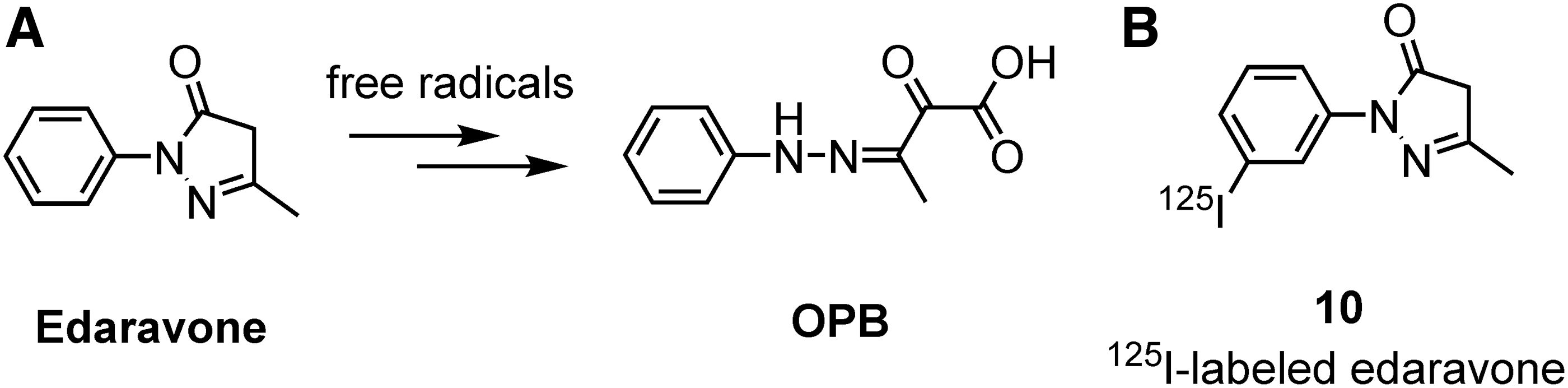
Radiolabeled edaravone derivative for free radical detection.
For this purpose, 1-(3′-[125I]iodophenyl)-3-methy-2-pyrazolin-5-one
The radiochemical purity of
The high specific activity compound was not stable and has not yet been investigated in vivo. Nevertheless, the applicability of this probe needs further investigation in a brain oxidative stress model.
Derivatives of spin trapping reagents
A radiolabeled spin-trapping reagent for the detection of free radicals in vivo has been reported by Fujibayashi et al. (14, 41). A spin-trapping reagent is a nitrone or a nitroso compound that reacts with radicals to form stable radical adducts that can be detected by ESR. In their study, one of the spin-trapping reagents, α-phenyl-N-tert-butylnitrone (PBN) (Fig. 7A), was labeled with radioiodine. Initially, in 1994, nitrone

Radiolabeled derivatives of spin trapping reagents for free radical detection.
In 1997, another nitrone
Nitroxide derivatives
Nitroxides, which are also called aminoxyls, are radical compounds whose spin density localizes on N–O atoms and are relatively stable as they can be handled at room temperature and normal pressure. They have potential reactivity toward one-electron oxidation and reduction as well as carbon radicals to form covalent bonds (26). Using these reactivities, nitroxides have been used for various applications such as catalysts in alcohol oxidation (30), antioxidants (33), and agents for nitroxide-mediated radical polymerization (29).
The reactivities of nitroxides vary according to the basic structure of the compounds, such as ring- or chain-type, and by electronic and steric substituent effects. It has been reported that six-membered ring nitroxides have various reactivities toward ascorbate, depending on the substituents proximal to the radical moiety (52). In addition, an inhibition effect toward lipid peroxidation was shown by using a series of nitroxides with alkyl groups as substituents (51).
This effect was enhanced by the increase in lipophilicity due to the alkyl substituent, which facilitated the trapping of carbon radicals in the lipid fraction. One fluorescent probe application involving lipid carbon radical detection through taking advantage of the off/on quenching of fluorescence before and after the reaction, which is a quenching effect derived from the diamagnetism of the radical moiety, has been reported to date (46).
Based on the reactivity of this nitroxide compound for lipid radical detection, we developed 125I-labeled nitroxide derivatives
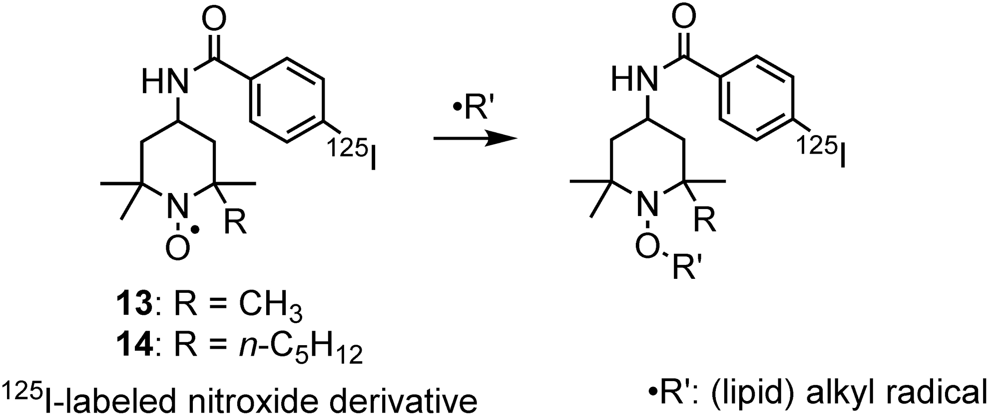
Radiolabeled nitroxide derivatives for free radical detection.
In a carbon tetrachloride-treated mouse model that showed enhanced lipid radicals from metabolic processes, radiolabeled nitroxide
Nitroxides are often used for in vivo redox monitoring probe (17, 35). It is known to be reduced mainly by ascorbate that exists in a relatively high concentration, even if its reaction rate constant is low in the order of 100–101 M−1s−1 (18, 20). On the other hand, nitroxides have a high reaction rate constant in order of ∼109 M−1s−1 for the coupling reaction with carbon radicals (10) although the amount of lipid carbon radicals generated in vivo is assumed to be quite low.
In addition, lipid peroxidation has been associated with a variety of diseases such as cancer, ischemia/reperfusion injury, and neurodegenerative disorders. And ferroptosis, a new type of cell death characterized by iron-dependent lipid peroxidation (34), is receiving a great deal of attention in recent years. To elucidate the mechanism of these disease generation and progression, it would be useful to clarify the localization of lipid radicals in vivo.
From the viewpoint of these requirements for sensitivity and imaging, the use of nuclear medical imaging is suitable for the development of the in vivo detection method for lipid radicals. Therefore, radiolabeled nitroxide
Redox state
Ascorbic acid derivatives
Ascorbic acid, also known as vitamin C, is a water-soluble antioxidant that is oxidized to dehydroascorbic acid (DHA) by extracellular ROS (24). In the process of H2O2 elimination, it is recycled via the glutathione-ascorbate cycle. In addition, ascorbic acid regenerates vitamin E, which is a lipophilic antioxidant. Therefore, the dynamics of ascorbic acid are believed to reflect the in vivo redox state.
One of the radiolabeled ascorbic acids, 6-deoxy-6-[18F]fluoro-L-ascorbic acid
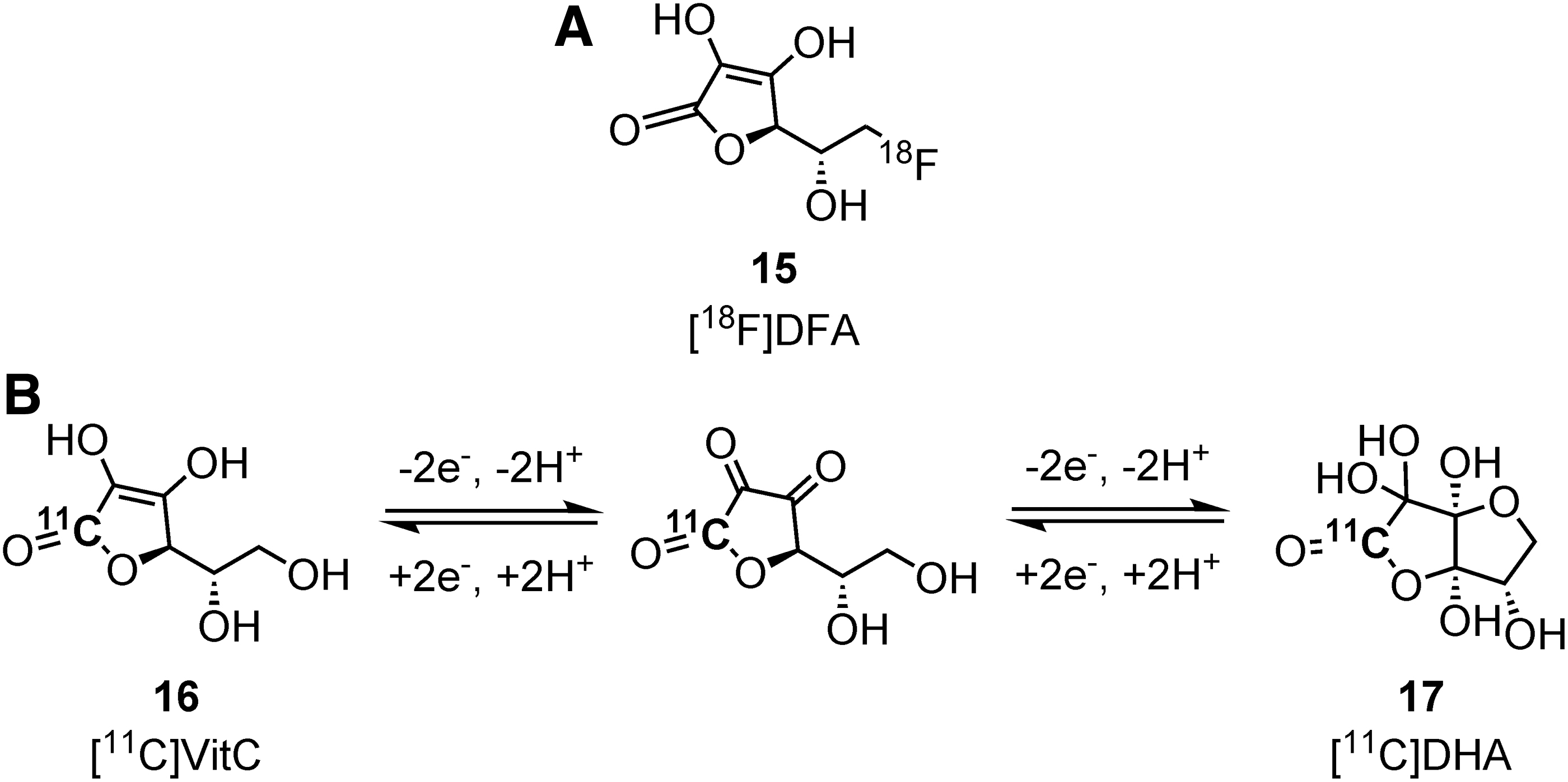
Radiolabeled ascorbic acid derivatives for redox state detection.
A lower accumulation of radioactivity was observed as glutathione content decreased. This suggested that cellular uptake of
Carroll et al. proposed that this principle could provide for the design of a probe that reflects extracellular ROS. They synthesized 11C-labeled ascorbic acid
To evaluate the cellular transport of this probe, DHA labeled with 11C (
Further, to investigate endogenous ROS-dependent accumulation in cells, the radioactivity accumulation of
In both cells, the radioactivity increased after the stimulation of PMA, and its accumulation was significantly decreased by cytochalasin B. This indicated that
As seen earlier, radiolabeled probe derivatized from ascorbic acid can monitor the in vivo redox status through the redox reaction. Especially, as seen in a probe
However, since this probe is oxidized extracellularly and trapped in the cell via GLUT transporter, not only the ROS production but also the expression level of GLUT would affect the results of probe accumulation. Therefore, the precise consideration would be required for the relationship of the ROS production and GLUT expression in each of the disease models.
Dihydroquinoline derivatives (NAD+/NADH)
The NAD+/NADH is a redox substance that is involved in redox reactions in vivo. Similar to NADH, its derivatives, 1,4-dihydropyridine and 1,4-dihydroquinoline, become quaternary ammonium salts when oxidized. In other words, it may be possible to detect the redox balance when the reduced form passes through the cellular membrane with relatively high hydrophobicity and accumulates in the cell after oxidation, depending on the redox state.
Okamura et al. reported 11C-labeled 1,4-dihydroquinoline
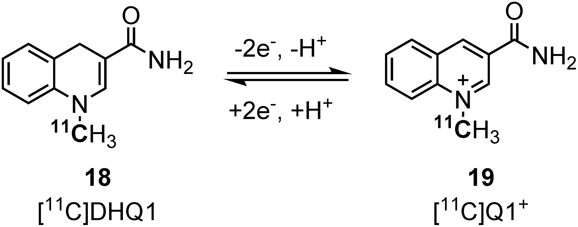
Radiolabeled dihydroquinoline derivatives for redox state detection
The radiochemical purity immediately after purification was less than 90%, even with the addition of ascorbate into the collecting flask. It was found that oxidation could be inhibited by adding ascorbate to the mobile phase during purification. This mixture was stable in phosphate-buffered saline at 37°C, whereas the fraction of
These results suggest that
Further, to investigate whether
Cystine-glutamate transporter substrate
During oxidative stress, the ubiquitin-ligase protein KEAP1 is oxidatively modified to form disulfide bonds, which inhibit the degradation of transcription factor nuclear factor erythroid 2-related factor 2 (Nrf2) and promote the transcription of xCT by binding to the antioxidant response element promoter region. xCT is a subunit of the cystine-glutamate transporter (system xc −), which imports cystine from the extracellular space to the cytosol and releases glutamate simultaneously.
The xCT subunit maintains low basal expression in normal tissues but is universally upregulated in tumors in response to oxidative stress. Therefore, cellular responses to oxidative stress induce xCT expression and promote cystine uptake for glutathione synthesis in tumors. 5-[18F]Fluoroaminosuberic acid

Radiolabeled cystine derivative for redox state detection.
Cellular uptake in human ovarian carcinoma cell SKOV-3 was inhibited by sulfasalazine, an inhibitor of system xc
-. In the SKOV-3 cancer transplantation model,
In addition, in three breast cancer models, MDA-MB-231, which has the highest xCT messenger RNA level, had the highest uptake of
Conclusions
Nuclear medical imaging probes that target ROS, free radicals, and redox substances related to redox fluctuations have been outlined in this review. Since these substances are highly reactive and in very small quantities, the high sensitivity of measurement in nuclear medical imaging is very vital. On the other hand, since radioactive material constantly emits a signal, it is difficult to distinguish between probes that have reacted with the target molecules and those that have not.
Therefore, it is necessary to design probes in which only the reacted probes remain where they are reacted, and the unreacted probes are quickly eliminated. In fact, as in the case of most of the probes presented in this review, the strategy of molecular design is to (1) increase cell membrane permeability by decreasing the molecular weight and increasing the lipophilicity of the probes before the intracellular reaction and (2) reduce permeability by making ionic compounds of probes on a reaction with the target molecule so that they remain in the cell.
Regarding to these requirements, fluorescein derivative
And for probes targeting other ROS such as hydroxyl and alkoxyl radicals, the specificity is not sufficient at this time. To elucidate the role of these species in pathology, the improvements of specificity or discovery of novel reacting groups are indispensable. In addition to the strategy mentioned earlier, another trapping method is useful in molecular design, for example, which is trapped in DNA by intercalation or in membrane by trapping lipid-derived radicals and trapped inside cells by utilizing the transporter.
At the same time, the ideal scaffold needs the abilities to be easily introduced in a reacting group combined with the trapping mechanism, and to be easily labeled by radioisotope with high radiochemical yield and radiochemical purity. In addition, if a probe is targeted to the brain disease, the BBB permeability is needed for a probe design. Namely, the scaffold should be chosen considering the molecular weight/size and moderate lipophilicity. Since these properties should be altered with the types of the reacting group, they need to be investigated precisely one by one for the target molecule and target disease.
As we have seen with DHE derivatives, as well as in nuclear medical imaging probes, it should be carefully considered what the probe reacts with and what reflects the produced result(s). This is because the unique problem with radiolabeled probes, especially ROS-targeting probes, is that the radiation from the radioisotope is known to cause radiolysis of water and generate ROS, which not only degrades the probe itself and reduces its purity before it is administered into the body, but may also reflect results other than the reaction with the targeted endogenous ROS.
Nonetheless, the addition of carriers or radical scavengers is an effective solution for degradation before administration into the body. To examine whether the reaction with endogenous ROS can be detected, it is necessary to confirm the estimated reaction products by comparison with standards or identification by liquid chromatography–mass spectrometry analysis or by utilizing specific inhibitors for target molecules.
Thus, although radiolabeled probes require precise molecular design that controls internal behavior and may cause problems specific to them, they have many advantages over existing imaging modality probes, such as superior sensitivity and capability of noninvasive measurements, including deep regions. Also, it would be possible to measure from cells to the living body with one common structure that has fluorescent/chemiluminescent moiety and radioisotope such as fluorescein (
The use of such radiolabeled probes is expected to promote translational research that can verify the contribution of redox fluctuations to diseases at the molecular level and enhance drug discovery as well as the development of diagnostic technologies based on the elucidation of redox alterations.
Footnotes
Author Disclosure Statement
No competing financial interests exist.
Funding Information
A part of this work was supported by JSPS KAKENHI [Grant Nos. 19K17283 (T.Y.), 19H03607 (T.M.)] and Hyogo Science and Technology Association (T.Y.).