




Abstract
Antibody microarrays are becoming frequently used tools for analytical purposes. A key factor for optimal performance is the stability of the immobilized (capturing) antibodies as well as those that have been fluorescently labeled to achieve the immunological test (tracers). This is especially critical for long-distance transport, field testing, or planetary exploration. A number of different environmental stresses may affect the antibody integrity, such as dryness, sudden temperature shift cycles, or, as in the case of space science, exposure to large quantities of the highly penetrating gamma radiation. Here, we report on the effect of certain stabilizing solutions for long-term storage of printed antibody microarrays under different conditions. We tested the effect of gamma radiation on printed and freeze- or vacuum-dried fluorescent antibodies at working concentrations (tracer antibodies), as well as the effect of multiple cycles of sudden and prolonged temperature shifts on the stability of fluorescently labeled tracer antibody cocktails. Our results show that (i) antibody microarrays are stable at room temperature when printed on stabilizing spotting solutions for at least 6 months, (ii) lyophilized and vacuum-dried fluorescently labeled tracer antibodies are stable for more than 9 months of sudden temperature shift cycles (−20°C to 25°C and 50°C), and (iii) both printed and freeze- or vacuum-dried fluorescent tracer antibodies are stable after several-fold excess of the dose of gamma radiation expected during a mission to Mars. Although different antibodies may exhibit different susceptibilities, we conclude that, in general, antibodies are suitable for use in planetary exploration purposes if they are properly treated and stored with the use of stabilizing substances. Key Words: Planetary exploration—Life Detector Chip—Gamma radiation—Antibody and fluorochrome stability—Antibody microarrays—Long-term storage. Astrobiology 11, 759–773.
1. Introduction
Antibodies are appropriate receptors because they can specifically bind to a broad range of molecular-sized biomarkers, from amino acids or other metabolites to whole cells or spores with high sensitivities and specificities (Fernández-Calvo et al., 2006; Parnell et al., 2007). Moreover, there is a tremendous wealth of knowledge within the biotechnological industry with regard to antibody production and performance (Rodriguez-Mozaz et al., 2004). Antibodies are relatively robust molecules when stored or displayed under appropriate conditions. Low temperatures and dryness usually do not affect their functionality; they are very frequently stored frozen in liquid nitrogen, at −80°C, or as a lyophilized powder. However, the three-dimensional structure of antibodies and, consequently, the binding properties to their antigens could be affected by several parameters, such as the nature and solvent ratio, temperature, lyophilization, freezing, ionic strength, light, or grafting step. The biotechnological industry has developed procedures by which to maintain the antibody stability either for storage or shipping. It is well known that frozen antibodies (between −20°C and −80°C) maintain their activity for years in a saline solution or when lyophilized and stored at ambient temperature (Breen et al., 2001). Usually, stabilizers such as sugars/polyols (sucrose, trehalose, polyethylene glycol, etc.) are added to reduce the degradation of the active biochemical components during processing and storage (Newman et al., 1993; Chang et al., 2005). Lyophilization in the presence of disaccharides like sucrose or trehalose is frequently used to provide long-term storage stability to protein pharmaceuticals. Drying at ambient temperature under atmospheric pressure in the presence of trehalose (United States Patent 4891319) or other formulations (Spanish Patent ES2180416) also renders functional antibodies that can be preserved for long periods of time.
The use of immunosensors in planetary exploration requires antibodies that are resistant against gamma radiation (most abundant during spaceflight as estimated by Le Postollec et al., 2009b), high-energy radiation (protons, electrons, and alpha particles), microgravity, sudden temperature shifts, and so on. In a trip to Mars, for example, high- and low-energy radiation or sudden changes in temperature would have to be considered. Thompson et al. (2006) found no significant alteration in the absorption and emission wavelengths or the quantum yields of two fluorescent dyes after high-energy proton and helium ion radiation. Recently, Le Postollec et al. (2009a) reported no significant effect of high-energy neutrons on a fluorescently labeled antibody reporter or fluorescein. Additionally, Maule et al. (2003) reported that reduced gravity would not present a barrier to a successful immunoassay. Here, we report other critical extreme conditions that antibodies must overcome for an optimal performance in space exploration, which include long-term storage, highly penetrating gamma radiation, the effect of sudden and prolonged temperature shift cycles, and the effect of high vacuum on immobilized antibodies. The study was performed with printed (immobilized) antibody microarrays and with fluorescent antibodies at the working concentrations for fluorescent sandwich immunoassays. All these antibodies (Table 1) are relevant components of the LDCHIP immunosensor and have been used in the different SOLID versions. In addition, we tested several stabilizing solutions for long-term storage and functionality of both the immobilized and fluorescent antibodies. Our results, using appropriate printing solutions, show that spotted antibodies kept their functionality after at least one year of storage under different conditions. Gamma radiation doses 1000 times higher than that expected on a mission to Mars did not substantially affect either the antibody or the fluorochrome. Also, antibodies subjected to multiple temperature cycles (−20°C to +25°C and +50°C) did not significantly lose their functionality.
2. Materials and Methods
2.1. Antibodies, microarray construction, and sandwich microarray immunoassays
The antibodies used in this work (except Anti-DsrB, see below), the microarray construction (i.e., antibody immobilization on microscope slides), and the sandwich immunoassay that uses fluorescently labeled antibodies have been described previously (Parro et al., 2005; Fernández-Calvo et al., 2006; Rivas et al., 2008; Parro and Muñoz-Caro, 2010). A139 to A186 are antibodies against chemolithotrophic acidophilic bacteria from the Río Tinto Mars analog environment whose metabolism is mainly based on iron and sulfur. IVH1C1 (Anti–Bacillus subtilis spores) is a representative antibody used to detect common biomarkers from Gram-positive low G+C content bacteria. Anti–E. coli and Anti-Salmonella are representative of enterobacteria that could indicate some kind of human contamination, and anti-proteins like GroEL, thioredoxin, and glutathione transferase are representative of proteins present in all prokaryotes involved in stress and oxi-reduction reactions. Streptavidin is an example of a biotin-binding protein related to secondary metabolism of broadly extended members of Actinobacteria, and Anti-DsrB, a key component of sulfur-reducing bacteria, is used as an example of a protein indicative of a specific metabolism. Anti-DsrB is a polyclonal rabbit antibody against a recombinant protein fragment of dissimilatory sulfite reductase subunit B from Archaeoglobus fulgidus. Primers GGAATTCCATATGGTGGTAATGGTAGTTGAGG as forward and CCGCTCGAGCCACTTGAACTGCGTTGAAGC as reverse were used for PCR amplification with Archaeoglobus fulgidus DNA as a template. The amplicon was cloned, and the protein was overproduced and purified from E. coli following standard procedures (AMS biotech Limited, Abingdon, UK). All tested antibodies were affinity purified with protein A and printed in a duplicate spot pattern on epoxy-activated glass slides (Arrayit Corp., CA) with a MicroGrid II TAS arrayer (Digilab, MA) as described previously (Rivas et al., 2008; Parro and Muñoz-Caro, 2010). In a typical sandwich microarray immunoassay (SMI), the sample was first incubated with the antibody microarray for 1 h, washed with incubation buffer to remove the non-bound material, incubated for 1 h with fluorescently labeled tracer antibodies, and then washed again to remove the excess tracer. Finally, the slides were dried out by a quick spin and scanned for fluorescence at 635 nm in a GenePix 4100A scanner (Genomic Solutions). The SMI experiments were repeated at least twice, and the scanned images were analyzed and quantified with GenePix Pro Software. The final fluorescent intensity (FI) for each antibody spot was calculated by applying the following equation: FI=(F635-B)sample − (F635-B)blank, where F635-B is the fluorescent intensity at 635 nm minus the local background (B) of each spot as quantified by the software (GenePix Pro), and the blank is the image obtained by using only buffer as antigenic sample. In a potential mission to search for biomarkers on the surface of Mars, the SMI would be automatically performed by the SOLID instrument, which bears its own micro fluidics, liquid deposits, and thermal control. In such a case, SOLID would be loaded with 0.5 to 1 g of dust or ground material (soil, ice, etc.) through an appropriately designed sampling port. Further sample processing (preparation of a liquid suspension) and SMI with LDCHIP would require a liquid water suspension or solution that would be provided by the instrument (see Parro et al., 2011a, for further details about how the SOLID-LDCHIP system works).
2.2. Effect of different printing buffers and long-term storage of printed microarrays
Several antibodies (protein A-purified IgG fraction of rabbit polyclonal antibodies) were diluted and printed at 1 mg mL−1 final protein concentration in different spotting solutions: 40% glycerol; stabilization mixture ME1 (Biotools, Madrid, Spain); 0.04% polyethylene glycol (PEG 200); 0.08% PEG 200; 0.04% PEG 6000; 0.08% PEG 6000; 0.04% PEG 600; 0.08% PEG 600; 0.5×ME1+0.5×CB1 (CB1 for commercial buffer 1, which is Protein Printing Buffer from Arrayit Corp.); 1×CB1 (Arrayit Protein Printing Buffer); 1×CB2 (commercial buffer 2, from Whatman Schleicher & Schuell, Sandford, ME); 0.04% trehalose; 0.08% trehalose. Antibodies were spotted by duplicate spot pattern as indicated above. Then, microarrays were stored under different conditions, as follows: at −80°C; +4°C; room temperature (RT: 23–25°C); 37°C; and frozen under liquid nitrogen, lyophilized, and stored at RT. Sandwich immunoassays were performed after 3 and 6 months with the corresponding antigen and fluorescently labeled (Alexa-647 fluorochrome) antibody mixtures. For the assay, all the slides were blocked on 2% bovine serum albumin (BSA), incubated with an antigenic mixture (5 ng mL−1 thioredoxin; 10 ng mL−1 GroEL; 5 ng mL−1 streptavidin; Leptospirillum ferrooxidans; Acidithiobacillus spp.; and Acidiphilium spp. bacterial lysates equivalent to 106 cells mL−1 each) for 1 h at RT, washed, and revealed with a 1/500 dilution of fluorescent antibody mixture (2–4 μg mL−1 of each antibody) during 2 h at 4°C. The slides were then washed again, dried, scanned, and analyzed as indicated above.
2.3. Effect of different blocking solutions on the stability of antibody microarrays
Microarrays were either directly stored without further treatment (non-blocked microarrays) or treated with several blocking solutions (BS), as follows: BS1 (10% BSA+2% BSA in 0.5 M tris-ClH pH 9.0), BS2 (10% BSA +2% BSA, 0.1 M trehalose, 0.1 M tris-ClH pH 9.0), or BS3 (10% BSA followed by 5 mg mL−1 BSA in stabilization mixture ME1; patented by Biotools S.A., Madrid, Spain). This process was implemented as follows: the slides were flipped onto a drop of each of the different blocking solution for 2 min and then fully immersed in the corresponding blocking solution, with gentle agitation for 1 h. After the chip was dried by short centrifugation, different sets of slides were stored under different conditions (−80°C, 4°C, RT, and 37°C) or lyophilized and then stored at RT. Microarrays were assayed by SMI at different times after storage with the same antigenic and antibody mixtures as above and following the procedure described previously (Rivas et al., 2008).
2.4. Effect of gamma radiation on printed antibody microarrays
Two sets of slides printed with antibody microarrays, as described in Section 2.1, were subjected to different doses of highly penetrating gamma radiation. The radiation process was done by a source of 60Co at the Unidad Náyade of CIEMAT (CSIC, Madrid, Spain) at 3 and 15 krad, respectively (100 rad=1 Gray, or Gy). These radiation doses correspond to 600 and 3000 times the theoretical global dose accumulated by a biochip during a 6-month travel period from Earth to Mars, as estimated by Le Postollec et al. (2009b). After irradiation (with 1.18–1.33 MeV gamma rays), the biochips were stored at 4°C to be assayed by SMI at different times with the next antigenic mixture (called Ag10): 10 ng/mL thioredoxin; 10 ng/mL GroEL; 10 ng/mL streptavidin; 10 ng/mL glutathione S-transferase; bacterial cell lysates equivalent to 106 cells mL−1 each of L. ferrooxidans, Acidiphilium spp., Acidithiobacillus thiooxidans, Salmonella bongorii, and E. coli. The sandwich assays were revealed with a cocktail of fluorescent antibodies (SM11): Anti-Thio, Anti-GroEl, Anti-Stv, Anti-GST, Anti-Salmonella, Anti–E. coli, A-139, A-183, A-184, A-185, and A-186 at 2–4 μg mL−1 each antibody (see Table 1). Assays were performed in triplicate microarrays and duplicate spots in each microarray. Scanning and analysis were performed as mentioned above.
2.5. Effect of buffer composition and the storage conditions on fluorescent tracer antibodies
In a fluorescent sandwich immunoassay, the antigen-antibody reaction is revealed by using a second incubation with a fluorescently tagged antibody (a tracer). All our antibodies had been dissolved at the appropriate working dilution and titrated. To check the effect of buffer composition and the storage conditions on the stability of fluorescent tracer antibodies, a new, SM11 fluorescently labeled (Alexa-647) cocktail of antibodies was prepared at the working dilution (each at 2–4 μg mL−1) in different buffers:
2.6. Effect of gamma radiation on fluorescent tracer antibodies
From each of the 3 sets of SM11 fluorescent antibodies described above (sets
2.7. Temperature shift cycles of fluorescent tracer antibodies
An extra subset
2.8. Effect of high vacuum on the functionality of immobilized and of fluorescent antibodies
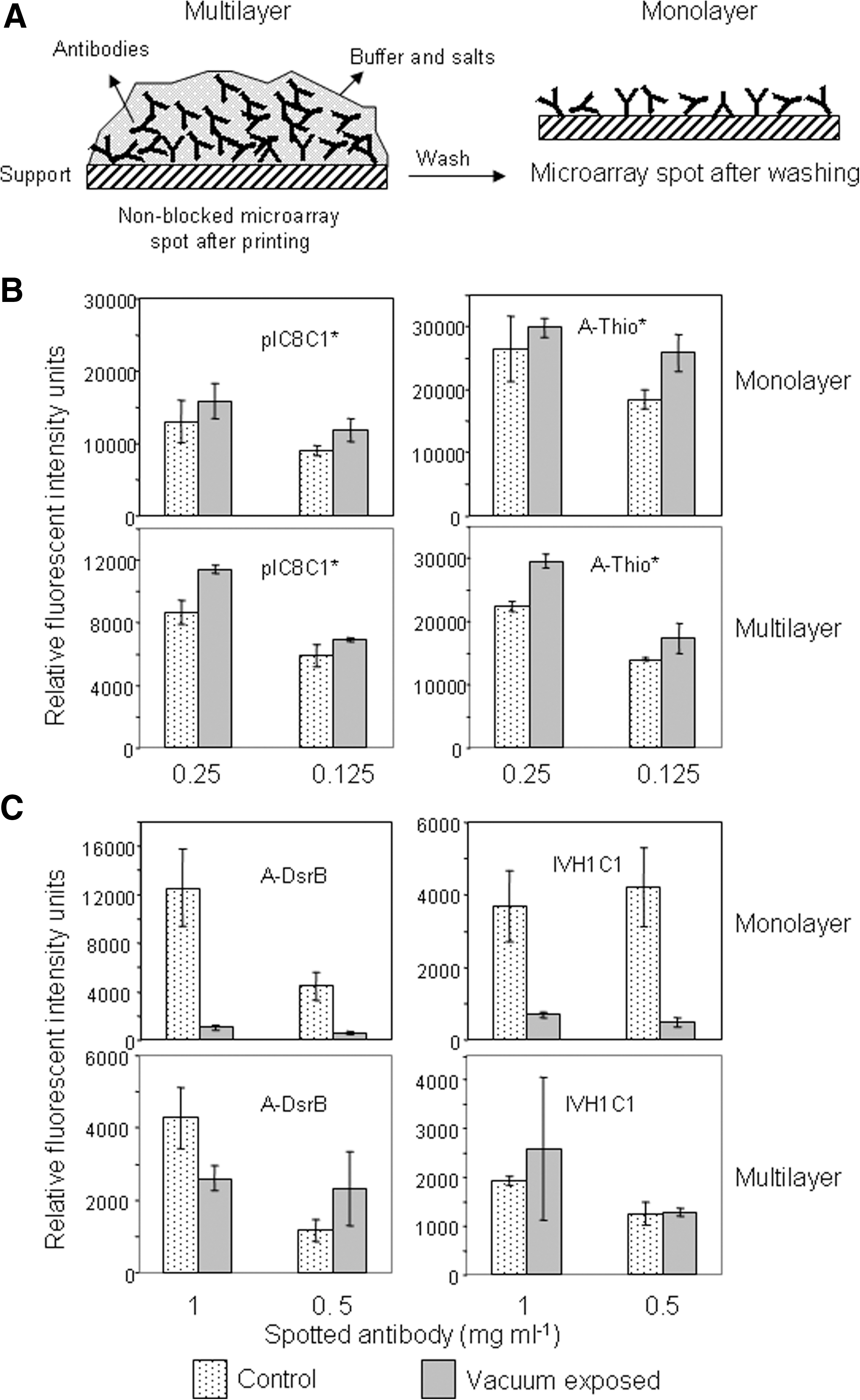
Two different rabbit polyclonal antibodies (IVH1C1, against Bacillus subtilis spores and Anti-DsrB protein from Geobacter metallireducens) were printed at two concentrations (1.0 and 0.5 mg mL−1) on epoxy-activated glass slides in 1× spotting solution buffer (Whatman Schleicher & Schuell) and by quadruplicate spots. Two additional fluorescently labeled antibodies (pre-IC8C1 and the commercial Anti-Thio, anti-thioredoxin protein from Sigma Cat. No. T0803 labeled with Alexa-647) were printed next to the formers at two different concentrations (0.25 and 0.125 mg mL−1). With a diamond pencil, the microarray slides were split into small pieces (1×1 cm), each containing one printed microarray. A set of microarrays was maintained as printed, and another was washed out with phosphate-buffered saline with 0.01% Tween 20 (1× PBST) and dried out. In the first case, the covalently bound antibodies to the glass support were considered to be covered by a “multilayer” of an excess of antibody molecules above them, while in the second one the immobilized antibodies were directly exposed to the environment. The arrays were put into the sampler holder inside a high-vacuum chamber at Centro de Astrobiología and maintained under 10−6 mbar for 3 h. A similar set was kept on the bench for positive control arrays. After high-vacuum conditions, the arrays were scanned for fluorescence and assayed by a SMI with the use of B. subtilis spores (107 spores mL−1) and DsrB protein (0.5 μg mL−1) mixtures as antigen and their respective fluorescent antibodies (2–4 μg mL−1) as revealing solution. The SMI, scanning, and image analyses were performed as described above. Antibodies were spotted by quadruplicate at two different concentrations (see above), and the experiments were repeated twice on different days. Fluorescent intensity was calculated as above, and the final value resulted from the average of different microarrays and spot replicates.
3. Results
3.1. Long-term antibody microarray storage: effect of stabilizing and blocking solutions
The use of antibody microarrays for environmental, medical, and biotechnological applications requires high stability of the immobilized reactants until used. An extreme of this is the use of antibody microarrays for space applications, where they are subjected to several environmental stresses. Consequently, antibodies and accessory reactants must be placed in a friendly environment to keep their functionality as intact as possible. In a typical SMI, antibodies are used in two formats and functions: (i) as immobilized (printed) on a solid support to act as a capturing component and (ii) as liquid solution/suspension to act as tracers to reveal whether the antigen-antibody reaction took place (Fig. 1). Therefore, the effect of external stresses on the antibodies under both presentations needs to be evaluated. In addition, if the tracer is fluorescently labeled, it is also obligatory to evaluate the stability of the fluorochromes to be used. We first tested the effect of several stabilizing and blocking solutions on the functionality of printed antibodies after long-period storage. Several rabbit polyclonal antibodies were printed on epoxy-activated glass slides by using different printing solutions (see Materials and Methods), processed, and stored under different conditions (Fig. 2). The post-printing processes consisted of no further treatment, or wash and block with the next blocking solutions (BS): BS1 (10% BSA+2% BSA in 0.5 M Tris pH 9.0), BS2 (10% BSA+2% BSA, 0.1 M trehalose, 0.1 M Tris pH 9.0), or BS3 (10% BSA followed by 5 mg/mL BSA in ME1). The microarrays were assayed after 3 or 6 months of storage by SMI with an antigenic mixture and then the corresponding fluorescently labeled antibody cocktail as tracer (see Section 2.2). The results indicate that all the tested antibodies were not equally affected and the composition of the printing buffer was critical to keep the antibody functionality (Fig. 2A). Antibodies in printing buffers containing only glycerol, PEG, or 0.04% and 0.08% trehalose clearly diminished their activity with increasing storage temperatures. Among the 13 tested printing buffers, those that contained formulated stabilizing solutions (ME1, 0.5×CB1+0.5×ME1, CB2) showed more uniform results for all storage conditions and performed similarly after 3 and 6 months of storage (Fig. 2B). Concerning the post-printing process, microarrays that received no further treatments after printing (non-blocked) showed the best performance (Fig. 3). The inclusion of stabilizing compounds like trehalose (in BS2) or ME1 (in BS3) considerably improved the performance of blocking solutions at ambient and 37°C storage temperatures (Fig. 3B). Only non-blocked printed microarrays maintained similar antibody functionality in all storage conditions. From the results, we inferred that antibody microarrays must be kept as printed until used or, alternatively, blocked in the presence of a preservative like trehalose.
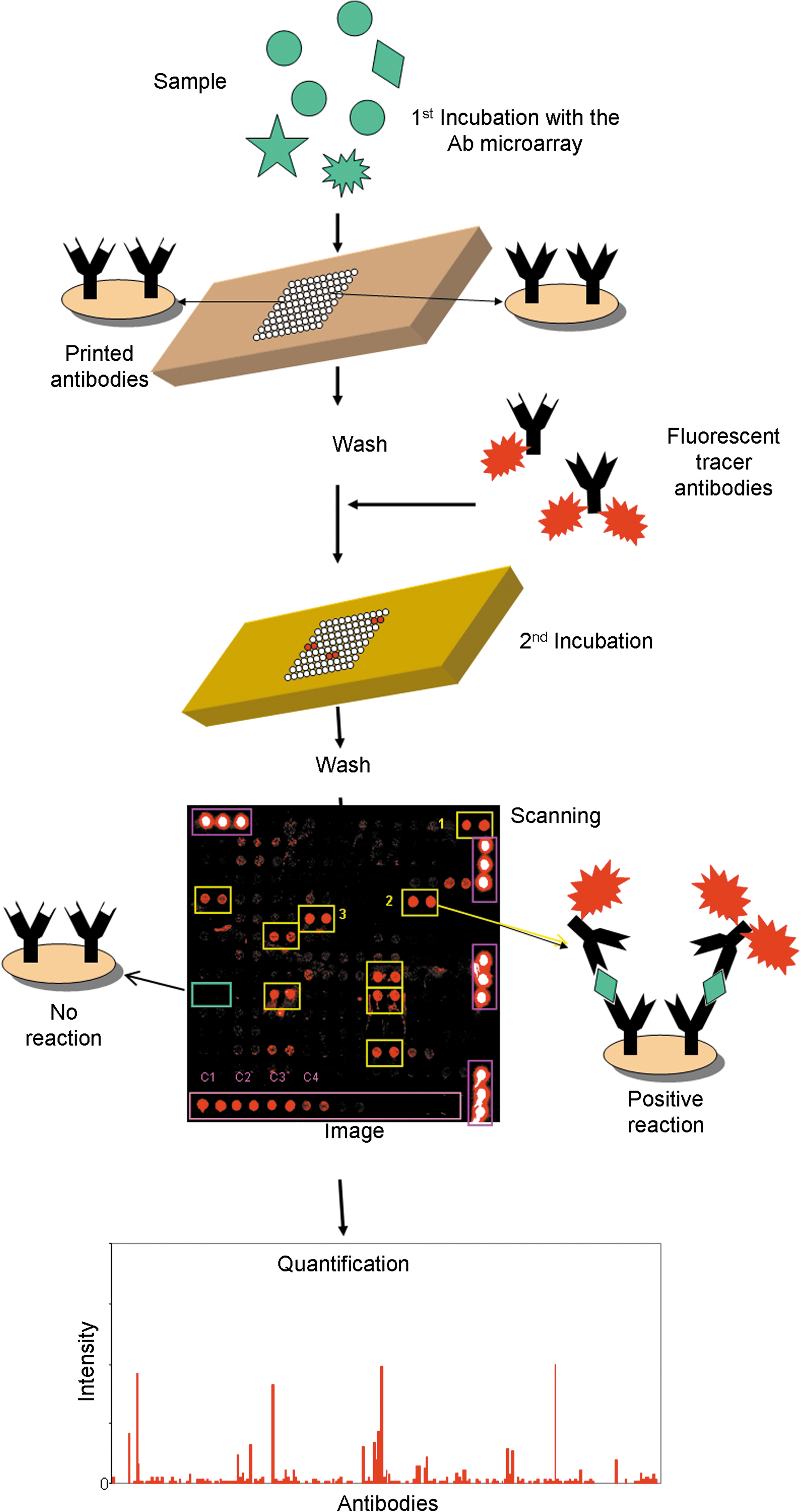
Fluorescent SMI. A scheme showing the fundamental steps of the whole procedure for the analysis of multianalyte-containing samples. Sample and tracer antibodies must always be in a liquid solution/suspension for a functional assay. During transportation, however, capturing and tracer antibodies must be maintained dried to avoid degradation. Color images available online at
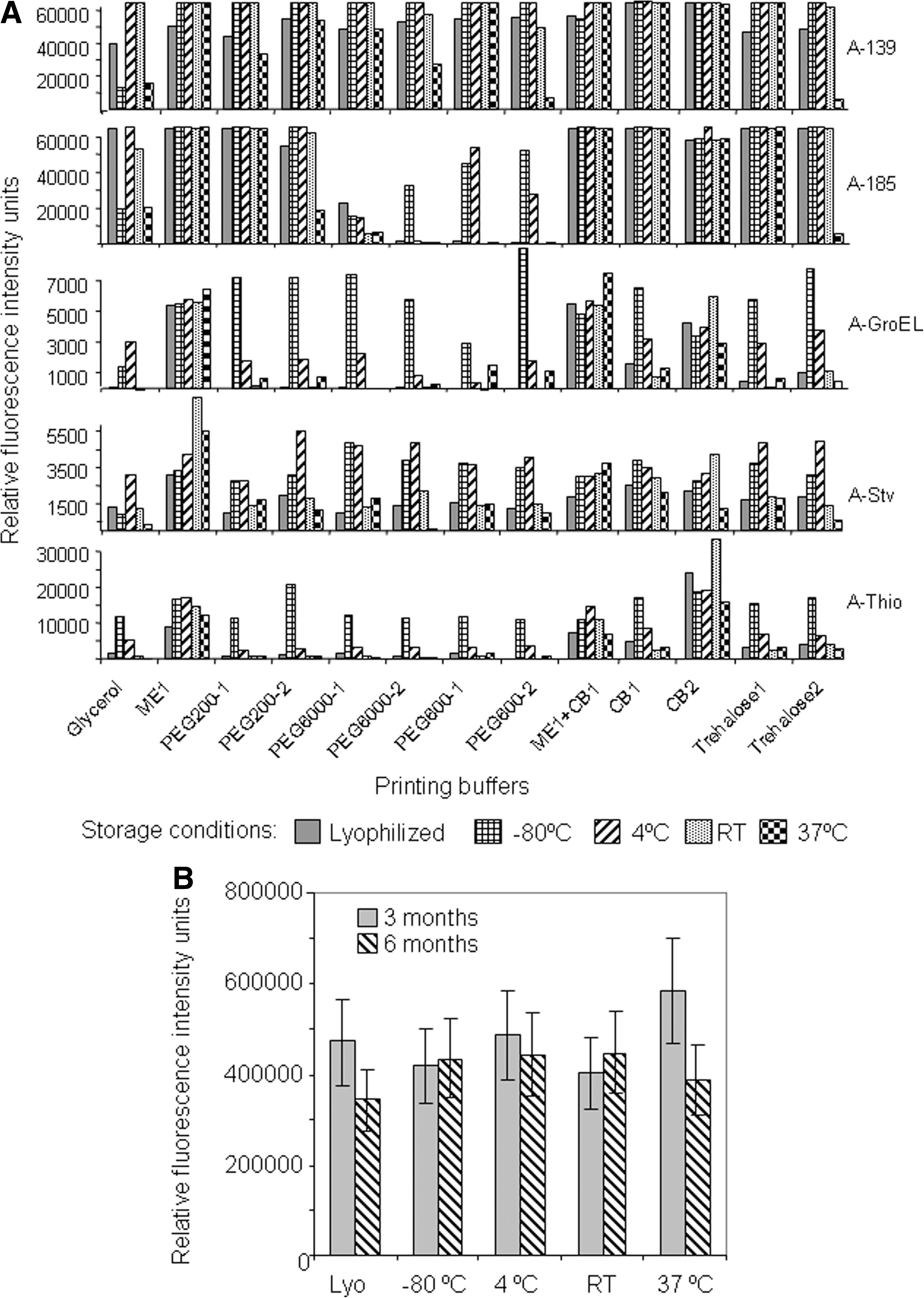
Effect of printing buffers and storage conditions on antibody stability for SMI. Several antibodies (see Table 1) were printed in different buffers (horizontal axis), and microarrays were stored at different conditions. (
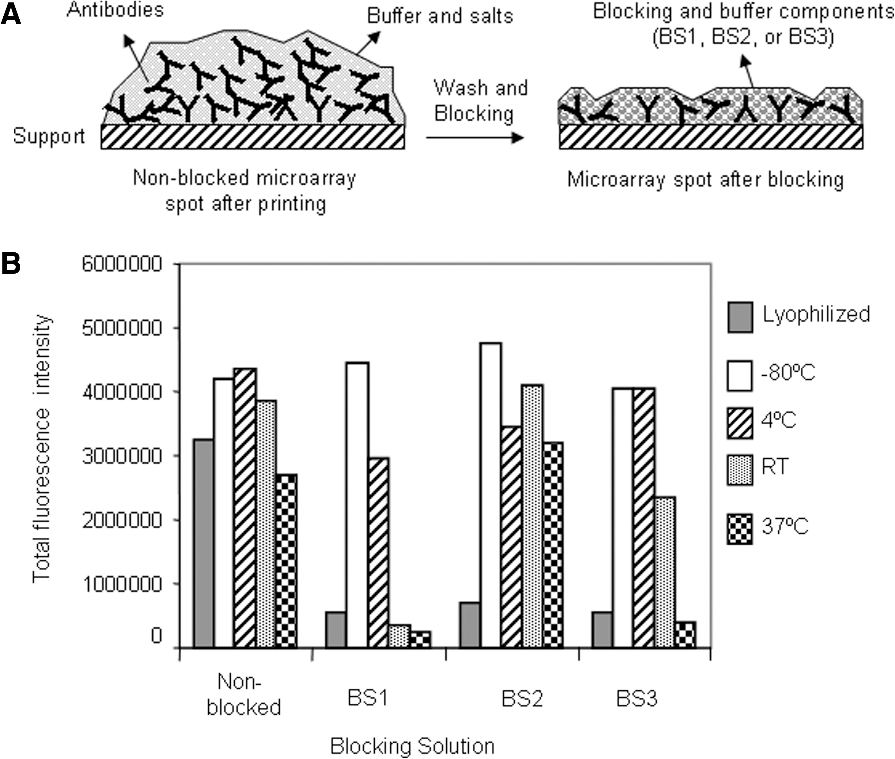
Effect of blocking solution and different storage conditions on printed antibody microarrays. (
3.2. Effect of high-penetrating gamma radiation on printed antibodies
One of the most abundant radiation types to which a biochip would be exposed in a planetary mission is the highly penetrating gamma radiation. Printed antibody microarrays were subjected to 30 and 150 Gy of gamma radiation, which corresponds to doses 600 and 3000 times, respectively, higher than the theoretical global dose accumulated by a biochip during a 6-month travel period from Earth to Mars as estimated by Le Postollec et al. (2009b). As a visible effect of the radiation, we observed that microscope slides turned slightly brown (Fig. 4A). We assayed the antibody functionality in each of the irradiated slides by SMI with several antibodies, and the results were compared to parallel assays with non-irradiated slides. After 3 months, we detected no significant differences between irradiated and non-irradiated slides. However, significant loss of functionality was detected when the assay was repeated after 4 years of microarray printing in the irradiated slides compared to the non-irradiated ones (Fig. 4B, 4C). The effect is clearly visible both in individual antibodies (Fig. 3B) and in the sum of all of them (Fig. 4C). This result indicates that gamma radiation produced deleterious effects that were only visible over time (compare upper plots with the bottom ones in Fig. 4B, 4C) and further indicates that this effect was dependent on the radiation dose. Nevertheless, about 50% of the antibody activity was retained in the 15 krad irradiated slides compared to the non-irradiated ones after 4 years.
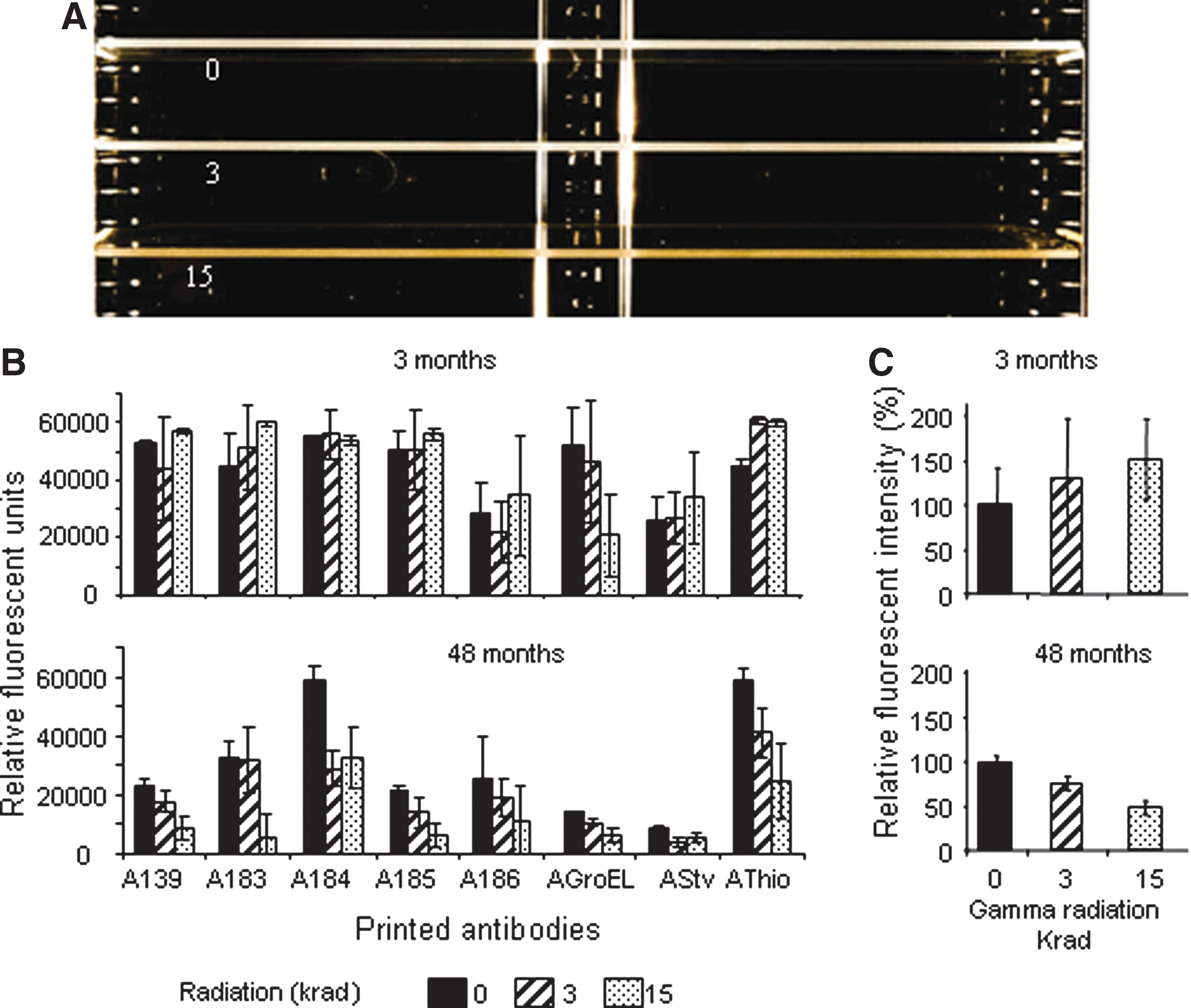
Effect of gamma radiation on the performance of printed antibody microarrays. (
3.3. Effect of high vacuum on the fluorescence and functionality of printed antibodies
To check the effect of high-vacuum conditions on printed antibodies, we used the high vacuum chamber at Centro de Astrobiología (Madrid, Spain). The sampler is designed for 1×1 cm sampling supports. Thus, we printed small microarrays with two different antibodies for functional assays and two fluorescently labeled ones for checking any effect on the fluorochrome stability (see Materials and Methods). A set of microarrays was introduced directly into the vacuum chamber after printing, and another set was washed with buffer solution, dried, and then introduced into the vacuum chamber. In the first case, the immobilized antibodies were under a multilayer of antibody molecules that were immersed in a protected buffered environment, while in the second, they formed an apparent monolayer of molecules that were directly exposed to the environment (Fig. 5A). Meanwhile, similar microarrays were kept on the bench as controls that were not exposed to a vacuum. After 3 h under high-vacuum conditions (10−6 mbar), the microarrays were recovered, scanned for fluorescence, and assayed for functionality by SMI. The results indicate that high-vacuum conditions did not affect the fluorescence intensity in the fluorescent antibody under a multilayer nor in an unprotected monolayer (Fig. 5B). However, the functionality of the monolayer antibodies was severely impaired after 3 h of high vacuum but showed similar performance to the positive controls when the antibodies were protected under a multilayer (Fig. 5C).
Effect of high vacuum on the fluorescence and functionality of printed antibodies. (
3.4. Effect of gamma radiation on the stability of fluorescent tracer antibodies
In a fluorescence sandwich microarray immunoassay, the fluorescent tracer is also critical for optimal performance of the assay. So we tested the effect of gamma radiation and long-term storage on printed and freeze- or vacuum-dried fluorescent tracer antibodies that were previously diluted at the working concentration (pre-diluted) (see Materials and Methods). A mixture of 1/500 dilution (2–4 μg mL−1 each) of several antibodies (A-139, A-183, A-184, A-185, A-186, A-Salmonella, A–E. coli, A-GroEL, A-Thio, A-Stv; Table 1) was prepared in seven different buffers and then subjected to different treatments (Fig. 6 and Section 2.5), as follows: (i) frozen under liquid nitrogen and then lyophilized, (ii) dried under vacuum at atmospheric pressure (speed-vac), and (iii) kept liquid at 4°C. Additionally, aliquots from each condition were subjected to 3 and 15 krad of gamma radiation, while one extra aliquot was subjected to multiple cycles of sudden temperature shifts (see Section 3.5). The performance of these fluorescent tracer aliquots was assayed at 3, 9 and 48 months after treatment by SMI with the so-called LDCHIP200 (a 200-antibody microarray, Rivas et al., 2008) as capturing antibodies and a cocktail of the corresponding antigens as antigenic sample. The results (Fig. 7) indicate that the effect of gamma radiation was similar, although less aggressive, to that on the printed antibodies, that is, there was negligible effect just after irradiation and a clear loss of functionality (to around 60%) after 4 years post-irradiation. The antibody functionality was around 80% or higher with most of the assayed buffers after 9 months of storage. Remarkably, fluorochrome (in this case Alexa-647) seemed not to be affected by gamma radiation or by the time elapses. Concerning the storage conditions, the vacuum-dried antibodies gave the most homogeneous results with all the tested buffers, while the lyophilized antibodies showed good performance but showed significant differences as well for some buffers. This may reflect a different behavior of buffers during lyophilization, while similar buffer composition may be less relevant for the vacuum-dried process.

Scheme showing the procedure to test the effect of gamma radiation, temperature shifts, and long-term storage on the stability of fluorescent tracer antibodies. (
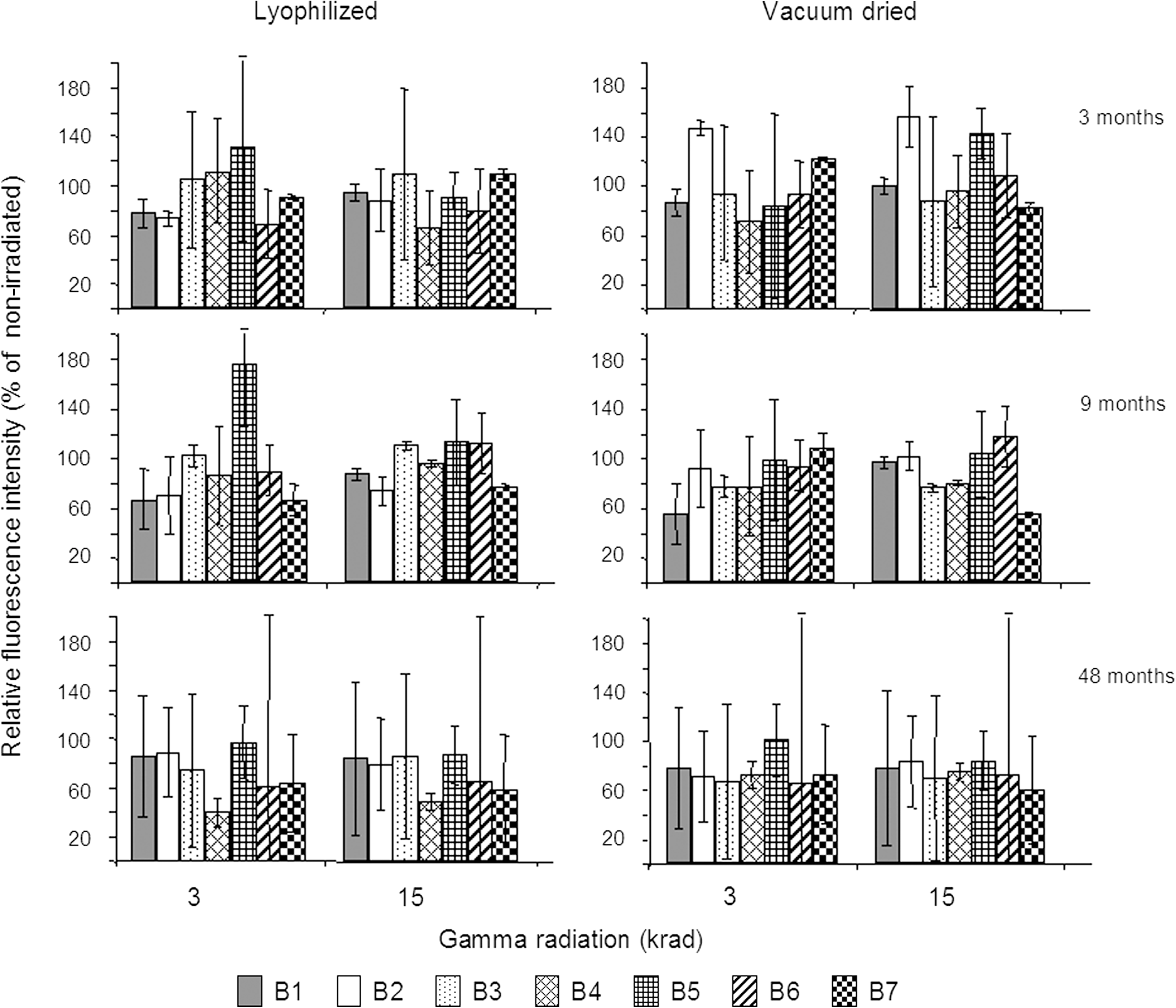
Effect of gamma radiation, buffer composition, and long-term storage on the performance of fluorescent tracer antibodies in sandwich microarray immunoassays. Relative fluorescent intensity refers to the sum of all the fluorescent signals of all the positive antigen-antibody reactions expressed as the percentage with respect to the assay control done with the non-irradiated antibodies (considered as 100%). B1–B7 different storage buffer, as in Materials and Methods and Fig. 6: B1 (ME1 from Biotools, 5% w/v BSA); B2 (ME1, 0.5× protein printing buffer from Arrayit Corp., 5% w/v BSA); B3 (Whatman Schleicher & Schuell protein printing buffer, 5% w/v BSA); B4 (1% w/v trehalose, 1× PBS, 5% w/v BSA); B5 (0.1% w/v PEG 200, 1× PBS, 5% w/v BSA); B6 (1% w/v sucrose, 1× PBS, 5% w/v BSA); and B7 (10% w/v trehalose, 1× PBS, 5% w/v BSA). Error bars represent the standard deviation of the fluorescent values from two different microarray slides with duplicate stop pattern.
3.5. Effect of sudden temperature shift cycles on fluorescent tracer antibody stability
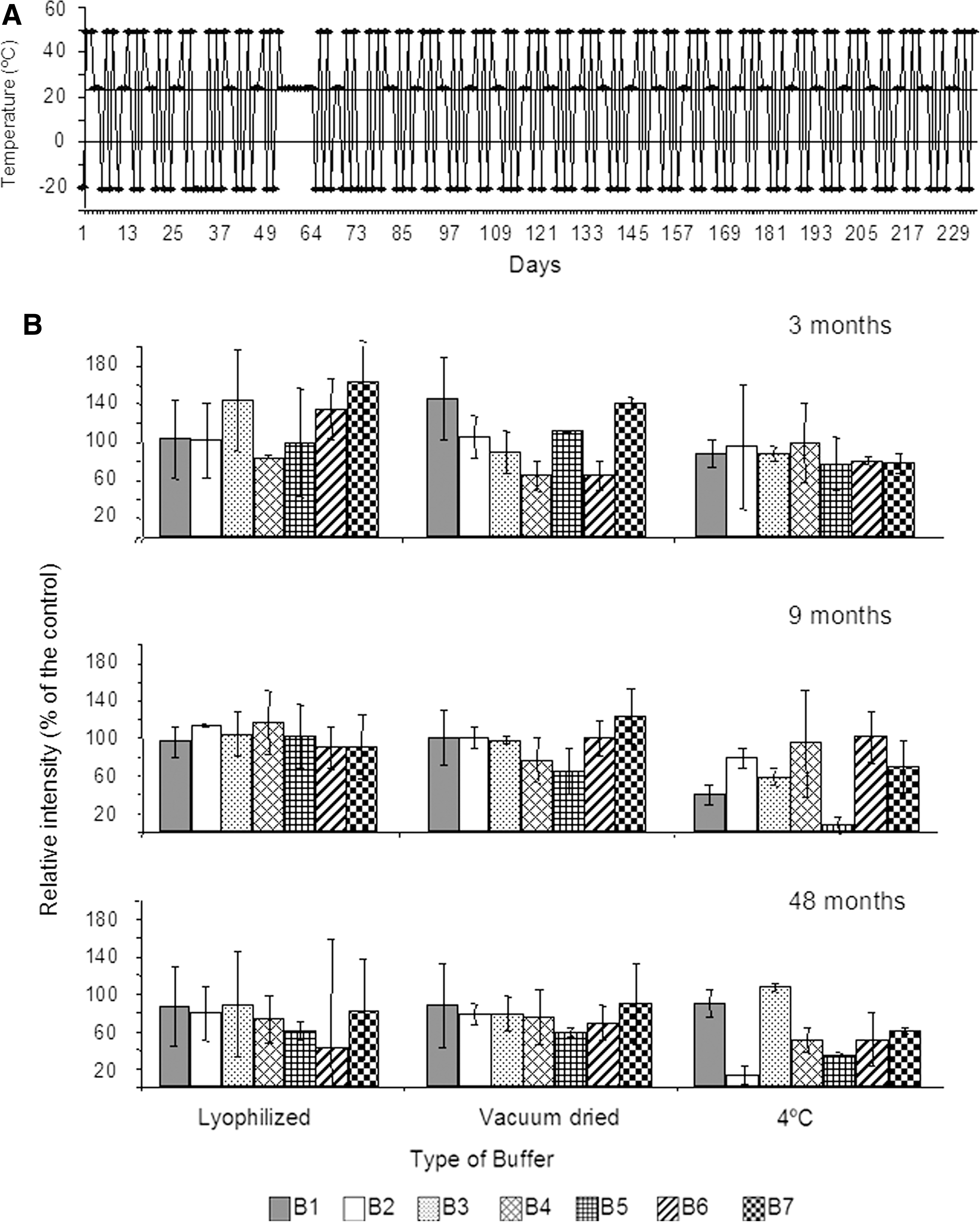
Additionally, one extra aliquot from each of the storage conditions described above was subjected to multiple cycles of sudden temperature shifts during 340 days (−20°C→25°C→50°C→− 20°C and so on) with a period of 24 h in each temperature (Fig. 8A). Again, the tracer antibodies were assayed by a SMI with LDCHIP200 as indicated above. The temperature-shifted fluorescent antibodies maintained similar functionality to their non-shifted counterparts both at 3 and 9 months when they were either freeze-dried or vacuum-dried (Fig. 8B). As expected, however, when the antibodies were kept in liquid suspension and subjected to temperature cycles, activity was clearly affected from the beginning. After 4 years of storage, the loss of activity was appreciable in all conditions, mainly in liquid suspension, but activity levels remained close to 70–80% in most of the buffers in the lyophilized and vacuum-dried samples. Among the buffers, those that contained 1×ME1 (B1), 1% trehalose (B4), and the commercial buffer (B3) gave the most homogeneous results for all storage conditions. Also, sucrose-containing buffer showed good performance mainly in the irradiation experiments.
Effects of sudden temperature shift cycles on the performance of fluorescent tracer antibodies. (
4. Discussion
4.1. Antibody resistance under extreme conditions: biochips for planetary exploration
The stability of antibodies is a critical aspect for the biotechnological industry. The transport to the final user very often requires medium- to long-term storage, temperature alterations, or the exposure to other environmental parameters that can affect the stability, functionality, and performance of the antibodies. This is the case for those applications that involve in-field analysis, such as environmental monitoring or veterinary uses that require a displacement to farms. Due to the intended use of immunosensors in planetary exploration and the space and planetary environments such instruments will encounter, antibody stability is an issue that will need to be addressed prior to deployment. In the present study, we addressed the effect of some of the most critical environmental parameters that can affect the functionality of fluorescent microarray immunoassays for a space mission to Mars. Long-term storage, sudden temperature shifts, vacuum, and gamma radiation are the most probable conditions that a biochip would be exposed during a space mission. We monitored the antibody functionality during a reasonable flight-to-Mars timescale (6–9 months) and a fraction of this time (3 months). The feasibility of samples in some cases allowed us to perform some experiments of up to 2–3 times the duration of a representative mission on Mars (4 years). Our results indicate that the storage of antibodies either in the printed version or the pre-diluted and store-dried version should not be a significant problem for approximately 4 years when using stabilizing solutions such as those used in the biotechnological industry. In fact, it is a general practice of biotech companies to store antibodies for years before they are used. Printing, that is immobilizing, the antibodies under appropriate buffer is probably the most important step for long-term antibody stability on microarray format. Our results show that adding appropriate amounts of substances, such as some disaccharides (trehalose, sucrose), to the printed solution increases antibody stability, and that several commercial buffers and solutions work fairly well. In our study, the best way to proceed after printing is to leave the printed microarrays untreated and then, prior to use, apply a blocking and washing step immediately before the assay This is true for a human, hands-on user, but it is true as well for automatic and remote processing such as would be the case for planetary exploration. Herein, we show that antibody microarray can be blocked after printing and then treated with a preservative solution or simultaneously blocked in a preservative-blocking solution (Fig. 3). The best performance was exhibited by the non-blocked slides. A feasible explanation is the protective effect of the buffer, preservatives, and the antibody molecules that are covering the layer of covalently bound antibodies. In printed/blocked/washed arrays both buffer and excess of antibodies are removed and substituted by a new layer of blocking agent. However, the blocking agent usually binds to free sites on the slides, rather than antibodies already bound on the slide surface, which allows for direct exposure of the bound antibodies to ambient conditions (Figs. 2A and 4A).
Especially relevant for space exploration are the results obtained after irradiation with 600 and 3000 times the gamma radiation dose an antibody microarray would receive during a trip to Mars. We did not detect any significant effect directly or immediately after irradiation even 3 months later, but a clear dose-dependent loss of functionality was observed after 4 years of storage (Fig. 4), which indicates that the irradiated antibodies were more sensitive with time. Bearing in mind that the average duration of a mission to Mars is much less than 4 years and that the potential gamma radiation a biochip can suffer would be considerably lower, we conclude that gamma radiation would not be a critical issue on a mission to the red planet. Similar conclusions can be drawn from the experiments performed with the fluorescently labeled tracer antibodies (Fig. 7). Furthermore, fluorochrome Alexa-647 seemed to be insensitive to the tested gamma radiation when bound to the tracer antibodies, which is fundamental for sandwich immunoassays.
Antibody microarrays and accessory reactants, like the pre-diluted fluorescent tracer antibodies, may be subjected to temperature shifts either during manipulation and transport for application on Earth or during preparation and development of a space mission. Our exhaustive temperature treatment of fluorescent antibodies with multiple temperature cycles indicated that antibodies are robust and can stand these extreme conditions when they are properly treated and stored. Dried fluorescently labeled antibodies either by lyophilization (freeze-dried) or dried at atmospheric pressure (speed-vacuum) are the most resistant to long periods of temperature cycling (Fig. 8).
The stability of printed and fluorescent tracer antibodies were improved by the addition of preservative agents such as the disaccharides sucrose and trehalose. Although the mechanisms by which the sugars and polyols improve protein stability are not well understood, these compounds are normally added to biopharmaceutical formulations to protect proteins against degradation during processing and storage (Pikal, 1990). Two main current interpretations are those based on a “glass dynamic mechanism” and a “water substitute mechanism” (Chang et al., 2005). The first one states that the excipients form rigid matrices in which the protein molecules are dispersed and the high viscosity glass slows down the protein mobility, which is necessary for protein degradation. The second assumes that the interaction of water molecules and protein (or antibody in this case) is critical for the thermodynamic stability of the latter. Once water is removed during drying processes, disaccharides or other stabilizers establish hydrogen bonds with the protein in much the same way water does and, consequently, preserve the native structure during desiccation by thermodynamic stabilization of the native conformation.
Our results under high-vacuum conditions indicate that fluorochromes are not affected by this parameter, but the three-dimensional structure of antibody molecules may be severely damaged if they are not protected by a buffered environment. High vacuum may cause irreversible deleterious effects if the printed antibodies are not conveniently protected by stabilizing substances and/or by a multilayer of an excess of non-immobilized antibody molecules. We hypothesize that the protective effect of the multilayer is mainly due to the physical barrier formed by the excess of protein (in that case the same antibody molecules) and so the fact that there are more available antibodies. The excess is always removed in the washing steps before the immunoassay. This is in agreement with the results shown in Fig. 3, which highlight the importance of post-printing processing of antibody microarrays and the need to use stabilizing printing and blocking solutions with other proteins, like BSA, as protecting agents. High vacuum further strengthens the negative effect observed at atmospheric pressure when antibodies are not under protective buffer.
The results shown herein, along with those previously reported by Parro et al. (2011a), which demonstrated that SMI is not affected by a perchlorate concentration 10 times higher than that measured on Mars, support the use of immunosensor biochips for the search for life on the red planet. This is a relevant issue after the discovery of relatively high perchlorate concentration at the Phoenix lander site on Mars (Hecht et al., 2009) and the reported destructive effect on the organic molecules at high temperatures in the presence of these anions (Navarro-González et al., 2010).
4.2. High-energy radiation from cosmic rays: some dimensional considerations
Theoretical calculations about the number of antibody molecules per spot in the printed microarrays allow estimations as to the number of effective deleterious high-energy impacts needed to compromise the functionality of each capturing antibody spot (Fig. 9). For example, a 120 μm diameter spot occupies an area of 11,309.76 μm2. An antibody 15 nm in diameter “nanosphere” will occupy an area of 1.7×10⁻⁴ μm2. In the best case, it is possible to form a monolayer with 6.6×10⁷ antibody molecules per spot. The relative molecular mass (Mr) for an antibody is 150,000 Da, which is an average of 1500 amino acids. An average of 15 atoms per amino acid would mean 22,500 atoms per molecule of antibody, that is, 1.48×10¹² atoms per spot. A total of 0.74×10¹² accelerated particles would be required to affect half the atoms in one spot. However, it must be considered that only a few atoms (probably less than 10%) are involved in binding the antigen or maintaining the antibody structure for correct antigen binding. How many radiation particles will impact a biochip area of 11,309.76 μm2 during a trip to Mars? Taking into account the theoretical calculations from Le Postollec et al. (2009b), 6 particles cm−2 s−1 (more than the sum of all types of radiation particles they calculated) could be considered the radiation dose received during a 6-month travel to Mars; then each spot on the array would receive 1.15×10⁴ radiation particles. This is 1000 times less than the number of antibody molecules per spot. Assuming that 10% of the antibody molecule is critical for the antigen binding, only 2.3×10³ particles per year will destroy antibodies, that is, 104 times less than spotted antibodies. This calculation indicates that the fraction of functional antibodies that survive cosmic rays on a trip to Mars is by far higher than those inactivated. During the operation at the martian surface, gamma, neutron, and proton radiation are the most abundant, and the first two do not seem to be significantly deleterious to polyclonal rabbit antibodies, as the present study demonstrated for gamma radiation and Le Postollec et al. (2009a) demonstrated for neutrons.
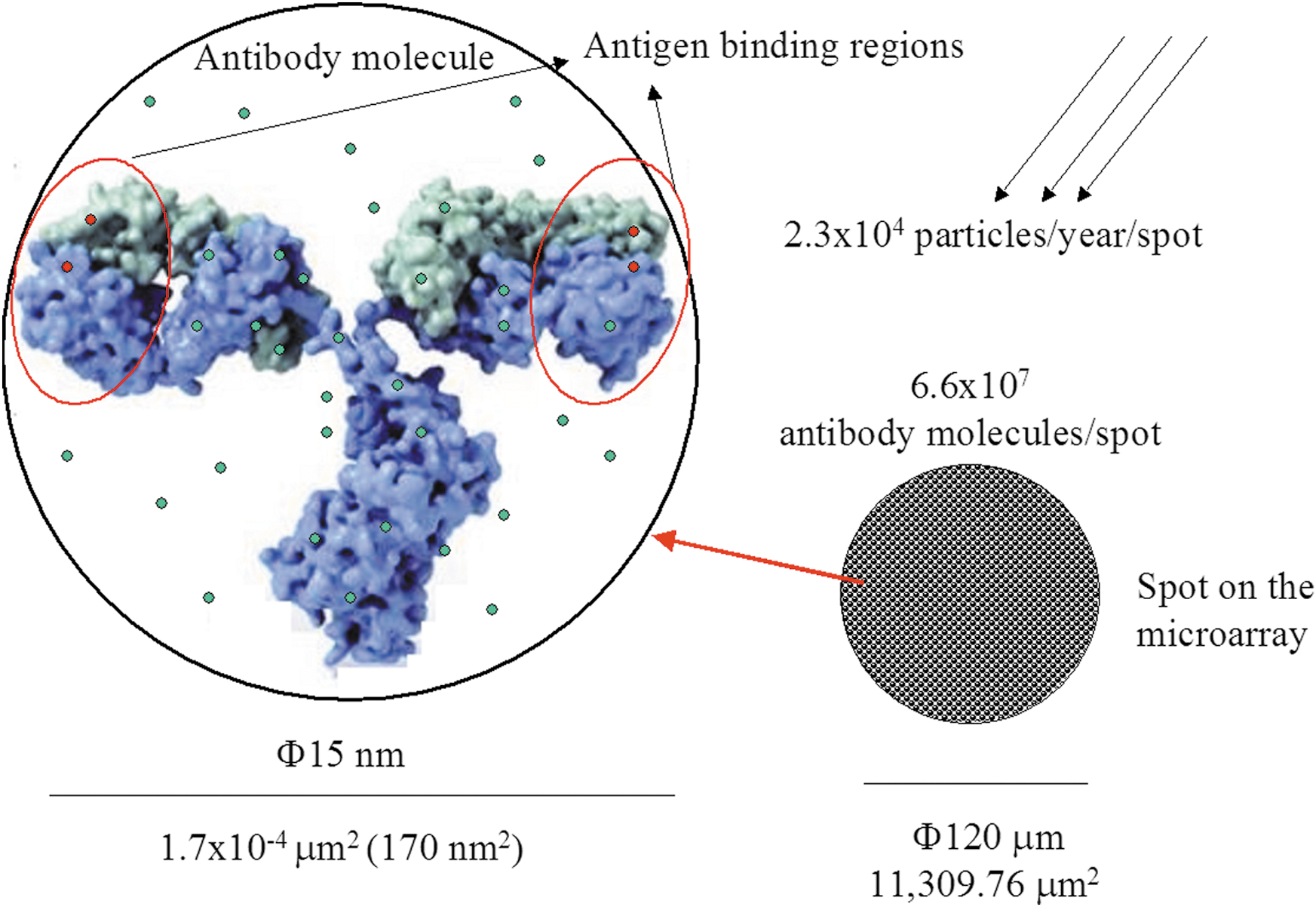
Some theoretical calculations about the dimensions of antibodies and antibody spots on the biochip. See text for explanation. Color images available online at
5. Conclusions
We have further demonstrated the robustness of antibody molecules and how they can withstand space-like environments when they are under appropriate buffer and storage conditions. They can maintain from 60% to 80% of their functionality both printed and pre-diluted at working concentration after 4 years of storage under stabilizing solutions. Exposure to high-vacuum conditions accelerates the negative effects on unprotected antibodies. Consequently, printed antibodies must be maintained under printing stabilizing buffer until use, or in those cases where blocking procedures must be done in advance, stabilizing substances must be applied to the blocking solution. Up to 3000-fold exposure to the gamma radiation dose that a biochip would receive during a trip to Mars did not result in an immediate and significant reduction in the functionality of the antibodies, printed or fluorescent. Theoretical estimations about the number of high-energy particles that an antibody spot would be exposed to during a 1-year mission to Mars indicates that, in the worst scenario, only a small fraction of antibodies would be affected by these events. Additionally, sudden and prolonged temperature shifts would have no serious effects on the antibodies and the fluorochromes if they were properly treated, that is, they were lyophilized or vacuum dried. In conclusion, antibodies are robust enough molecules such that, if properly treated, they can survive extreme environmental conditions and long-term storage for terrestrial and planetary exploration applications. Therefore, antibodies are stable, reliable, and promising tools in the search for life in the context of planetary exploration.
Footnotes
Acknowledgments
We thank Miriam García-Villadangos for technical work. This work has been funded by the Spanish Ministerio de Ciencia e Innovación (MICINN) granted projects N°. ESP2006-08128 and AYA2008-04013, entitled “Detection of Biomolecules in Planetary Exploration (DBEP).”
Abbreviations
BS, blocking solution; BSA, bovine serum albumin; CB, commercial buffer; LDCHIP, Life Detector Chip; LMC, Life Marker Chip; PEG, polyethylene glycol; RT, room temperature; SMI, sandwich microarray immunoassay; SOLID, Signs Of LIfe Detector.