
Abstract
Dissimilatory iron-reducing bacteria are able to enzymatically reduce ferric iron and couple to the oxidation of organic carbon. This mechanism induces the mineralization of fine magnetite crystals characterized by a wide distribution in size and irregular morphologies that are indistinguishable from authigenic magnetite. Thermoanaerobacter are thermophilic iron-reducing bacteria that predominantly inhabit terrestrial hot springs or deep crusts and have the capacity to transform amorphous ferric iron into magnetite with a size up to 120 nm. In this study, I first characterize the formation of hexagonal platelet-like magnetite of a few hundred nanometers in cultures of Thermoanaerobacter spp. strain TOR39. Biogenic magnetite with such large crystal sizes and unique morphology has never been observed in abiotic or biotic processes and thus can be considered as a potential biosignature for thermophilic iron-reducing bacteria. The unique crystallographic features and strong ferrimagnetic properties of these crystals allow easy and rapid screening for the previous presence of iron-reducing bacteria in deep terrestrial crustal samples that are unsuitable for biological detection methods and, also, the search for biogenic magnetite in banded iron formations that deposited only in the first 2 billion years of Earth with evidence of life. Key Words: Biosignatures—Magnetite—Iron-reducing bacteria—Deep subsurface biosphere—Banded iron formation. Astrobiology 12, 1100–1108.
1. Introduction
A
On the other hand, dissimilatory ferric iron–reducing bacteria (FeRB) are able to enzymatically reduce amorphous ferric iron and induce mineralization of magnetite (e.g., Lovley, 1991; Roden, 2008). Under laboratory conditions, respiration of Fe(III) by FeRB facilitates mineralization of Fe2+-excess magnetite (Li et al., 2011a) in a time span much shorter than that required by any geological process (Vali et al., 2004). For example, Geobacter and Shewanella are both mesophilic or psychrophilic FeRB that generally produce superparamagnetic (SPM) magnetite crystals characterized by crystal sizes <35 nm and irregular shapes (Sparks et al., 1990; Moskowitz et al., 1993; Hanzlik et al., 1996; Li et al., 2009). Although this mineralization mechanism is much more efficient than the assimilation of Fe(III) by magnetotactic bacteria (Lovley, 1991), the biogenic magnetite produced by the former is rare in natural environments (e.g., Maloof et al., 2007). It has been shown experimentally that these SPM magnetite nanoparticles can either transform to ferro-carbonate (Kukkadapu et al., 2005) or degenerate to a stoichiometric composition indistinguishable from the abiotic magnetite (Frankel and Bazylinski, 2003; Faivre et al., 2005; Li et al., 2009). These experimental results suggest that extracellularly produced magnetite is unsuitable as a biosignature for the previous existence of FeRB.
Ferric iron–reducing bacteria can also induce mineralization of SD magnetite under laboratory conditions at elevated temperatures. Zhang et al. (1998) demonstrated that Thermoanaerobacter spp. strain TOR39 can produce magnetite of >120 nm at 65°C. Roh et al. (2003) reported that Shewanella produces magnetite with a small fraction of SD particles when incubated at 40°C, the upper temperature limit of these bacteria. Vali et al. (2004) reported a very rare case of SD magnetite mineralization through multiple-stage mineral phase transformations in the culture of Geobacter metallireducens strain GS15, in which a variety of magnetite morphology, including rod, pseudohexagonal, and the common octahedral shapes, was observed. Thermoanaerobacter is uniquely connected to terrestrial hot springs or deep subsurface microbial communities (e.g., Gold, 1992; Szewzyk et al., 1994; Liu et al., 1997; Wagner and Wiegel, 2008); its ecophysiological requirements are similar to physicochemical features of the early Precambrian oceans with the deposition of banded iron formations (BIFs). However, its evolutionary significance has not been demonstrated through the biomineralization of magnetite. In this study, I report the formation of unique hexagonal platelet-like magnetite crystals from the prolonged incubation of TOR39 cultures and suggest that this particular crystal habit can be used as the biosignature of thermophilic FeRB inhabiting the modern deep, hot crust of Earth and tracing their emergence and geological evolution in ancient times.
2. Experiments and Methods
Thermoanaerobacter spp. strain TOR39 was isolated from >2652 m deep sedimentary rock with an in situ temperature of >65°C (Liu et al., 1997). TOR39 is able to ferment glucose that couples to the reduction of amorphous ferric iron (ferrihydrite or akaganeite). Details of experimental conditions are similar to those of Roh et al. (2003). Briefly, the culturing medium contained 10 g/L PIPES [piperazine-N,N′-bis(2-ethanesulfonic acid)], 0.08 g/L CaCl2, 1.5 g/L NH4Cl, 0.2 g/L MgCl2·6H2O, 10 g/L NaCl, 0.4 g/L K2HPO4·3H2O, 1.0 mL/L 0.01% resazurin solution, 0.5 g/L yeast extract, 10 mL/L trace minerals, and 1 mL/L vitamin solution (Phelps et al., 1989). The media were heated to 70°C under a stream of flowing pure N2 for 10 min to remove O2 and dispensed 10 mL to each 26 mL pressure tube with N2 as the headspace gas. The tubes were capped with butyl rubber stoppers and sealed by Al crimp for autoclave at 121°C for 12 min. The pH of the media was adjusted to 7.2 before incubation. Glucose and ferrihydrite from stock solutions were injected into the tubes to bring the final concentrations of glucose to 2.0 mM and Fe(III) to ∼50 mM, respectively. Generally, precipitation of magnetite could be observed after 1 week of incubation at 55°C. Some tubes were stored at 55°C for more than 2 years to examine the ongoing growth of crystals after cell lysis. The precipitated magnetite was extracted with a permanent magnet and briefly washed with anhydrous methanol for dehydration in an anaerobic chamber. To measure possible changes of chemical stoichiometry of magnetite crystals by aging, magnetite from tubes stored at room temperature for more than 7 years along with the freshly prepared magnetite was measured for Mössbauer spectra. The Mössbauer spectra were collected at Mount Holyoke College at room temperature, protected with helium gas, and analyzed with the methods described in Li et al. (2009).
The morphological and structural characteristics as well as chemical composition of the magnetite samples were characterized with a Hitachi Field Emission S4800 scanning electron microscope (SEM) and FEI Tecnai G20 scanning transmission electron microscope (STEM) equipped with selected area electron diffraction (SAED) at the Electron Microscope Center of the University of Hong Kong. For SEM observation, the low voltage (5 kV) and secondary electron mode were used to enhance signals from the surface.
3. Results
The mineral product after 1 week of incubation of TOR39 was an assemblage with magnetite as its major component and goethite and the precursor ferrihydrite as minor components, as indicated by the Mössbauer spectroscopic results (Fig. 1A). The external and internal sextets represent Fe3+ on the A site and (Fe2++Fe3+) on the B site of the magnetite structure (Fig. 1 and Table 1). Mössbauer hyperfine parameters of the newly formed magnetite (Fig. 1A, Table 1) are the same as for magnetite stored at room temperature for more than 7 years (Fig. 1B, Table 1). However, the relative abundance of magnetite increased from 77.8% in the 1-week culture to 97.8% in the 7-year-old culture at the expense of ferrihydrite (Fig. 1B), which suggests ongoing nucleation of magnetite during the prolonged incubation. Similarly, the prolonged incubation of the TOR39 culture at 55°C for 2 more years also showed further mineralization of magnetite and the increase of crystal size. The SEM examination of the resultant precipitates reveals that the magnetite crystals are the largest biogenic magnetite crystals ever reported. Both octahedral- and hexagonal-shaped magnetite crystals were observed (Fig. 2A–B). Particularly, Fig. 2C–D shows the shape and dimensions of individual platelet-like crystals. The whisker-like crystals on the surface or between the magnetite crystals (Fig. 2) are goethite needles determined by STEM imaging (Fig. 3A), SAED analysis (Fig. 3B), and the Mössbauer spectroscopic hyperfine parameters (Table 1 and Fig. 1A).

Mössbauer spectra of magnetite produced by Thermoanaerobacter. (
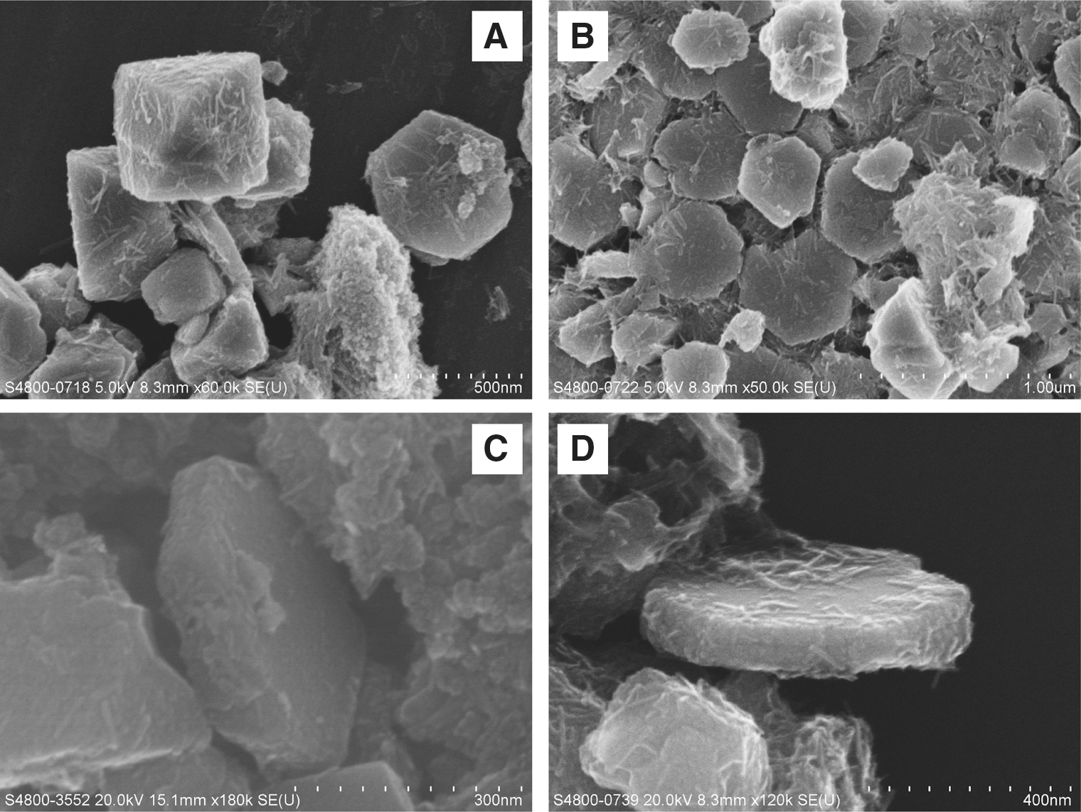
SEM observation of platelet-like magnetite produced in the culture of TOR39. (
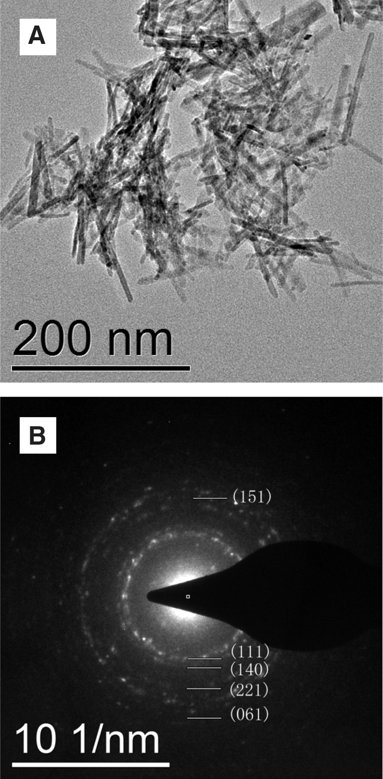
Characterization of small particles on the surface of magnetite crystals. (
After 1 week of incubation, TOR39 cultures showed magnetite crystals clustered by biological materials and the ferrihydrite precursor (Fig. 4A–B). The average size of the magnetite crystals was 91±44 nm. Magnetite crystals after more than 2 years of incubation showed significantly increased crystal size (292±51 nm) (Fig. 4C–D). STEM observation showed the coexistence of octahedral and platelet-like magnetite crystals at different projection angles (Fig. 4). We did not rotate the crystals as Thomas-Keprta et al. (2001) did to get the multiple 2-D projections because the SEM observations (Fig. 2C–D) clearly show platelet-like shapes already. The platelet-like crystals account for a small fraction of total crystals in the TOR39 culture (16%). The hexagonal particles aligned with a/b planes perpendicular to the electron beam display perfect hexagonal symmetry morphology (Fig. 4E), and those with a/b planes aligned parallel to the incident electron beam show the actual thickness of these crystals (Fig. 2C–D). The platelet-like magnetite crystals have an average size of 448±99 nm of hexagonal planes perpendicular to {111} and 89±18 nm of c parallel to {111}. As these crystals are larger than the conventional biogenic SD magnetite, the dimension perpendicular to the {111} crystallographic direction places them in the multiple domain region of the Butler-Banerjee diagram (Butler and Banerjee, 1975). When, however, dimensions of a cross section through the crystal parallel to the {111} are considered (Fig. 4F), they fall within the SD field of the diagram.
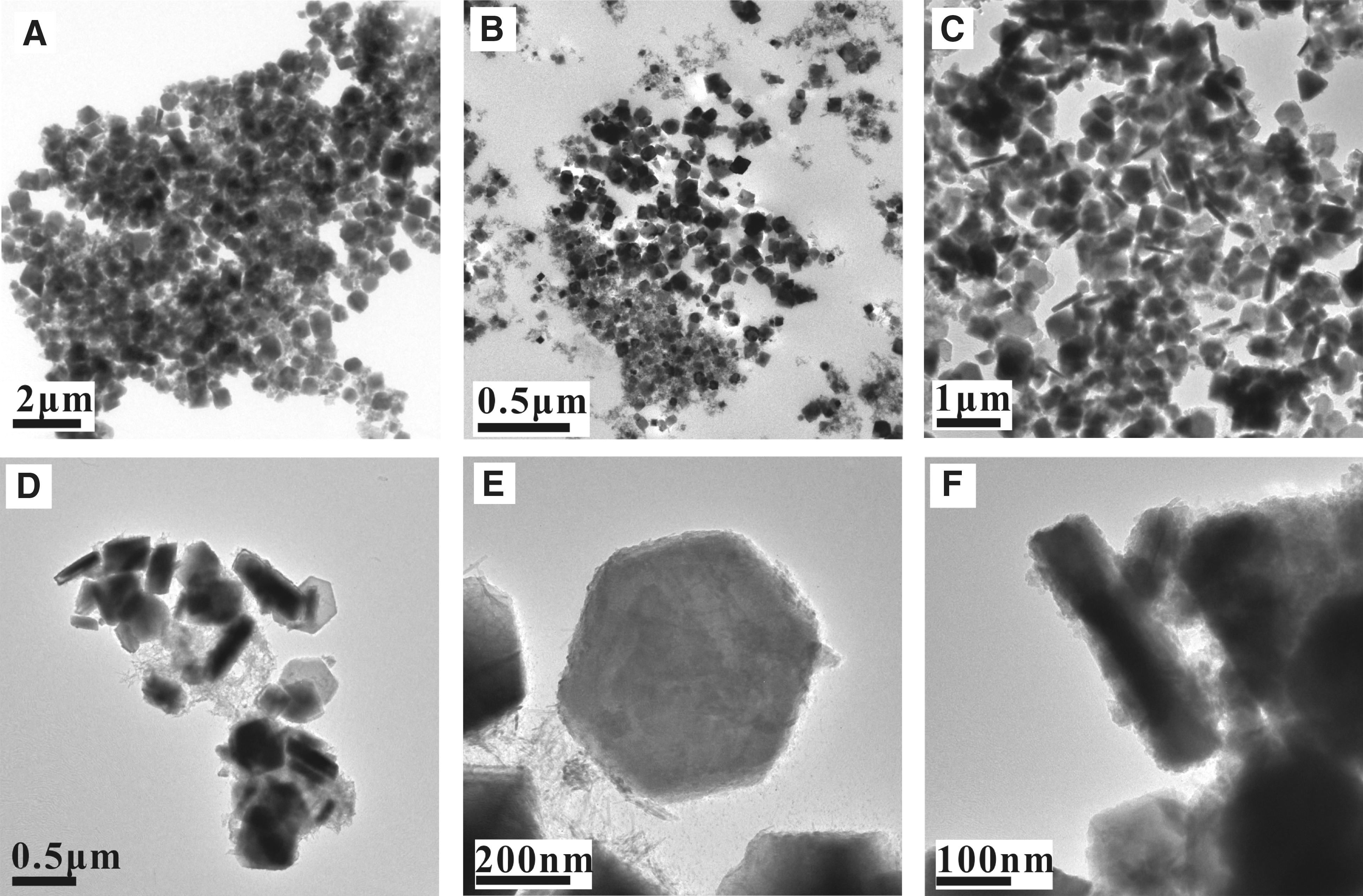
STEM observation of hexagonal magnetite from the culture of TOR39. (
Scanning transmission electron microscope–based SAED analysis of the platelet-like crystals reveals that they are indeed single crystals of magnetite (Fig. 5). Figure 5A shows the hexagonal projection of a crystal under STEM. The SAED from {111} direction yielded a set of diffraction indexes typical for magnetite (Fig. 5B). Figures 5C–D are direct observations of {200} and {100} d-spaces, respectively. The d-space measured from Fig. 5C is 0.48 nm. Figure 5E is the Fourier transform of the area in Fig. 5C, which gives the same structural information as Fig. 5B. Figure 5F draws the lattice planes of the hexagonal magnetite according to the combined morphology and the diffraction direction, which shows the actual angle (120°) between these planes.
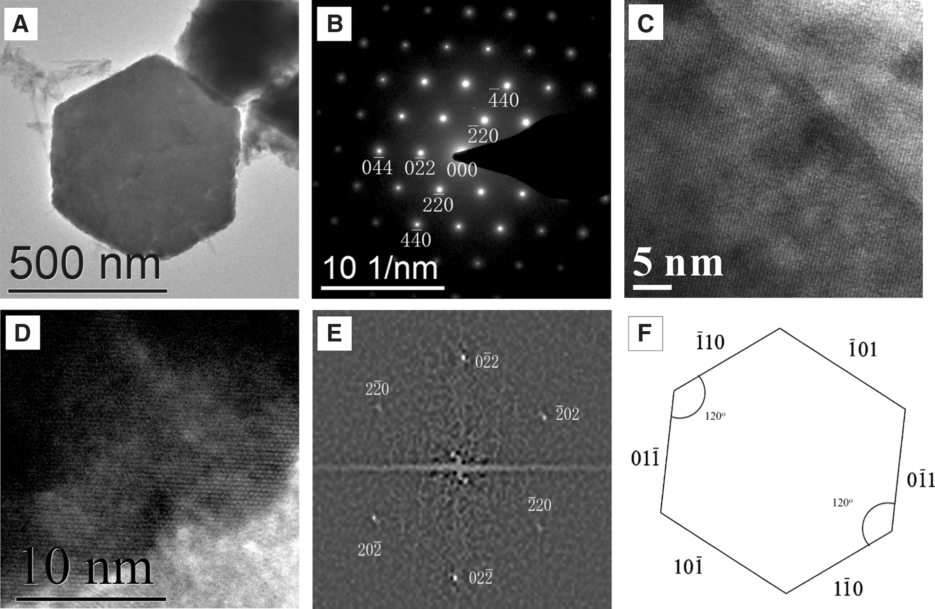
Characterization of the crystallographic habits of the platelet-like magnetite. (
4. Discussion and Applications
Consistent with previous results (Li et al., 2011b), the highly soluble Fe(II) and the unreacted ferrihydrite precursor provide reactants for the ongoing magnetite mineralization during prolonged incubation. Similar Mössbauer hyperfine parameters of magnetite from cultures before and long after cell lysis (Table 1) also reveal that the mineralization has a chemistry-controlled mechanism (e.g., Frankel and Bazylinski, 2003). The result also indicates the high stability of these magnetite crystals when compared to SPM magnetite produced by Shewanella or Geobacter at relatively low temperatures (e.g., Sparks et al., 1990; Kukkadapu et al., 2005; Li et al., 2009). However, the Mössbauer hyperfine parameters do not tell whether the increased percentage of magnetite results from the enlarged size of the existing crystals or the nucleation of new magnetite particles. Though the soluble Fe(II) in the cultures can be high enough (Li et al., 2011b) to inhibit further mineralization of magnetite by the recrystallization mechanism (Roden, 2006), the ultrafine goethite on the surface of magnetite observed in this study may serve as the Fe(III) source for the continuous mineralization of magnetite.
In the past, such a unique morphology has not been observed in SPM magnetite produced from the culture of Geobacter or Shewanella (e.g., Li et al., 2009) or the SD magnetite produced from that of Thermoanaerobacter (Zhang et al., 1998). SPM magnetite crystals produced by Geobacter or Shewanella do not have any particular morphology (Sparks et al., 1990; Hanzlik et al., 1996; Li et al., 2009), though the newly precipitated magnetite ultrafine particles have Fe2+-excess stoichiometry that makes them distinct from abiotic crystals (Li et al., 2011a, 2011b). The instability of SPM magnetite (Kukkadapu et al., 2005; Li et al., 2009) makes it difficult to detect the previous presence of FeRB in modern or past geological environments based on the crystal morphology (Maher and Taylor, 1988; Stolz et al., 1990; Li et al., 2009). The hexagonal magnetite observed in this study is the first characterization of biologically induced mineralization of magnetite that displays unique crystal morphology. Vali et al. (2004) reported observation of platelet-like magnetite briefly, in which the pseudohexagonal magnetite was not well characterized because the described multiple-stage mineralization pathway could not be repeated afterwards. SD magnetite crystals produced by magnetotactic bacteria (e.g., cuboid, octahedral, or hexaoctahedral) only have hexagonal projections when viewed from some particular directions under STEM (e.g., Thomas-Keprta et al., 2001; Li and Pan, 2012), which can only be called pseudohexagonal magnetite according to Vali et al. (2004). Bradley et al. (1996) also mentioned platelet-like magnetite from the martian meteorite ALH84001, which, however, appeared irregular in morphology, and the euhedral crystal plane was not well characterized. However, if the martian hexagonal magnetite could also be confirmed to be platelet-like by direct SEM observation, as observed for the TOR39-magnetite in this study (Fig. 2C–D), its putative biogenicity with an extracellular forming mechanism is also possible. Comparable to the ∼120 nm magnetosome preserved in sedimentary rocks for as long as 1 or 2 billion years (Chang et al., 1989; Kopp and Kirschvink, 2008), these much bigger crystals should also be stable enough to survive the long geological evolution. As the platelet-like magnetite is produced by FeRB dwelling in particular environments only (terrestrial deep crust or hot spring) with thermophilic temperature domains (>45°C, Liu et al., 1997), it can be used as a potential biosignature for thermophilic FeRB in the deep, hot biosphere of modern Earth and to search for the origin of ferric iron metabolism in the Archean to Palaeoproterozoic oceans' precipitated BIFs.
4.1. Application to the rapid detection of biogenic magnetite in the deep terrestrial crust and the existence of the deep, hot biosphere
Since the identification of tons of biogenic magnetite from the >5278 m deep crust (Gold, 1992), the concept of a deep, hot biosphere on Earth was eventually formulated (e.g., L'Haridon et al., 1995; Pedersen, 2000; Kashefi et al., 2004). However, the extremely high cost and lack of effective measures for avoiding contamination from the surface biosphere are two factors that significantly hinder the exploration of the deep, hot biosphere as a major research purpose (Szewzyk et al., 1994; Stevens, 1997; Pedersen, 2000; Kieft et al., 2007). The high temperature of the rocky crust for the growth of thermophiles (45–75°C, L'Haridon et al., 1995; Liu et al., 1997) allows only prokaryotes to thrive (Kristjánsson and Hreggvidsson, 1995; Rothschild and Mancinelli, 2001). Indeed, most of the reported terrestrial deep-dwelled microbial ecosystems have simple community structures but have the common existence of Thermoanaerobacter. For example, genera Thermoanaerobacter or Thermoanaerobacterium were detected in deep terrestrial environments such as terrestrial deep crusts (Szewzyk et al., 1994; Liu et al., 1997; Roh et al., 2003; Zhang et al., 2006), deep oil reservoirs (e.g., L'Haridon et al., 1995; Slobodkin et al., 1999; Birkeland, 2004; Fardeau et al., 2004), and terrestrial hot springs that serve as channels between the deep, hot biosphere and the surface biosphere (Cook et al., 1996; Slobodkin et al., 1999; Bao et al., 2002; Wagner and Wiegel, 2008; Bonch-Osmolovskaya, 2010). However, despite quite a few other types of thermophilic metal-reducing bacteria detected (e.g., Slobodkin, 2005), genera Thermoanaerobacter and Thermoanaerobacterium have rarely been reported from deep-sea hydrothermal vents where diverse thermophilic microorganisms thrive (e.g., Parkes et al., 1994; Reysenbach et al., 2000; Sokolova et al., 2001; Holden and Adams, 2003; Huber et al., 2007; Nakagawa and Takai, 2008; Wagner and Wiegel, 2008; Tsai et al., 2011). All these geological and microbial ecological observations suggest that Fe(III) respiration is the key component of the deep, hot biosphere (Gold, 1992; Kashefi and Lovley, 2003). The strong magnetic properties and the unique hexagonal platelet-like morphology of magnetite produced by Thermoanaerobacter provide the possibility of rapid screening of deep-crust rock samples for the previous presence of thermophilic FeRB, especially when samples are unsuitable for molecular detection. The optimized growth temperature of Thermoanaerobacter with the capability of magnetite mineralization is equivalent to a terrestrial crust depth of about 850–5500 m, depending on the geothermal gradients (Gold, 1992; L'Haridon et al., 1995; Liu et al., 1997; Roh et al., 2003; Zhang et al., 2006). Deep-drilling core samples of terrestrial crust for petroleum exploration are abundant and well preserved in core repositories all over the world (e.g., Core Research Center of the United States Geological Survey; Geological Cores and Samples Center of China). After long exposure to the surface environments, these core samples are unsuitable for molecular detection of FeRB because of contamination and destruction of genetic materials. Applying a magnet to the ground sample may effectively extract the magnetic material, and a conventional SEM examination is sufficient to characterize the morphology and size of magnetite crystals for possible biogenesis.
4.2. Application to the origin and evolution of dissimilatory iron-reducing bacteria
Recent studies have indicated that dissimilatory ferric iron reduction is a highly conserved characteristic of thermophiles (Slobodkin et al., 1999; Kashefi et al., 2004), which implies its antiquity (Vargas et al., 1998), but there is so far no mineralogical evidence of its emergence and evolution. Iron is the most widely used transition metal by microbial life over the course of evolutionary history (e.g., Poulton and Canfield, 2010; Konhauser et al., 2011). It has been observed that, throughout the long BIF-depositional history (Klein, 2005), Fe(II) was the most abundant energy source for chemotrophs, and Fe(III) oxide was the most abundant electron acceptor for heterotrophic metabolism. Though the ecophysiological requirements for Thermoanaerobacter are quite similar to those of the BIF-deposited ocean (e.g., Knauth, 2005), the biogenicity of magnetite in BIFs has received only limited support from studies of the crystallochemical structure (Li et al., 2011a), Fe- and light-elementary isotopes (e.g., Johnson et al., 2008; Heimann et al., 2010), and the biogeochemistry of enzymatic Fe(III) reduction (Nealson and Myers, 1990; Weber et al., 2006). Many BIFs only experienced low-grade metamorphism since their deposition 1.8 billion years (or longer) ago (Klein, 2005). The method suggested for the examination of deep crustal samples can also be used for screening BIF samples to trace biogenic magnetite and, accordingly, the emergence of iron-reducing bacteria in early Precambrian oceans.
5. Conclusions
Thermoanaerobacter is capable of ferric iron reduction at high temperature, which uniquely limits its dwelling environments to the deep crust or hot springs. The ferric iron metabolism by thermophilic dissimilatory iron-reducing bacteria induces crystallization of magnetite of a few hundred nanometers in size. These magnetite crystals have much higher stability than the superparamagnetic magnetite crystals produced by Geobacter or Shewanella, which are widespread in surface terrestrial and marine biospheres. The hexagonal platelet-like magnetite of several hundred nanometers observed only from the growth of Thermoanaerobacter can be considered a mineral biosignature of thermophilic iron-reducing bacteria because of its large size, unique morphology, and high forming temperature. These magnetite particles can be easily extracted and characterized to indicate the existence of the deep, hot biosphere of modern Earth and the emergence of ferric iron metabolism from BIFs deposited planetwide on Earth only from Archean to Paleoproterozoic eons.
Footnotes
Acknowledgments
I appreciate valuable comments from Tommy Joe Phelps of Oak Ridge National Laboratory, Hojatollah Vali of McGill University, and two anonymous reviewers. I thank Professor M. Darby Dyar of Mount Holyoke College for her kind help in measuring the Mössbauer spectra of magnetite samples. This study is supported by General Research Fund (HKU703609P) from Research Grants Council of Hong Kong.
Author Disclosure Statement
No competing financial interests exist.
Abbreviations
BIFs, banded iron formations; FeRB, ferric iron–reducing bacteria; SD, single domain; SAED, selected area electron diffraction; SEM, scanning electron microscope; SPM, superparamagnetic; STEM, scanning transmission electron microscope.