
Abstract
A portable, rapid, microbial detection unit, the Lab-On-a-Chip Application Development Portable Test System (LOCAD-PTS), was launched to the International Space Station (ISS) as a technology demonstration unit in December 2006. Results from the first series of experiments designed to detect Gram-negative bacteria on ISS surfaces by quantifying a single microbial biomarker lipopolysaccharide (LPS) were reported in a previous article. Herein, we report additional technology demonstration experiments expanding the on-orbit capabilities of the LOCAD-PTS to detecting three different microbial biomarkers on ISS surfaces. Six different astronauts on more than 20 occasions participated in these experiments, which were designed to test the new beta-glucan (fungal cell wall molecule) and lipoteichoic acid (LTA; Gram-positive bacterial cell wall component) cartridges individually and in tandem with the existing Limulus Amebocyte Lysate (LAL; Gram-negative bacterial LPS detection) cartridges. Additionally, we conducted the sampling side by side with the standard culture-based detection method currently used on the ISS. Therefore, we present data on the distribution of three microbial biomarkers collected from various surfaces in every module present on the ISS at the time of sampling. In accordance with our previous experiments, we determined that spacecraft surfaces known to be frequently in contact with crew members demonstrated higher values of all three microbial molecules. Key Words: Planetary protection—Spaceflight—Microbiology—Biosensor. Astrobiology 12, 830–840.
1. Introduction
T
Previous studies conducted on the Russian space station Mir attributed surface degradation of multiple materials to microorganisms collected and isolated from space-grade aluminum-magnesium and multiple polymers (Novikova, 2004). Such results underscored the need for continuous monitoring of surfaces, air, and water aboard the ISS. Therefore, a repetitive cleaning and microbial monitoring protocol based upon the use of a standard agar-based culture system has been in place aboard the ISS before, during, and after construction. In this protocol, contact slides (Biotest, Rockaway, NJ) are briefly placed on a flat surface, incubated for 5 days, and then visually inspected for colonies. Interestingly, promoting growth of bacteria with these contact slides actually creates potential biohazards that then require appropriate containment, disposal, and in some cases archiving for transfer to ground if further analysis is required. In order to improve upon the current protocol, multiple assay platforms, including the LOCAD-PTS, are currently being considered for future environmental monitoring of the ISS and possible long-range human flight projects. To date, no flight-qualified system has proven to be all-inclusive in the detection, quantification, and characterization of surface bacteria on the ISS, primarily due to the sheer diversity of microbial species.
When using available techniques, the vast majority of environmental bacteria cannot be cultured (Kaeberlein et al., 2002), and more inclusive methods should be analyzed for future use. At present, the best scenario may be a set of assays that would work in coordination with one another to conform to crew health and safety standards. The LOCAD-PTS suite of cartridges, based upon the Limulus Amebocyte Lysate (LAL) assay, detects live as well as dead Gram-negative and Gram-positive bacteria, mold, and fungi regardless of their ability to be cultured (Morris et al., 2010). The ability of the LOCAD-PTS to detect dead bacterial cells is a major advantage over current and proposed methods for microbial monitoring on the ISS in that it provides a more comprehensive picture of the surface bioburden. However, from a crew health perspective, it is the “live” or viable signatures that are of concern to the flight surgeons.
Herein, we report the results obtained during the continuation of our technology demonstration aboard the ISS from June 2008 through August 2009. Upon the successful completion of our first round of technology demonstration tests, our team's primary objective was to expand the capabilities of the equipment that we currently had available on the ISS by developing low-mass, swift time-to-flight cartridges specific for additional microbial molecules. The molecules beta-glucan, a component of fungal cell walls, and lipoteichoic acid (LTA), a molecule found in Gram-positive bacterial cell walls, were selected for ease of assay development. They were developed, produced, and launched to the ISS for testing within 2 years. The expanded capabilities afforded by these additional cartridges allowed us to design a set of experiments to determine the ability of the LOCAD-PTS to quantify three microbial molecules both separately and in parallel on ISS surfaces. Therefore, our new objectives for this second round of testing were as follows: • Perform side-by-side analysis of a location with beta-glucan or Gram-positive (LTA) cartridges and contact media slides; • Conduct parallel analysis of at least one site with all three cartridges and contact media slides.
2. Materials and Methods
The essential elements to the LOCAD-PTS are the reader, cartridges, swab kits, and the swab unit. For a complete description of each element, refer to the descriptions listed in Maule et al. (2009).
2.1. Reader
The LOCAD-PTS reader is a handheld spectrophotometer with built-in heater and pump, integrated with electronics and software. A LOCAD-PTS cartridge is inserted into an opening at the front of the instrument. Liquid samples are introduced into a sample well exposed on the cartridge end. The onboard pump draws the sample into the channels of the cartridge where dry reagents are hydrated and the assay is performed. At the completion of each test, the results are displayed on the LED screen, which indicates the endotoxin concentration of the sample in Endotoxin Units per milliliter (EU/mL), the amount of glucan in the sample in nanograms per milliliter, or the quantity of LTA in the sample in milligrams per milliliter. The reader was manufactured by Charles River Laboratories and modified for spaceflight in conjunction with Marshall Space Flight Center.
2.2. Cartridges
For each cartridge, all reagents are fully contained within each of four channels; therefore, gloves are not required for handling. Liquid samples are directly pipetted with the use of the swab unit (see below) to a sample well at the end of each channel, then pumped down the channel and mixed with dried LAL reagent and a chromogenic substrate. The absorbance of the reacted sample is measured and compared to an internal standard curve. Following analysis, the used cartridges are sealed in a plastic bag and placed in a down-mass disposal bag. The three types of cartridges used are as follows: • • •
2.3. Swab unit and swab kits
The swab unit and swab kits were designed and manufactured by Marshall Space Flight Center's LOCAD team. The swab unit is similar in size and function to a standard single-channel laboratory pipette. The swab unit is utilized during both the sample swabbing and sample dispensing steps. The swab kit consists of a water cartridge, swab tip, and dispensing tip.
2.4. Sample collection, processing, and analysis
The preferred sampling site was one that was previously sampled with the LOCAD-PTS. If a previously tested site was not available, suggestions from the LOCAD science team were conveyed to the crew. The crew were also allowed to choose several locations at their discretion, most opting for locations that they hypothesized may have a high microbial presence. To begin a sampling session, a Bacterial Contact Slide and a Fungi Contact Slide were placed on the selected surface. The slides were placed on the surface for 1–2 seconds and then removed. After removal, the slides were sealed and incubated.
The surface for each swabbing procedure covered an area of 25 cm2, a 5×5 cm square. To prepare the swab unit for surface swabbing, a water cartridge is attached to the lower end. Then a swab tip is attached to the other end of the water cartridge. Both ends of the water cartridge are identical, so orientation is irrelevant. In this configuration, the swab unit is held perpendicular to the surface to be swabbed while swabbing in a back-and-forth motion, covering the entire area. Following swabbing, a dispensing tip is applied over the swab tip. The swab unit is then manipulated such that the collected sample on the swab tip is mixed with the sterile water in the water cartridge. After completing a standardized mixing protocol, four 25 μL samples are placed in a cartridge for testing. Please refer to Maule et al. (2009) for a more detailed explanation of the sampling apparatus and procedure and for photos of the sampling process on the ISS.
3. Results
This paper presents results for the Phase 3 and Phase 4 set of experiments spanning 2008 and 2009, which continued the Phase 1 and 2 experiments reported previously (Maule et al., 2009). During Phases 3 and 4, the Gram-positive cartridges detecting the molecule LTA and beta-glucan cartridges for detecting yeast and mold were tested, expanding the repertoire of the LOCAD-PTS. Using the Gram-positive, beta-glucan, and LAL cartridges in parallel, we analyzed a total of 52 samples. As was observed for the LAL cartridges in the Phase 1 and Phase 2 testing, the highest beta-glucan readings were from the Cycle Ergometer with Vibration Isolation and Stabilization (CEVIS), a stationary exercise bicycle. However, only one site sampled registered a significant level of LTA; this was the Waste Hygiene Compartment (WHC) in the Russian Functional Cargo block (FGB or Zarya).
3.1. LOCAD-PTS cartridges have surveyed surfaces from many modules of the ISS
The onset of these experiments coincided with the arrival, attachment, and opening of the Japanese Exploration Module (JEM), providing a unique opportunity to be one of the first detection technologies utilized in that space. Because of the rigorous cleaning and contamination prevention protocols in place prior to launch, we expected to observe signals close to the background levels of the cartridges used. As listed in Table 1 and Fig. 2B, the values obtained from all four surfaces swabbed and sampled in the JEM and analyzed with the Gram-positive cartridges were at or near background. We were unable to test enough surfaces to analyze samples, using all three cartridges, but these results do provide one set of baseline measurements for a new module.
The background levels for LAL, beta-glucan, and Gram-positive cartridges are <0.05 EU/mL, 1 ng/mL, and 0.05 mg/mL, respectively. Bacterial and fungal colony-forming units (CFUs) are also presented when culture-based tests were performed.
As depicted in Fig. 1, almost every module of the ISS at the time of the experiments has now been sampled. Since the values listed in Fig. 1 are averages, they are in no way intended to be used to interpret the relative cleanliness of the various modules. However, from the results obtained in Phase 1 testing and the values listed in Table 1, we observed consistently elevated levels of microbial molecules in the Zarya module and Node 1 (Unity), the two oldest modules, using all three cartridges. Some of the readings obtained from surfaces in relatively newer modules (e.g., 27.03 ng/mL beta-glucan in the U.S. Destiny lab or 11.70 ng/mL beta-glucan in ESA's Columbus lab) were higher than those observed in Node 1 but still lower than the values obtained from Zarya. These data could be attributed to the variation in the texture, material, and crew contact of the surfaces sampled in different modules, even though most sites chosen were from instruments or panels with frequent crew member contact. The data listed in Table 1 and depicted in Fig. 1 provide the first complete data set summarizing the distribution of microbial molecules throughout the ISS as determined by an onboard, rapid detection device.
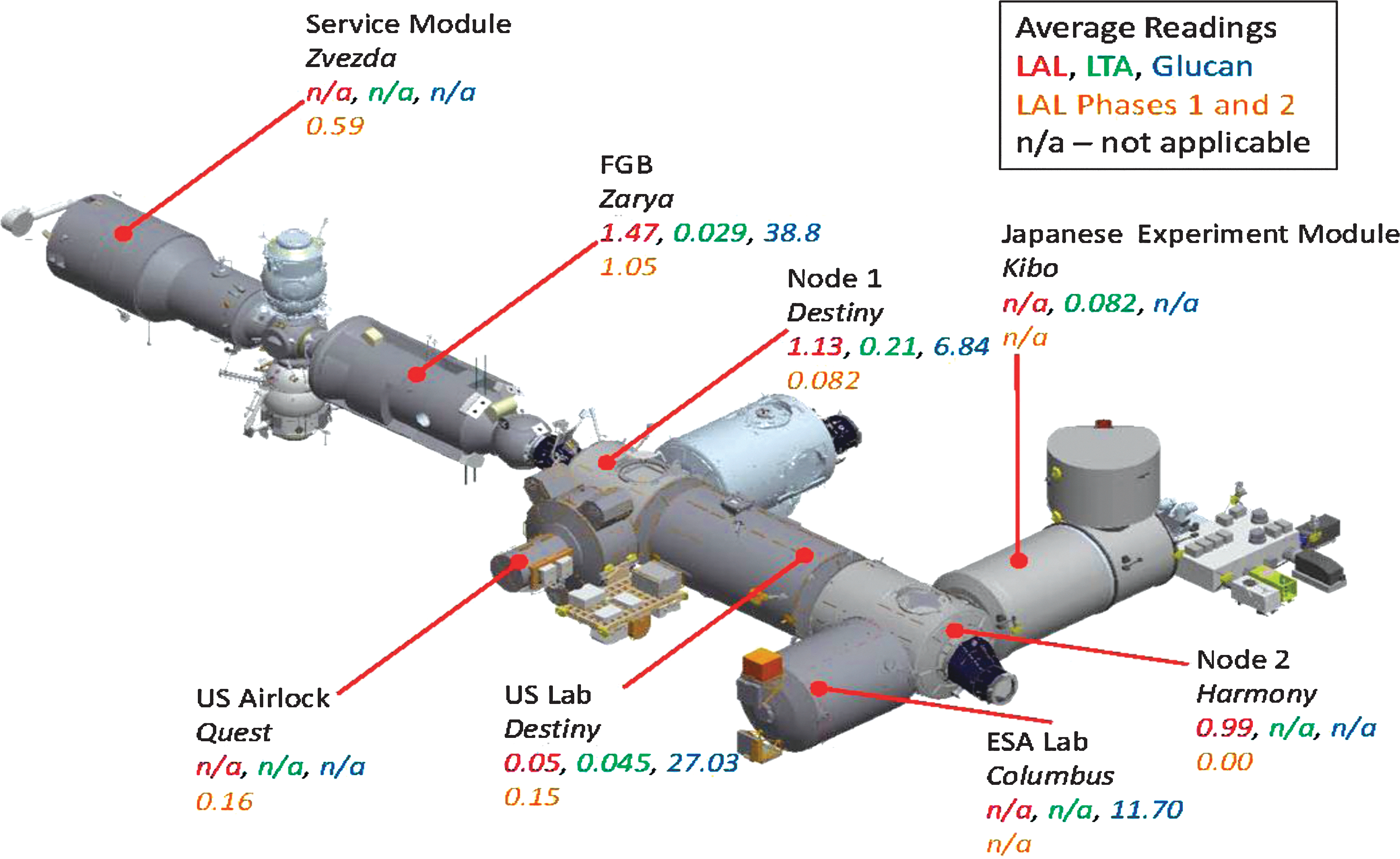
A diagram of the ISS depicting levels of microbial molecules for each module. LOCAD-PTS cartridges have been used to sample surfaces from most modules of the ISS. Values listed for LAL, Gram-positive (LTA), and beta-glucan cartridges are averages of all the values obtained for each of the types of cartridge from each module. LAL results from Phases 1 and 2 are taken from Maule et al. (2009). Color images available online at
3.2. Microbial molecule values can vary significantly even on the same surface
As observed in Phase 1 and 2 testing of the LAL cartridges, the values obtained from different sections of the same surface returned markedly diverse values for both the beta-glucan and Gram-positive cartridges (Table 2). Similar to the methods performed during the first rounds of testing the LAL cartridges, a surface was selected with an area large enough to place a fungal contact media slide and a bacterial contact media slide side by side. Four areas—north, south, east, and west—of the contact slides were then swabbed and analyzed with four separate beta-glucan cartridges or four Gram-positive cartridges. As listed in Table 2, the values returned by the beta-glucan cartridges varied significantly between each area sampled. Although the level of beta-glucan detected on Swab Area 3 is higher than the other three swab areas (18.1 ng/mL), it is lower than some values obtained elsewhere in the space station (e.g., 66.4 ng/mL in the WHC). The remaining three swab areas reported values at or very near background (1 ng/mL), which is in agreement with the lack of colonies that grew on either contact media slide.
Values obtained from beta-glucan and Gram-positive cartridges are given in ng/mL and mg/L, respectively.
We observed consistently low values when using the Gram-positive cartridges, reflected in the numbers listed in Table 2. However, these results were not unexpected. We attempted to produce a very sensitive cartridge for detecting Gram-positive cells, using a colorimetric-based assay, the basis for our instrument platform. Our ground tests conducted prior to flight demonstrated reproducibility but not sensitivity (Fig. 2). We determined that it would take approximately 104 bacterial cells from laboratory culture to yield a signal on the low end of the standard curve, which is a large number of cells compared to the sensitivities of both the LAL and beta-glucan cartridges (Fig. 2). The low reactivity of whole cells may be due to the lack of bioavailability of the LTA, meaning the LTA molecules may be buried in the bacterial cell wall and therefore be unable to react with the test reagents. Since these assays were developed postlaunch to take advantage of existing equipment on orbit, and due to time constraints, we elected to launch and test the Gram-positive cartridges, realizing the constraints imposed by the relatively low sensitivity.

Standard curves for all three LOCAD-PTS cartridges. These cartridges demonstrated reproducible standard curves, allowing for accurate data interpretation. Purified LPS, glucan, or LTA was diluted in endotoxin-free, ultrasterilized water to yield the following concentrations: 0.05 EU/mL, 0.5 EU/mL, 5 EU/mL for LPS; 1 ng/mL, 10 ng/mL, 100 ng/mL for glucan; or 0.05 mg/mL, 0.5 mg/mL, 5 mg/mL for LTA. These dilutions were analyzed with LAL, beta-glucan, and Gram-positive cartridges, respectively. Onset time is given in seconds and increases as the amount of microbial molecule in a sample decreases. Color images available online at
In light of the relative insensitivity of the Gram-positive cartridges, the fact that we obtained a positive signal from the foot restraint is significant (0.119 mg/mL vs. 0.05 mg/mL, the background value). This result was observed in conjunction with a relatively large number of colonies grown on the contact slides [14 colony-forming units (CFUs)], which were probably composed of both Gram-positive and Gram-negative species. The background values returned by the samples from other areas of the foot restraint could be due to a concentration of the bacteria to one area of the foot restraint.
3.3. Microbial molecules were elevated at sites frequently contacted by crew members
Similar to the experiments performed in Phase 2 of LAL cartridge testing documented previously (Maule et al., 2009), we requested that the operating ISS crew member select multiple locations to sample with either beta-glucan or Gram-positive cartridges. As shown in Fig. 3, most sites returned background values for both cartridges (1 ng/mL for beta-glucan and 0.05 mg/mL for Gram-positive). However, both cartridges detected elevated levels of their respective microbial molecules at sites contacted frequently by crew members.
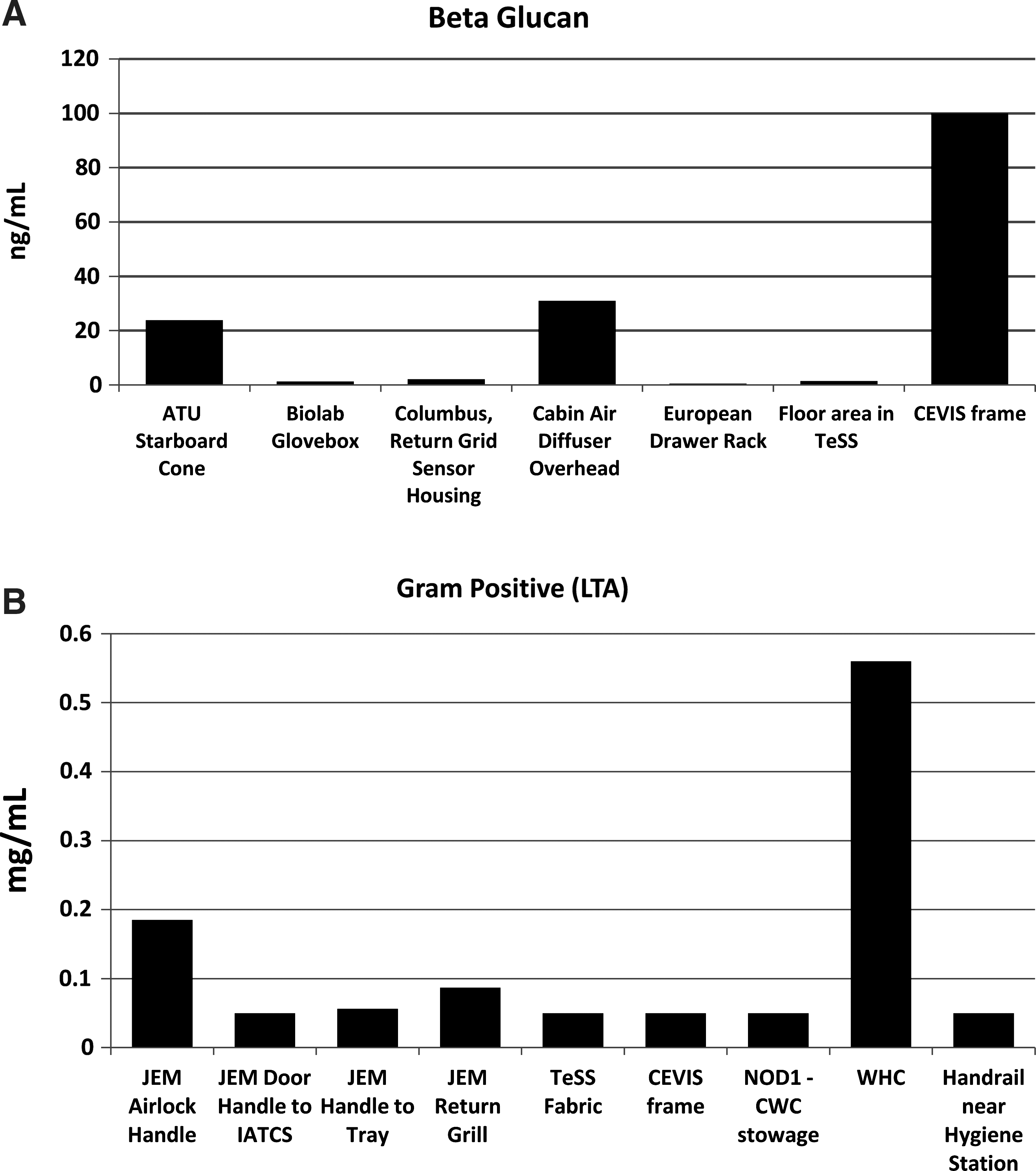
Graphical representation of beta-glucan and LTA signals detected at sites frequently contacted by ISS crew members. (
The majority of the sites surveyed by crew members were those frequently handled, such as airlock handles, drawers, and audio terminal units (ATUs). Other sites surveyed with the beta-glucan cartridges, such as the Biolab Glovebox and the floor area in the Temporary Sleep Station (TeSS), returned background values, even though they are frequently contacted by crew members (Fig. 3A). The TeSS fabric sampled with Gram-positive cartridges also returned a background value (Fig. 3B). Crew members frequently clean the TeSS with stringent protocols, which may have contributed to these low values. Alternatively, the texture of the surface may have prevented an adequate sampling of the surface using our current swab tool. Similar results have been observed using sterile cotton swabs, our swab tool, and LAL cartridges for analysis on fabric surfaces in the laboratory (Morris et al., unpublished data).
The CEVIS (the cycle exercise machine), the back wall of the WHC, and the ATU demonstrated the highest levels of beta-glucan recorded during this series of experiments (>100, 66.4, and 23.8 ng/mL, respectively; Table 1). By comparison, measurements of beta-glucan taken at the Shuttle Launch Facilities at Kennedy Space Center gave results ranging from 0.010 to 0.160 ng/mL on exposed exterior surfaces of the space shuttle just prior to launch and 0.021–0.688 ng/mL in samples taken from adjacent ocean and beach surfaces (Maule et al., unpublished data). The data obtained from sampling surfaces at Kennedy was collected by more sensitive (pg/mL sensitivity) beta-glucan cartridges than those available at the time of these ISS experiments; however, they underscore the possibility that more microbial molecules may be present on certain ISS surfaces than previously expected. The beta-glucan results for the CEVIS and the ATU had no contact media slides used in parallel on the same surfaces while samples were collected, but contact media slides for both bacteria and fungi were used in conjunction with all three cartridges in sampling the back wall of the WHC. No fungal colonies were recovered, despite the elevated levels of beta-glucan (Table 1). This apparent discrepancy could be due to the stringent disinfecting/cleaning protocols performed on the ISS, which would kill all the microbes on a surface, potentially exploding the cells and releasing contents but not removing all the microbial molecules. Therefore, no viable organisms remain on the surface, but the build-up of microbial particles could account for the elevated numbers seen on the CEVIS, in the WHC, and on the ATU relative to exposed surfaces on the ground.
Interestingly, the CEVIS returned only background-level results when using the Gram-positive cartridges (Fig. 3). Again, this result is possibly due to the significant difference in sensitivity between the two cartridge types. Alternatively, different surfaces of the machine may have been sampled, since the tests were performed by different crew members at different times. Two significant readings from the Gram-positive cartridges were obtained from the JEM airlock handle (0.185 mg/mL) and from the WHC (<0.56 mg/mL). Taken together, these data confirm that microbial molecules can be detected on surfaces frequently contacted by crew members.
3.4. Standard culture-based methods and molecular monitoring yield different results from most surfaces
The second objective of this study was to perform a parallel analysis of a single surface by using all three cartridges and contact media slides. The contact media slides were placed on a surface, and four adjacent sites were swabbed and analyzed. These four samples were then analyzed with one LAL, two beta-glucan, and one Gram-positive cartridges. In keeping with the LAL results from Phase 1 (Maule et al., 2009) and the beta-glucan and Gram-positive results described previously (Table 2), each of the three surfaces sampled demonstrated differences between the results obtained from one type of cartridge to another and between the results from the same type of cartridge (beta-glucan). These results are listed in Table 3 and depicted graphically in Fig. 4.

Variation in the quantities of microbial molecules occurs across different sections of the same surface. Endotoxin, LTA, and beta-glucan were detected with LAL, Gram+, and beta-glucan cartridges, respectively. Swab samples were collected from four areas of the same surface (north, south, east, and west). Sites include the Waste Hygiene Compartment (WHC), a panel in Node 1 (D5), and the cycle ergometer handle (CEVIS). Units for LAL, glucan, and Gram+ cartridges are given in EU/mL, ng/mL, and mg/mL, respectively. Color images available online at
Averages, where applicable, and CFUs collected from the site are also included. Values of LAL, beta-glucan, and Gram-positive cartridges are given in EU/mL, ng/mL, and mg/mL, respectively.
Interestingly, we observed a large number of CFUs (13 CFUs) on a handle of the CEVIS. However, all the cartridges returned values at or near background levels (Table 3). Conversely, 3 CFUs were cultured from the surface of a wall of the WHC, and elevated levels of LAL and beta-glucan were observed from adjacent sampling sites (1.47 EU/mL and 66.4/11.2 ng/mL, respectively). The third site tested, a panel in Node 1 that is frequently handled, returned the same number of CFUs as the WHC (3 CFUs), yet all the cartridges demonstrated background-level signals.
These data are very similar to the observations made in Phase 1 and 2 of LAL cartridge testing. Relatively poor correlation could be drawn between the two methods—molecular and culture-based (Maule et al., 2009). Multiple laboratory and field studies performed by different investigators have borne this out for multiple molecular assay platforms, though sometimes overall trends (more “dirty,” more “clean”) can be established (Cooper et al., 2011; Wainwright et al., 2005). These two methods measure different characteristics—the ability to grow and survive on media versus the mere presence of microbial molecules—leading to the difficulty in establishing an exact correlation between the two methods (Morris et al., 2010). Molecular methods could detect the presence of dead microbes or those that are viable but nonculturable (Barer, 1997). Regarding the results from the CEVIS handle, microbial density varying across a surface or differences in sampling (contact slide vs. swab tool) could account for the discrepancies observed in the elevated CFU levels and the lower molecular values.
4. Discussion
Herein, we present the final data set, which completes the data collected from all the LOCAD-PTS operations performed aboard the ISS from 2007 through 2009. These data are the first to provide molecular data on the distribution of three microbial molecules in every habitable module of the ISS during that time frame. From these results, we have demonstrated that most surfaces on the ISS are relatively free of microbial molecules and that the LOCAD-PTS provides a rapid method of detecting these molecules on a variety of surfaces.
Our previously published data demonstrate that 31 samples gave an endotoxin level classified as “low” or “absent.” An additional 11 samples returned values of endotoxin that we classified as “moderate” or “high.” All the sites from these 11 samples were those that had frequent crew contact. The site with the highest LAL readings was the CEVIS training hardware (Maule et al., 2009).
On selected sampling locations on the ISS, we were able to record results with traditional culture methods and LOCAD-PTS cartridges measuring Gram-negative (LAL), Gram-positive (LTA), and fungal (beta-glucan) contaminants simultaneously. Rapid assessment of the specific type of contamination could enable ameliorative actions specific for bacterial or fungal contamination events aboard the space station. As a result of the relatively clean environment on the ISS, the number of locations showing elevated positive microbial results is small. The example of an elevated LTA measurement coincident with 14 CFUs detected on the starboard aft food restraint highlights the utility of the rapid nonculture method, validated by the multiday, traditional culture-based method. Unfortunately, we were unable to perform any Gram staining or other differential tests on the colonies grown on the flight media slides in order to discern whether the majority of the colonies were indeed Gram-positive.
Currently, researchers are unable to perform any sort of Gram stain or other basic microbial identification assay onboard the ISS. Therefore, it is impossible for the crew in space, or environmental control personnel, or medical supervisors on Earth, to know what species of organisms are present on a given surface without analyzing samples on the ground many weeks later. Due to these limitations, we were also unable to discriminate between Gram-positive and Gram-negative colonies grown on the culture media plate, simply lumping them together as total CFUs. Recently, a “Gram ID” cartridge has been developed for the LOCAD-PTS, which can determine whether a culture is Gram-positive or Gram-negative. Similar to the other cartridges, it uses a modified version of the LAL colorimetric assay. A rapid completion of the test indicates the presence of a Gram-negative organism, while a Gram-positive sample remains blank due to the paucity of lipopolysaccharide present in the sample. Adding Gram ID cartridges to further flight tests would enhance researchers' ability to analyze environmental, microbial data.
Although we did not obtain many positive readings with the Gram-positive cartridges, we can easily attribute that to the lack of sensitivity mentioned previously. Therefore, the fact that we observed three significant positive values when using these cartridges is worth noting (JEM airlock handle, 0.185 mg/mL; WHC, <0.56 mg/mL; and aft foot rest, 0.119 mg/mL). As mentioned previously, whole cells—viable, viable but nonculturable, or dead—may render LTA molecules invisible to this detection method by burying them inside the cell wall. Therefore, any means of extracting the samples from a surface may expose more LTA by rupturing cells or simply disturbing the cell wall, which would thereby improve subsequent LTA detection. The LTA recovered by our swab tool and the LTA-positive samples on the ISS may differ significantly from environmental samples, where cells may be live, dead, or in various stages of degradation. In such cases, a positive LTA result may indeed be caused by far fewer cells than observed in controlled laboratory experiments or those we performed on the ISS.
When using the LOCAD-PTS, for data to be considered valid, the test results must fall within the standard curve range, sample volume must be sufficient to fill the optical read areas of the cartridge, and the positive spike value must be recovered within 50% to 200%. Spike values outside that range may be caused by substances in the sample that could inhibit or enhance the assay. Also, because each cartridge has four channels (two with positive spikes and two without spikes), the reported results are an average of each of the two channels in those groups. The coefficient of variation (CV) must be within 25%. In normal ground operations, the occurrence of invalid test data is relatively rare. A possible cause of an invalid test result could be the presence of a detergent on space station surfaces that inhibits the assay enzymes. The solution would be to dilute the sample and run it again.
The number of invalid assay results on the ISS was higher than those during ground operations. Especially prevalent were instances of CV values that were too high (>25%), or a result labeled with a “?”, meaning that one of the two channels to be averaged did not have an expected value. The most likely cause of this type of error was that the liquid sample was not sufficient to fill the optical read area, returning a zero or infinity value for that sample.
Crew members observed that the amount of volume in the water cartridge of the swab tool, used to extract the swab and fill the sample chamber, was diminishing with storage time aboard the ISS. The likely cause of this was water loss through an improper sealing of the water cartridge. Observing the liquid as it filled each sample, crew members noted that less than a full water cartridge sometimes caused air bubbles to be introduced into the sample. When an air bubble gets pulled into a reaction channel, the leading edge may enter the optical area and cause a very high (or infinity) reading. If no sample reaches the optical area, zero absorbance is recorded.
One solution proposed to the crew was to observe the presence of bubbles in the swab tool and shake the bubbles out of the liquid path. After implementing this solution, significantly fewer invalid test results were obtained. However, we have begun planning more permanent solutions to this issue. On newer PTS units, as the sample is drawn into the channel, there is an infrared LED and photodiode that senses the leading and trailing edge of a sample. Through a programming change, we can count the number of pump steps between those edges and calculate the volume of a sample. In the future, implementation of this change will result in “flagged” samples—those that could have returned invalid results based on sample volume ranges.
A redesign of the water cartridge will also be necessary to remove the source of this problem. We have begun to focus on how water is sealed in the swab tool. One potential design contains the water in a “bulb” with a valve that requires the seal to be broken before water is released. Initial attempts on the ground have produced promising preliminary results. Stability testing at room temperature and 37°C has shown minimal water loss (<5%) over 12 months. We are confident that further improvements along these lines will greatly improve simplicity of the assay and the integrity of results.
When quantifying live bacteria, a limitation in the LOCAD-PTS system has been the fact that the method does not discriminate between live or dead microbes. While some space missions, such as those in astrobiology or planetary protection, may require extreme cleanliness (i.e., the absence of all biological markers), others may have a greater emphasis on eliminating all live organisms. For instance, potential pathogens that are dead may pose no health risk to crew, yet the LOCAD-PTS method would report a positive reading. This “uncertainty” could make setting requirements difficult. The tentative “requirements” suggested previously (Maule et al., 2009) would indeed be useful for a general cleaning indicator but inadequate to address crew health requirements if the positive were triggered by predominately dead organisms.
5. Conclusions and Implications
By incorporating cartridges that could detect all three classes of microorganisms, this study significantly expands the usefulness of rapid microbial assessment by LOCAD-PTS, which had previously only been able to detect Gram-negative contamination and fungal contamination. The report of assay results in minutes, instead of the days required for culture-based methods, gives information to the crew that will allow for near real-time decisions concerning the need to clean a particular surface. Additionally, information provided regarding the type of microorganism present helps determine possible causes for the contamination. For instance, fungal contamination could indicate excess moisture or poor ventilation that could be improved.
While LOCAD-PTS is a nonculture method that shows promise and fulfills a partial need, more capability is needed. We have begun to seek additional hardware capability in the LOCAD-PTS handheld format that can discriminate live cells versus dead and measure pathogen-specific levels when desired. A more relevant name for such hardware may be Real Time Bioburden Assessment (RTBA). Problems encountered with water loss have already resulted in a swab tool redesign with encouraging preliminary results. Future technological improvements upon the LOCAD-PTS platform will target the need to discriminate live versus dead microorganisms. To achieve this goal, multiple technical approaches are being investigated, including ATP content of the cells, selective PCR amplification, and live/dead fluorescent staining. Eventually, the ability to detect specific species of interest, such as potential pathogens, will be available with the use of specific antibodies (or pools of antibodies) and species-specific DNA probes. We also plan to expand the capabilities of sample collection and analysis to include air and water monitoring.
New rapid microbial testing methods are certainly needed while going forward to missions beyond low-Earth orbit. Crews must have the autonomy to collect routine monitoring information and make critical decisions without the need for material support or even real-time communication with the ground. Instrumentation development and testing, such as we have reported here, is integral to maintaining crew member health and equipment whether in low-Earth orbit or on long-duration space missions.
Footnotes
Author Disclosure Statement
Dr. Norman Wainwright is the Director of Research and Development at Charles River Laboratories, which manufactures the Endosafe-PTS upon which the LOCAD-PTS instrument is based. No other competing financial interests exist.
Abbreviations
ATU, audio terminal unit; CEVIS, Cycle Ergometer with Vibration Isolation and Stabilization; CFUs, colony-forming units; CV, coefficient of variation; EU/mL, Endotoxin Units per milliliter; FGB, Functional Cargo block; ISS, International Space Station; JEM, Japanese Exploration Module; LAL, Limulus Amebocyte Lysate; LOCAD-PTS, Lab-On-a-Chip Application Development Portable Test System; LPS, lipopolysaccharide; LTA, lipoteichoic acid; TeSS, Temporary Sleep Station; WHC, Waste Hygiene Compartment.