
Abstract
This is a case report of apparent thyroid structural and functional alteration in a single mouse subjected to low Earth orbit spaceflight for 91 days. Histological examination of the thyroid gland revealed an increase in the average follicle size compared to that of three control animals and three animals exposed to hypergravity (2g) conditions. Immunoblotting analysis detected an increase in two thyroid gland enzymes, sphingomyelinase and sphingomyelin-synthase1. In addition, sphingomyelinase, an enzyme confined to the cell nucleus in the control animals, was found in the mouse exposed to hypogravity to be homogeneously distributed throughout the cell bodies. It represents the first animal observation of the influence of weightlessness on sphingomyelin metabolism. Key Words: Gravity—Lipid—Spaceflight—Sphingomyelin—Thyroid. Astrobiology 12, 1035–1041.
1. Introduction
P
2. Materials and Methods
2.1. Reagent
Anti-SMase, anti-SM-synthase, and anti-Bax primary antibodies, horseradish peroxidase–conjugated, tetramethyl rhodamine isothiocyanate (TRITC)–conjugated, and fluorescein isothiocyanate (FITC)–conjugated secondary antibody were obtained from Santa Cruz Biotechnology, Inc. (CA, USA). SDS-PAGE molecular weight standards were purchased from Bio-Rad Laboratories (Hercules, CA, USA). Chemiluminescence kits were purchased from Amersham (Rainham, Essex, UK). Thin layer chromatography plates (silica Gel G60) were from Merck (Darmstadt, Germany); the radioactive SM (choline-methyl 14C, 54.5 Ci/mol), PC (L-3-phosphatidyl N-methyl-3H choline 1,2 dipalmitoyl, 81.0 Ci/mmol) were from Amersham Pharmacia Biotech (Rainham, Essex, UK); Ecoscint A was from National Diagnostic (Atlanta, GA, USA).
2.2. Experimental design and animal care
All experimental procedures were authorized by the Public Veterinary Health Department of the Italian Ministry of Health. The experiment was also conducted in accordance with regulations for the care and use of laboratory animals and with the guidelines of the Japanese Physiological Society. Furthermore, this study was also approved by the Committee on Animal Care and Use at Graduate School of Medicine, Osaka University (No. 22-071). Finally, the protocol utilized in the study has been authorized by the Public Veterinary Health Department of the Italian Ministry of Health. All experiments were carried out with male C57BL/10J mice (8 weeks old).
Hypogravity experiment
Three male C57BL/10J mice were individually housed in the MDS, a 11.6×9.8×8.4 cm payload developed by Thales-Alenia Space Italy (Cancedda et al., 2002). Food and water were supplied ad libitum. The MDS was launched on the Space Shuttle Discovery, within the Space Transport System (STS)-128 mission, on August 28, 2009. It was then housed in the Japanese Experimental Module (Kibou) on the ISS until its return to Earth via Space Shuttle Atlantis (STS-129 mission) on November 27, 2009 (Masini et al., 2012). Only one mouse returned to the Earth alive after 91 days of spaceflight. It was very difficult to establish the cause of death because this was the first long-term animal experimentation in space. It was not possible to perform an autopsy on the two dead animals because they were not frozen immediately after death.
After the spaceflight experiment, the on-ground experiment was also carried out at the vivarium of the Advanced Biotechnology Center in Genova, Italy. One group of mice with the same species, sex, and age were housed in a normal vivarium cage as the laboratory control (vivarium 1). The amount of food and water supplementation and the environmental conditions were simulated for the control group to approximate those of the flight group. All analyses were conducted on the thyroid gland of one animal alive. Thyroids were sampled bilaterally from each mouse killed by inhalation of carbon dioxide at the Life Sciences Support Facility of Kennedy Space Center within 3–4 hours after landing. The thyroid samples were either processed or frozen immediately, according to the various experimental protocols.
Hypergravity experiment
Three mice of the same strain as those used in hypogravity experiments were maintained in hypergravity, with conditions similar to the MDS experiment, in a 2g centrifuge in the laboratory of Dr. Y. Ohira at the Osaka University, Osaka, Japan. Control mice were similar to those reported in the hypogravity experiment (vivarium 2). Animals were treated, and thyroids were obtained and processed with the same procedures used in the hypogravity/space experiments.
2.3. Thyroid tissue treatment
The thyroid lobes in part were homogenized and used for protein dosage, immunoblotting analysis, and enzyme activity assay, and in part fixed in 4% neutral phosphate-buffered formaldehyde solution for 24 h. Thyroids were dropped with essentially random orientation in paraffin. The paraffin blocks were sectioned into 4 μm thick sections. All sections were mounted on silan-coated glass slides. Each slide contained a pair of sections at a distance equal to 140 μm. Between 7 and 14 pairs of sections were sampled, excluding the first and the last, and 7, 9, 11, and 13 sections were used for morphological analysis whereas 8, 10, 12, and 14 sections were used for immunofluorescence analysis. Tissue sections were deparaffinized and rehydrated through a series of xylene and ethanol washes.
2.4. Morphological analysis
The sections were treated with the hematoxylin-eosin (Chroma-Gesellschaft, Germany) staining method and investigated by using inverted microscopy EUROMEX FE 2935 (ED Amhem, the Netherlands) equipped with a CMEX 5000 camera system (40× magnification). The analysis of the tissue section size was performed by ImageFocus software.
2.5. Protein study
The proteins were quantified according to Lowry et al. (1951). The immunoblotting analysis to detect SMase, SM-synthase, and Bax content was performed as previously reported (Albi et al., 2011). Enzyme activity of SMase and SM-synthase1 was assayed according to Albi et al. (2008). For immunofluorescence analysis, sections were incubated, after three washes with phosphate-buffered saline (PBS), with 2 μg/mL anti-SMase or anti-SM-synthase1 primary antibodies diluted in a 0.5% solution of bovine serum albumin in PBS overnight at 4°C. The slides were washed three times with PBS and incubated with fluorochrome-conjugated secondary antibodies (TRITC for SMase and FITC for SM-synthase), diluted as primary antibodies, for 1 h at room temperature. After three washes with PBS, the slides were mounted with glycerol and coverslips. The samples were examined under a fluorescence microscope (OLYMPUS IX 51) equipped with an OLYMPUS DP 50 camera system and analyzed at 40× magnification.
2.6. Statistical analysis
The experiments were conducted on the thyroid of one animal for the hypogravity experiment (the only one returned alive from the mission), three control animals for the hypogravity experiment (vivarium 1), three animals for the hypergravity experiment, three control animals for the hypergravity experiment (vivarium 2). The immunoblotting analysis and enzyme activity assay were repeated three times on each thyroid. Means±SD of three results for hypogravity animals and of nine results for each other experiment are given. The significance of the differences between the data was checked by the Student t test.
3. Results
Microscopy analysis performed on four histological microsections, as reported in “thyroid tissue treatment,” which were subjected to hematoxylin-eosin staining, showed that the thyroid gland of the animal that had been in space has a greater number of large follicles, which supports recent observations (Masini et al., 2012). The differences of the follicle surface area are shown in Fig. 1 and Table 1.
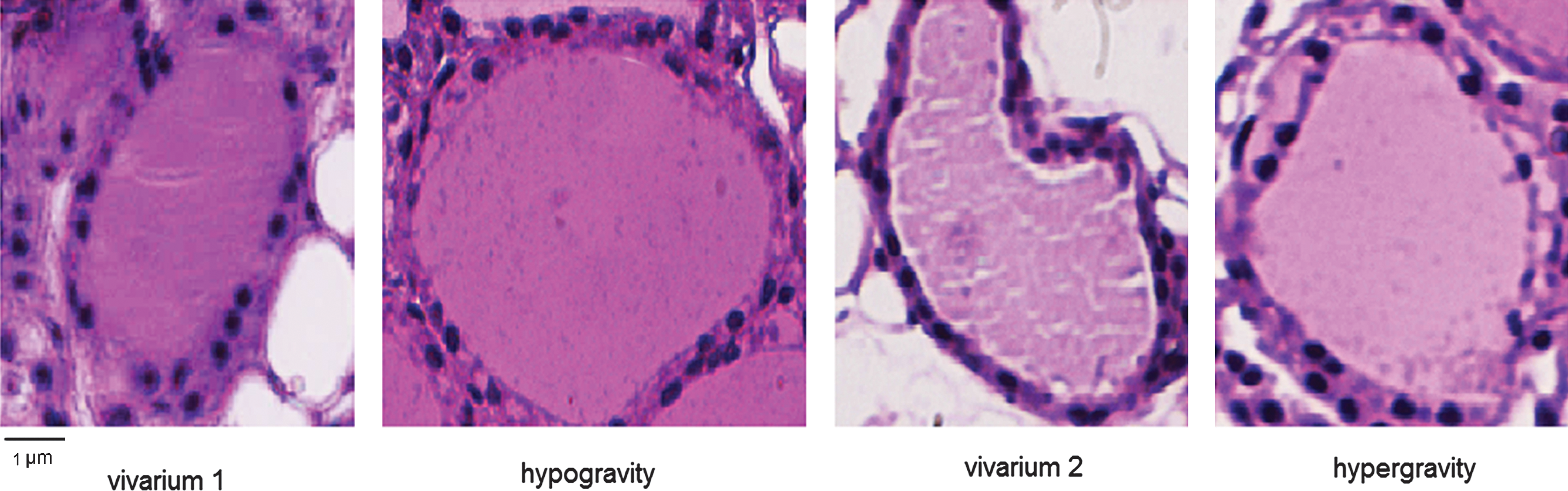
Morphology analysis of thyroid follicles of mice. “vivarium 1,” maintained in vivarium cages (control for experiment in hypogravity); “hypogravity,” experimental animal in space; “vivarium 2,” control for experiment in hypergravity; “hypergravity,” experimental animals in 2g centrifuge. Hematoxylin-eosin staining, 40× magnification, 1 μm scale bar. Color images available online at
The surface area of large follicles was analyzed by ImageFocus software. Data represent the mean±SD of three analyses performed in thyroid gland of three mice maintained in vivarium and used as controls for experiment in hypogravity (Vivarium 1), of three analyses performed in thyroid gland of the mouse maintained in hypogravity, of three mice maintained in vivarium and used as controls for experiment in hypergravity (Vivarium 2), and of three mice maintained in hypergravity. Significance: ** P<0.001 hypogravity versus vivarium 1.
Experiments of immunoblotting demonstrated that the long-term exposure to a real microgravity environment increased the levels of SMase and SM-synthase1 (Fig. 2a). The band density of SMase, corresponding to 42 kDa apparent molecular weight, was 2.18-fold higher than that of the vivarium 1 control, whereas the hypergravity reduced the enzyme 1.79 fold with respect to the vivarium 2 control (Fig. 2b). Immunofluorescence analysis confirmed these data and highlighted a different distribution of the enzyme with gravity change in comparison with control samples. In fact, in vivarium 1 and vivarium 2 the enzyme appeared particularly localized inside the cells, as specific round images, whereas in hypogravity the enzyme had a more uniform distribution and the hypergravity sample showed a peripherical localization (Fig. 3). The immunoblotting of SM-synthase1 showed a strong immunopositivity of band, corresponding to 49 kDa apparent molecular weight, only in the sample in hypogravity (Fig. 2a). The band area analysis showed an enrichment of about 20-fold in hypogravity in comparison to vivarium 1, whereas no variations were present in hypergravity with respect to its control (Fig. 2b). These data were confirmed with the immunofluorescence technique (Fig. 4).
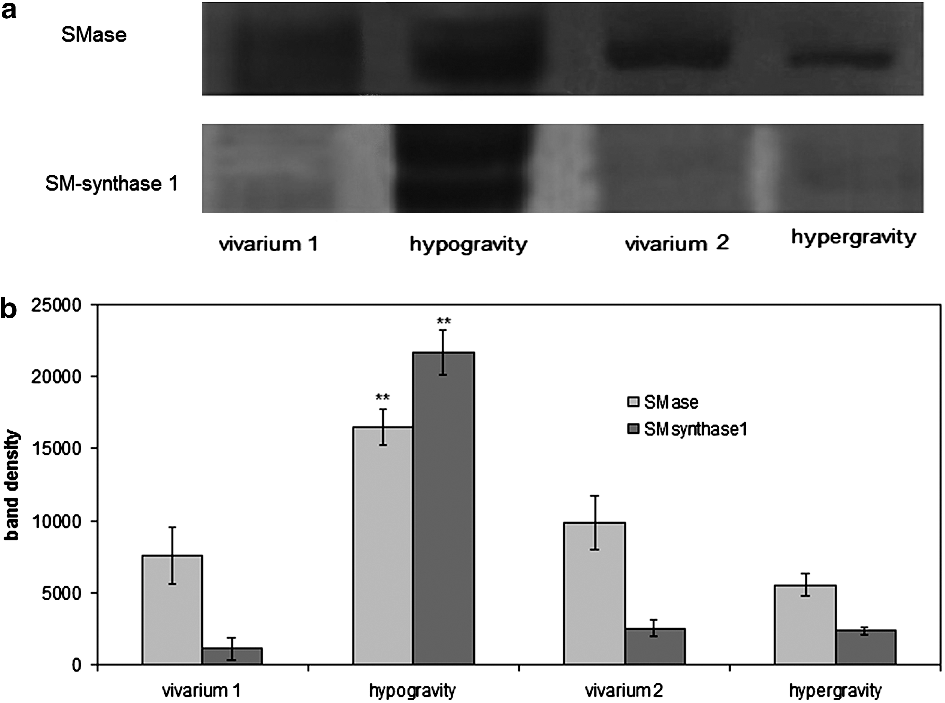
Comparison of SMase and SM-synthase1 content by immunoblotting analysis. The study was performed by using specific antibodies. The position of the 43 kDa apparent molecular weight for SMase and 49 kDa for SM-synthase1 is indicated comparing the position of molecular size standards. The area density was evaluated by densitometry scanning and analyzed with Scion Image program. Data represent the mean±SD of three analyses performed in the thyroid gland of three mice maintained in vivarium and used as controls for experiment in hypogravity (vivarium 1), of three analyses performed in the thyroid gland of the mouse maintained in hypogravity, of three mice maintained in vivarium and used as controls for the experiment in hypergravity (vivarium 2), and of three mice maintained in hypergravity. Significance: **P<0.001 hypogravity versus vivarium 1.

Fluorescence immunostaining of SMase in thyroid tissues. Analyses were performed using anti-SMase primary antibody and TRITC-conjugated secondary antibody. 40×magnification, 3 μm scale bar. The arrows indicate particulars at 100×magnification, 1 μm scale bar. (“vivarium 1,” control for the hypogravity experiment; “vivarium 2,” control for the hypergravity experiment.) Color images available online at

Fluorescence immunostaining of SM-synthase1 in thyroid tissues. Analyses were performed by using anti-SM-syntase1 primary antibody and FITC-conjugated secondary antibody. 20×magnification. Color images available online at
To analyze whether the SMase and SM-synthase1 in hypogravity and hypergravity were active, the enzyme activities of both enzymes were assayed. Hypogravity resulted in a 5.45- and 17.66-fold increase in SMase and SM-synthase activity, respectively, in comparison to the controls, while hypergravity resulted in a 1.95 and 10.55 increase in SMase and SM-synthase (Fig. 5a). The hypogravity increased 5.45- and 17.66-fold, whereas hypergravity resulted in a 1.95- and 10.55-fold increase in SMase and SM-synthase activity, respectively, in comparison with their controls (Fig. 5a). However, with regard to the enzyme activity in relation to the band density, it was evident that the increase of SMase activity was similar in hypogravity and hypergravity, and no variation was found for SM-synthase1 activity (Fig. 5b).

Activity of SMase and SM-synthase1. The enzyme activity was evaluated as (
The band of Bax, corresponding to 23 kDa apparent molecular weight, showed a higher immunopositivity either in hypogravity or in hypergravity with respect to control animals (Fig. 6a). The band area density analysis demonstrated that the value was 2- and 4-fold higher than that observed in vivarium 1 and vivarium 2, respectively; by comparison, the values in hypogravity and hypergravity were very similar (Fig. 6b).
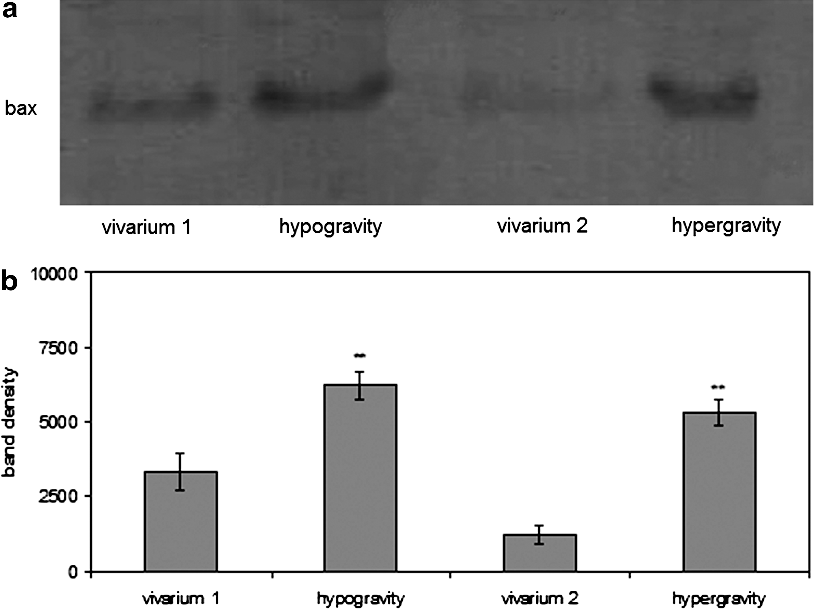
Comparison of Bax content by immunoblotting analysis. The study was performed by using a specific antibody. The position of the 23 kDa apparent molecular weight was indicated in comparison to the position of molecular size standards. The area density was evaluated by densitometry scanning and analyzed with Scion Image program. Data represent the mean±SD of three analyses of one experiment for the hypogravity sample and of three analyses of three independent experiments for the other samples. Significance: **P<0.001 hypogravity versus vivarium 1. (“vivarium 1,” control for the hypogravity experiment; “vivarium 2,” control for the hypergravity experiment.)
4. Discussion
Although we were able to discern by way of FRTL-5 thyroid cells in culture the effect of the space environment on the involvement of SM metabolism in the change of cell function (Albi et al., 2010), there have been no reports of this effect in thyroid glands in vivo. Our data show clearly that in the control sample the SMase was localized specifically in the nucleus, whereas in hypogravity the enzyme moved from the nucleus and was homogeneously distributed in the cells. Hypogravity induced an overexpression of both enzymes with a diffuse localization of SMase. Apparently, the activity of SM-synthase1 increased more than that of SMase, but considering the activity in relation to the enzyme content, only SMase activity increased. Therefore, only the SMase of mouse thyroid was more active in hypogravity, which is similar to results obtained in vitro in FRTL-5 cells. This represents the first animal observation of the influence of weightlessness on SM metabolism. Out of the three animals sent to space inside the MDS on board the Shuttle Discovery and then transferred to the ISS, only one returned to Earth alive after the 91-day space mission. Usually, such a low number of experimental animals would be considered inadequate for conducting scientific analyses. But because of the exceptionality of the experimental substrate (animals kept in space for the longest time, to date) and the improbability that such an experiment will be repeated within a reasonable amount of time, we present the results as a case report. Given that we recently demonstrated that the changes in the thyroid cell membrane during spaceflight are due to weightlessness (Albi et al., 2011), we conjectured that the modifications of SM metabolism enzymes shown in this case could be due to the hypogravity and were therefore interested in a comparison of the results with those obtained under hypergravity conditions. It would seem that hypogravity and hypergravity would generate opposite results, but some key results of our study were surprisingly similar. In fact, the most relevant distinctions to be made are that in hypergravity no variation of expression of SMase and SM-synthase1 were found. The SMase translocated, however, from the nucleus to the cytoplasm as occurs in hypogravity; and its activity, calculated in relation to the band density of the enzyme, had similar values. This implies that, although the SMase was present in thyroid with different content in hypogravity and hypergravity, the change of gravity could be responsible for a molecular remodeling that could influence the cell fate. It is possible that the change of gravity induces functional proteins to trigger modifications in cellular response and therefore significantly modifies thyroid function. It is interesting to note that, in analogy to SMase, the level of Bax is similar in hypogravity and hypergravity as well. Thus, as the associated behavior of the two proteins was previously observed when FRTL-5 thyroid cells entered into a pro-apoptotic state at the reentry of a space mission (Albi et al., 2010), we speculate that the cross-talk between the two proteins was stimulated by the change of gravity. On the other hand, morphological analysis of thyroid follicles also revealed large differences in comparison to the control samples but significant similarities between the hypogravity and hypergravity conditions. This supports our hypothesis of the significant influence of gravity on the partitioning of relevant molecules within the cell membrane. Looking ahead, the quest will now be to understand the interplay between the lipid environment and protein activities to identify possible countermeasures that are effective in preserving the integrity of the thyroid structure and function.
Footnotes
Acknowledgments
This work has been partially supported by grants from Agenzia Spaziale Italiana (ASI).
Glossary
Apoptosis: process of programmed cell death.
Bax: pro-apoptotic Bcl-2 protein.
Cyclic adenosine monophosphate (cAMP): second messenger involved in signal transduction.
Lipid raft: liquid-ordered microdomain, formed by sphingomyelin with a high affinity for cholesterol, in a liquid-disordered phase of cell membrane.
Signal transduction: the process by which an extracellular molecule activates a membrane molecule, that in turn induces a response by acting on intracellular molecules.
Sphingomyelin metabolism: sphingomyelin, a major lipid constituent of cell membranes, important in cell signaling, is degraded by sphingomyelinase and produced by sphingomyelin-synthase.
Thyrotropin: or thyroid-stimulating hormone (TSH) released by the anterior pituitary gland, located at the base of the brain, that regulates the activity of the thyroid gland.
Abbreviations
FITC, fluorescein isothiocyanate; ISS, International Space Station; MDS, Mouse Drawer System; PBS, phosphate-buffered saline; PC, phosphatidylcholine; SM, sphingomyelin; SMase, sphingomyelinase; STS, Space Transport System; TRITC, tetramethyl rhodamine isothiocyanate; TSH, thyrotropin; TSHR, TSH receptor.