
Abstract
The Kanab Wonderstone is sandstone (Shinarump Member, Chinle Formation) that is cemented and stained with iron oxide. The iron-oxide cementation and staining in these rocks have been considered examples of the Liesegang phenomenon, but we will show that they comprise a microbially induced structure. The spacing of bands of iron-oxide stain follow the Jablczynski spacing law (wherein the spacing between bands of iron-oxide stain increases as one traverses a series of bands) characteristic of Liesegang. Bands of iron-oxide cement exhibit more variable spacing and exhibit a weak but significant correlation between band thickness and distance between bands of cement. The pore-filling cement contains morphotypes that are similar in size and habit to those exhibited by microaerophilic iron-oxidizing bacteria. Other disseminated iron-oxide mineralization occurs as rhombohedra interpreted to be pseudomorphs after siderite. We interpret the cement to be produced by microbially mediated oxidation of siderite (a typical early diagenetic mineral in fluvial sandstones). Iron-oxidizing bacteria colonized the redox interface between siderite-cemented sand and porous sandstone. Microbes oxidized aqueous Fe(II), generating acid that caused siderite dissolution. The iron-oxide cement is the microbial product of a geochemical drive for organization; whereas the iron-oxide stain is true Liesegang. Together, they comprise a distinctive microbially induced structure with high preservation potential. Key Words: Biosignatures—Iron oxides—Diagenesis—Iron-oxidizing bacteria—Shinarump. Astrobiology 15, 616–636.
1. Introduction
I
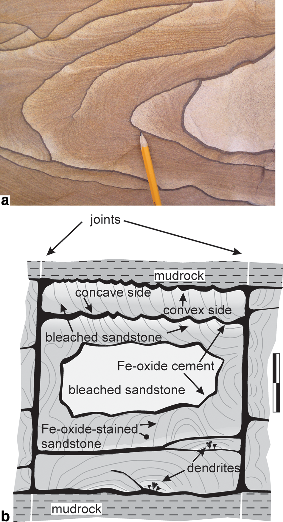
Characteristics of wonderstone. (
Ornate patterns of IOC and IOS in porous rocks, referred to here as wonderstone (Fig. 1), are widespread. Among the best-known examples are exposures of the Breathitt Sandstone (Middle Pennsylvanian, Kentucky, USA) (Fu et al., 1994), the Sharon Sandstone (Early Pennsylvanian, Ohio, USA) (Wells et al., 2003), the Hawkesbury Sandstone (Permian, New South Wales, Australia) (Hunt et al., 1977), the Umm Ishrin Sandstone (lower Paleozoic, Jordan) (Seilacher, 2001), and “wonderstone” (Fig. 1) from the Shinarump Member of the Chinle Formation (Triassic, Utah and Arizona, USA) (Hintze, 1986; Kettler et al., 2010). These features are not limited to sandstones; similar patterns of iron-oxide cementation and staining have been described in Cretaceous carbonate rocks (Marko et al., 2003; Serra et al., 2010), Miocene andesite sills (Jamtveit et al., 2011), and Cretaceous basalt (Singer and Navrot, 1970).
We have three reasons for studying the wonderstone phenomenon. (1) These features have long been considered to be enigmatic. The traditional explanation for these patterned rocks is that they are products of a geochemical self-organization process known as the Liesegang phenomenon (e.g., L'Heureux, 2013, and references therein; Hintze, 1986; Fu et al., 1994; Frankie and Jacobson, 2001; Seilacher, 2001; Marko et al., 2003; McBride, 2003; Pickett, 2003; Serra et al., 2010; Jamtveit et al., 2011; Wells et al., 2003; Wells et al. acknowledge but dispute the traditional Liesegang interpretation). Geochemical self-organization refers to the conversion of a geochemical system (e.g., rock and aqueous solution; silicate melt) from “an unpatterned to a patterned state without the intervention of an external template” (Ortoleva et al., 1987). Examples of geochemical self-organization include dissolution fingering at reaction fronts, oscillatory zoning in crystals, and orbicular granites (Ortoleva, 1994). The Liesegang phenomenon (Liesegang, 1896) is an example of chemical self-organization wherein bands of precipitate form in response to a migrating chemical front (e.g., Feeney et al., 1983; Antal et al., 1998). The interpretation of these patterned rocks as products of the Liesegang phenomenon does not, however, address the source of chemical components, the timing of pattern formation, or the fluid chemistry responsible for mineral precipitation at specific localities. (2) Mineral cements are archives in which records of fluid-mineral interaction are stored, a fact that has prompted considerable interest in the origin of carbonate and silicate mineral cements (e.g., Harwood et al., 2013; Tomkinson et al., 2013). IOCs have not, with recent exceptions (Chan et al., 2012, and references therein; Weber et al., 2012; Vasconcelos et al., 2013; Reiners et al., 2014), been subjected to the detailed study that is commonplace for carbonate or silicate cements. We are not aware, for example, of any attempts to test the Liesegang hypothesis of iron-oxide cementation in a specific formation. (3) The search for life on other planets has prompted an examination of lithological features that might be indicative of preexisting life. Ferric oxide minerals are highly insoluble and precipitate rapidly in oxidizing systems. They have been proposed as ideal recorders of ancient life and primary targets for sample return missions (Allen et al., 2001). Hofmann and Farmer (2000) presented images of filamentous fabrics collected from a number of different terrestrial environments and proposed that evidence of microbial activity could be preserved in a wide variety of settings on other planets. Krepski et al. (2013) examined the morphology of a number of iron-oxidizing bacteria. They noted that a number of morphological characteristics associated with these microbes, taken collectively, were strong indicators of biogenicity and were, therefore, biosignatures. Weber et al. (2012) were able to link biosignatures to some unusual iron-oxide mineralization in the Navajo sandstone and proposed that a variety of unusual concretions were the product of microbial activity. Among the more successful attempts to find large-scale biosignatures has been the description of microbially induced sedimentary structures (MISS) (e.g., Noffke, 2009). MISS are large (centimeter- to meter-scale) features produced by the interaction of microbial mats with unconsolidated sediment. The size of MISS allows them to be identified at the outcrop scale. MISS are, however, limited to sediments and sedimentary rocks by definition. MISS also can be vulnerable to erosion; many of the features form at the sediment surface and shallow subsurface. If the universe of microbially induced structures could be expanded to other rock types and to subsurface environments, exobiologists could greatly expand the range of rock types and past environments in which they might search for evidence of life profitably.
This paper focuses on the Kanab Wonderstone (or “Picture Rock”) from the Shinarump Member of the Triassic Chinle Formation. We selected this material because it is sold widely as a decorative stone (Doelling and Davis, 1989); the sandstone coasters are a familiar sight at geology conventions, rock and mineral shows, and household goods retailers. We were, therefore, able to gain access to both quarried sites and large blocks of quarried Shinarump sandstone that provided fresh exposures of the iron-oxide mineralization. We will present evidence to support three conclusions. (1) Wonderstone and similar-patterned IOC (iron-oxide mineralization that occludes pore space) and IOS (iron-oxide mineralization that coats mineral grains but does not occlude pore space) are products of evolving fluid-rock-microbe systems in aquifers. (2) A small amount of the iron oxide in the wonderstone pattern (the IOS) occurs as true Liesegang. This Liesegang forms as an integral part of the evolution of the fluid-rock-microbe system. (3) The Kanab Wonderstone can, therefore, be considered to comprise microbially induced structures. These microbially induced structures consist of thick bands of IOC precipitated as a consequence of microbial oxidation of iron and apparently abiotic Liesegang. They are similar to MISS in that they are centimeter- to meter-scale features interpreted to be evidence of preexisting microbial life but differ from MISS in that the mineral precipitates produced by the microorganisms are the distinctive features (cf. Noffke, 2009).
Those in search of extraterrestrial life must recognize that patterns in terrestrial rocks considered previously to be abiotic Liesegang include microbially mediated iron-oxide cementation. Moreover, when interpreted properly, these cements carry evidence of earlier diagenetic environments, directions of groundwater flow, and records of uplift and climatic conditions during uplift.
2. Geological Setting
The Shinarump Member of the Triassic Chinle Formation consists of conglomerates and sandstones that range from less than 1 m to 65 m in thickness (Hintze, 1986). The member is well known because it forms a resistant cap on the Moenkopi Formation in the Chocolate Cliffs of the Grand Staircase north of the Grand Canyon (Fig. 2) and the photogenic sandstone buttes and mesas of Monument Valley. It has also been explored extensively for uranium mineralization (Evensen, 1958). The detrital material that comprises the Shinarump was deposited in a northwest-flowing channel system that originated in the Grenville highlands in Texas (Dickinson et al., 2010). The Shinarump is underlain by the non-marine siltstones and shales of the Moenkopi Formation and is overlain by Chinle Formation mudstones.

Map of Shinarump Member exposures (black) in study area in southern Utah and northern Arizona. One portion of the Chocolate Cliffs east of Fredonia, Arizona, is referred to as the Shinarump Cliffs. Outcrop pattern after Billingsley et al. (2004, 2008), Sable and Hereford (2004), Hemphill (1956), and Morris (1957).
Wonderstone within the Shinarump Member (Fig. 1) consists of jointed blocks of sandstone that have been cemented and stained with iron oxide. The IOC (Fig. 1) occurs as bands as thick as 2 cm. The bands of IOC are undulatory and when viewed in detail comprise a series of cusps. We use the orientation of these cusps in combination with the orientation and distribution of the IOS to define convex and concave sides to the bands of IOC (Fig 1). The curved bands of IOS typically abut the convex side of the IOC and are asymptotic to the concave side of the IOC (Fig. 1). The convex sides of the IOC are notable for the occurrence of manganese oxide dendrites and sandstone bleaching, whereas sandstone abutting the concave side of the IOC is stained pervasively with iron oxide (Fig. 1). The sandstone bleaching may include apparent removal of IOS. The cores of some jointed blocks are completely enclosed by a single thick rind of IOC and bleached. Such bleached cores are noted only when the bands of IOC are convex toward the interior of the jointed block (Fig. 1).
3. Materials and Methods
Quarried blocks, exposures, and samples from a variety of locations in southern Utah and northern Arizona (Fig. 2) were examined. Fragments obtained by fracturing sandstone were cleaned with purified water and methanol and allowed to dry overnight at 314 K. The dried fragments were placed on a scanning electron microscope (SEM) stub, sputter-coated with a gold-palladium alloy, and stored in a desiccator. Samples were imaged on a Hitachi S4700 field-emission SEM at 5 and 10 kv at the Morrison Microscopy Core Research Facility at the University of Nebraska-Lincoln. Samples were also examined at 15 kV on the JEOL 5910LV SEM equipped with an energy-dispersive X-ray spectroscopy system at the Johnson Space Center.
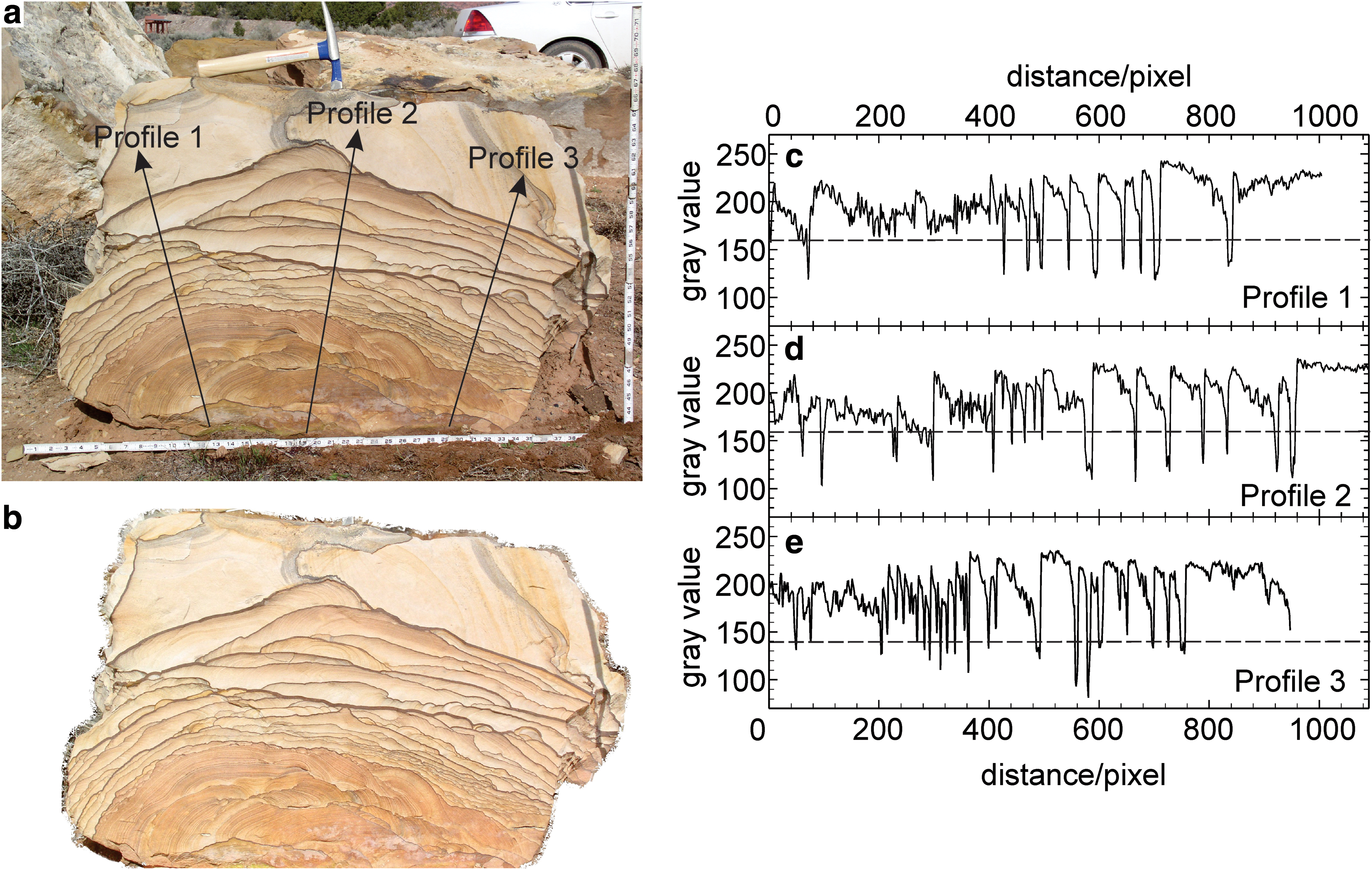
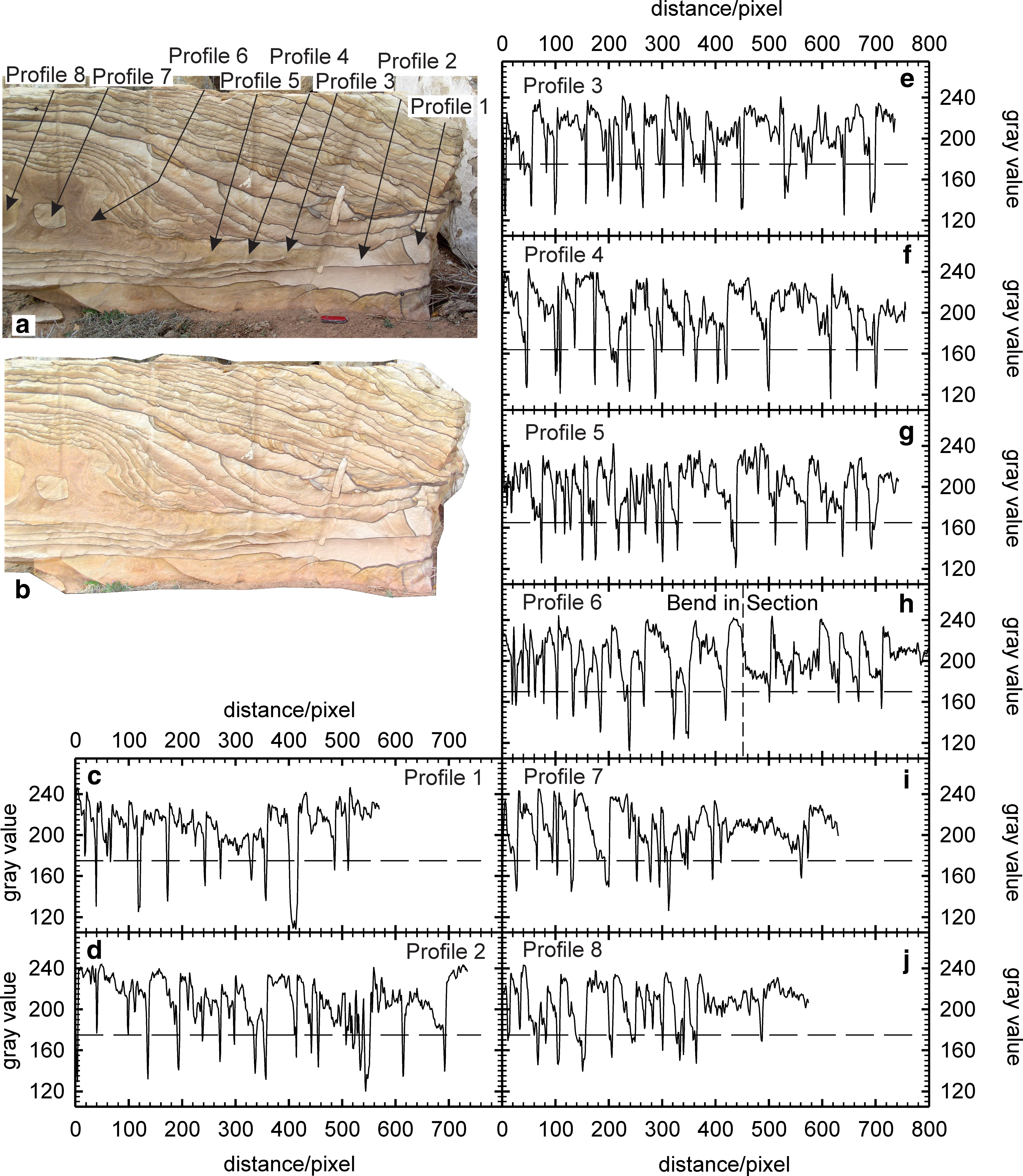
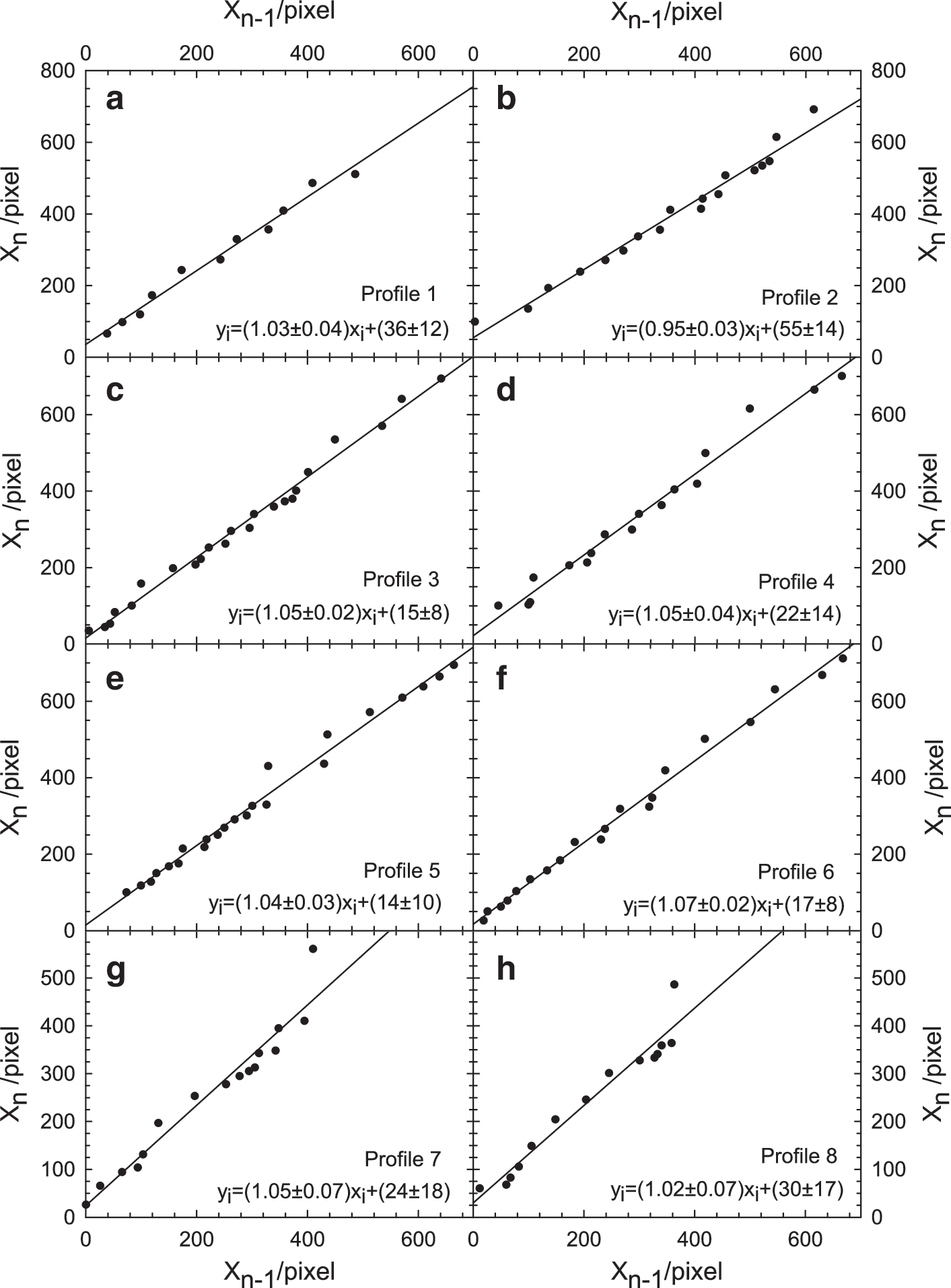
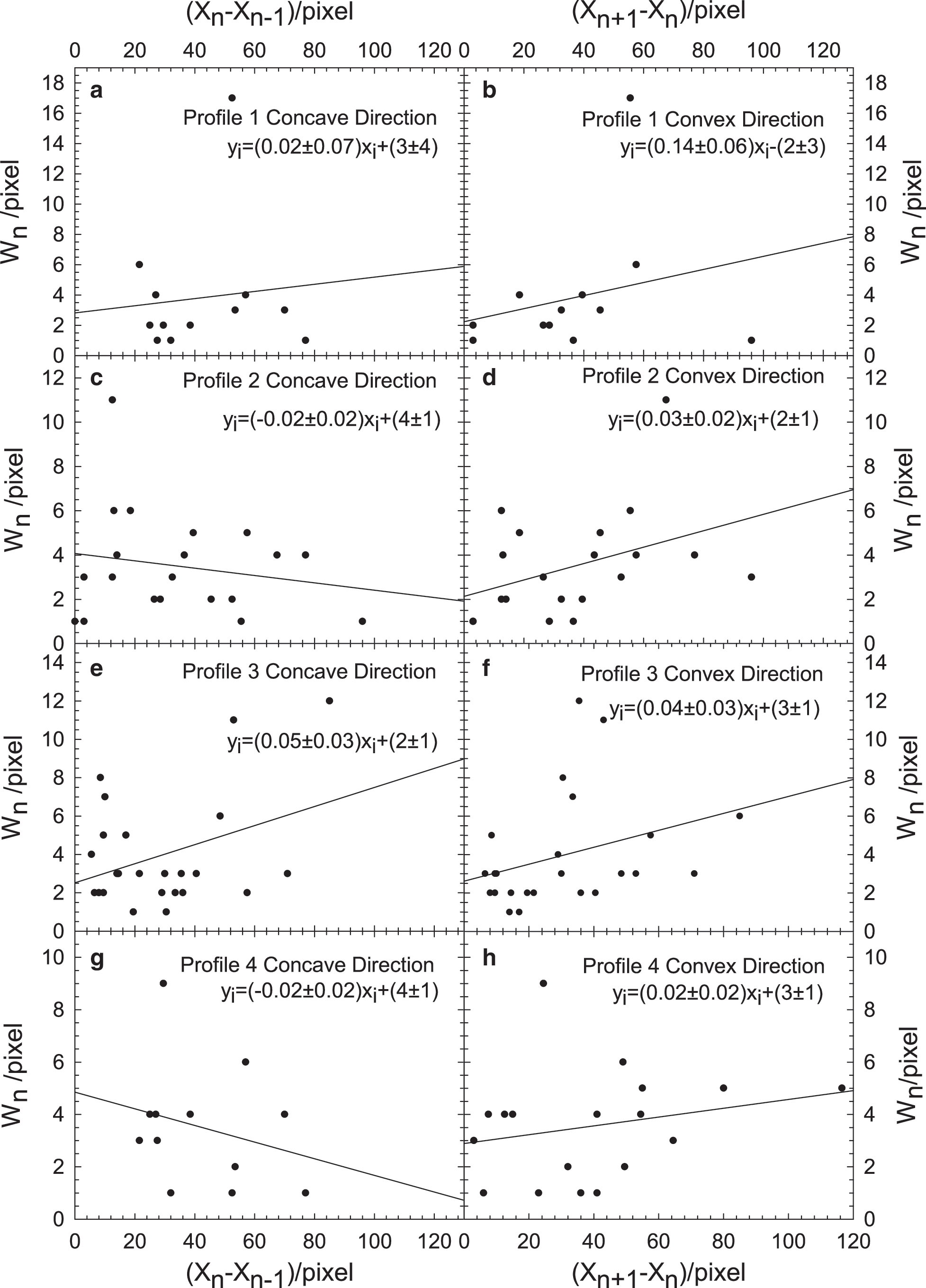
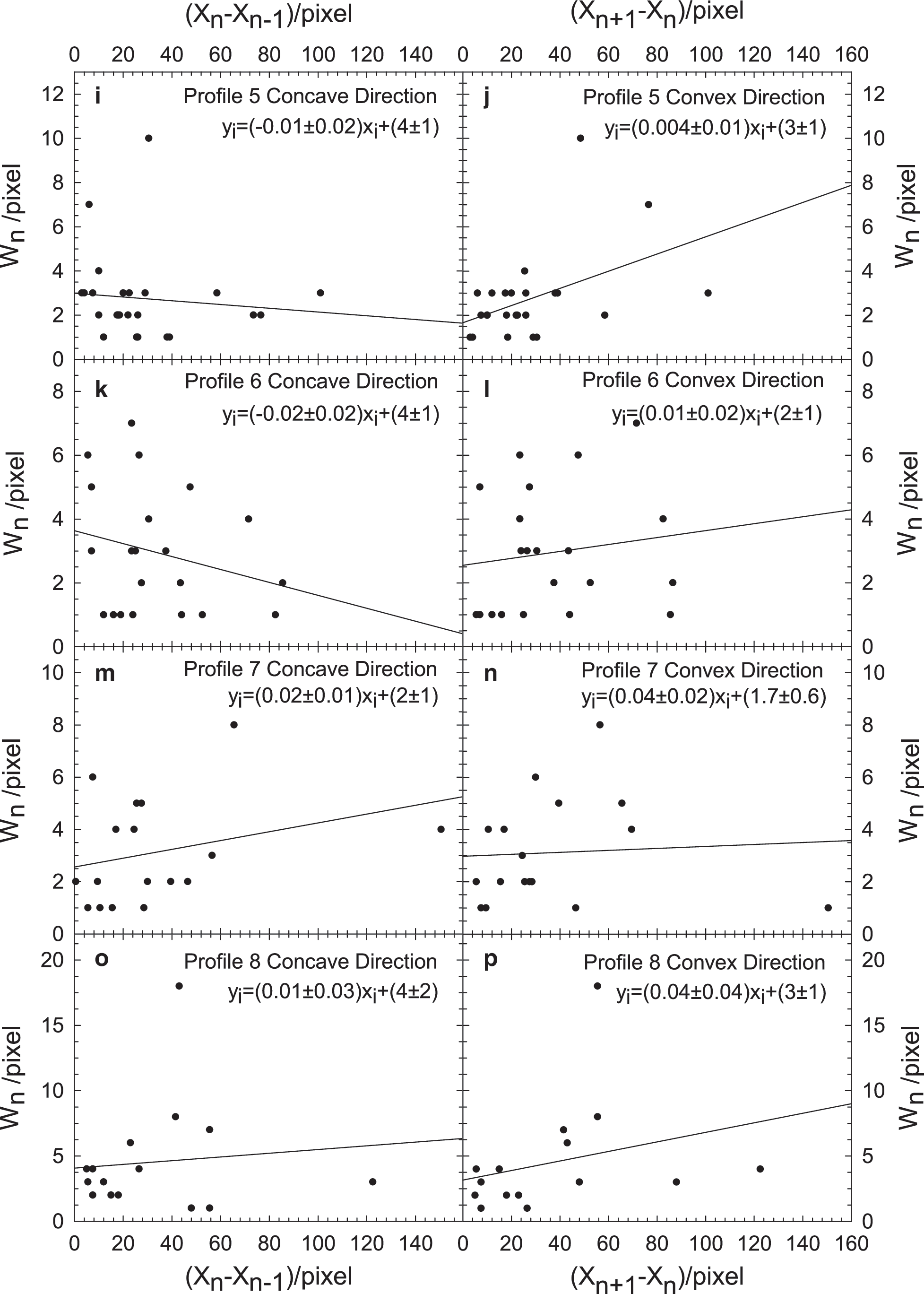
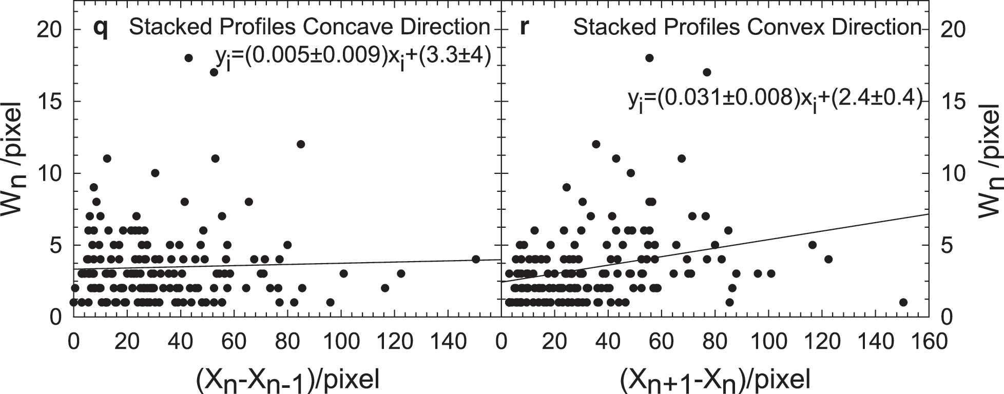
Images of quarried blocks of the Shinarump Member (Figs. 3a and 4a) were processed using Image J software (Schneider et al., 2012). A series of profiles were struck across each examined image. The profiles were oriented so that the pixel number increased in the “convex direction.” Images were processed by subtracting background using the light background option and rolling ball radius algorithm with radius set to 50 pixels (Figs. 3b and 4b). It was these processed images over which the profiles (Fig. 3c–3e and Fig. 4c–4j) were collected. A threshold gray value was then set for each profile by inspection to distinguish bands of IOC from areas of sandstone stained with iron oxide.
Analysis of Image P3180044. (
Analysis of Image P3190192. (
The value of Xn is taken to be the distance from the beginning of the profile to the center of the n th band. The width of the n th peak (Wn ) is defined as the number of pixels for which the gray value is less than the designated threshold (Fig. 3c–3e and Fig. 4c–4j).
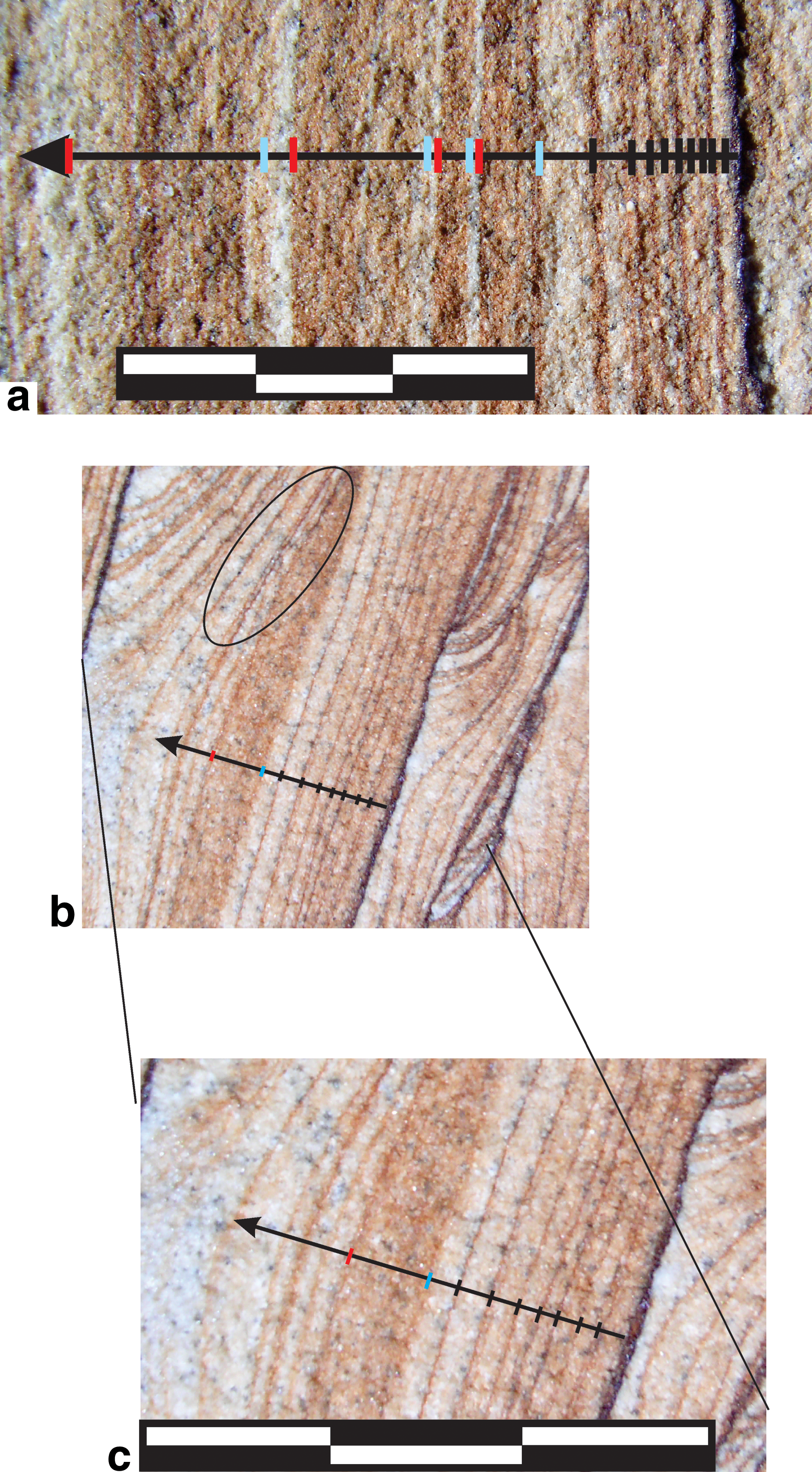
Profiles through bands of IOS (Fig. 5) were oriented to be orthogonal to bands of IOC. The bands of IOS were identified by inspection and the distance (Xn ) from the start of the profile to the middle of each band recorded.
(
The retrieved values of Xn and Wn were processed using least-squares linear regression. The analysis of variance (ANOVA) tables derived from this exercise are presented in Tables 1 –3. The column labeled DoF contains the number of degrees of freedom for the regression (1 independent variable), for the error (number of observations—2), and the total (the number of observations—2+1). The label SS designates the “sum of squares” and refers to the sum of squared deviations of the dependent variable about the mean (SS Total), the sum of squared deviations of each value held by the dependent variable from its predicted value given the value of the paired independent variable (sum of squares about the regression or residual sum of squares; SS Error), and the difference between SS Total and SS Error (sum of squares due to regression; SS Regression). The label MS designates “mean square” and is obtained by dividing each sum of squares value by the corresponding number of DoF. The t statistic (see Eq. 1.4.4 of Draper and Smith, 1966) is designated by T, whereas the F statistic (Eq. 1.4.10 of Draper and Smith, 1966) is designated by F. Guides to interpretation of ANOVA tables generated by linear regression can be found in the work of Draper and Smith (1966) or comparable resources.
Degrees of freedom.
Sum of squares.
Mean square.
T statistic for H 0: slope=1, H 1: slope>1.
See Table 1 for explanation of column headings.
H 0: slope=1, H 1: slope>1.
See Table 1 for explanation of column headings.
F statistic for H 0: slope=0, H 1: slope>0.
4. Results
4.1. Petrography
Examination with scanning electron microscopy revealed iron-oxide mineralization with a variety of habits. Pore-filling IOC comprises predominantly abundant plates with a crude hexagonal appearance that may be as large as 10 μm in long dimension (Fig. 6a). Locally, the iron-oxide grains have an acicular to lath-like habit and align to form platy aggregates (Fig. 6b). Spherical aggregates of iron-oxide needles comprise local discontinuous coatings on the surface of quartz grains and all other forms of iron-oxide mineralization (Fig. 6b–6c).
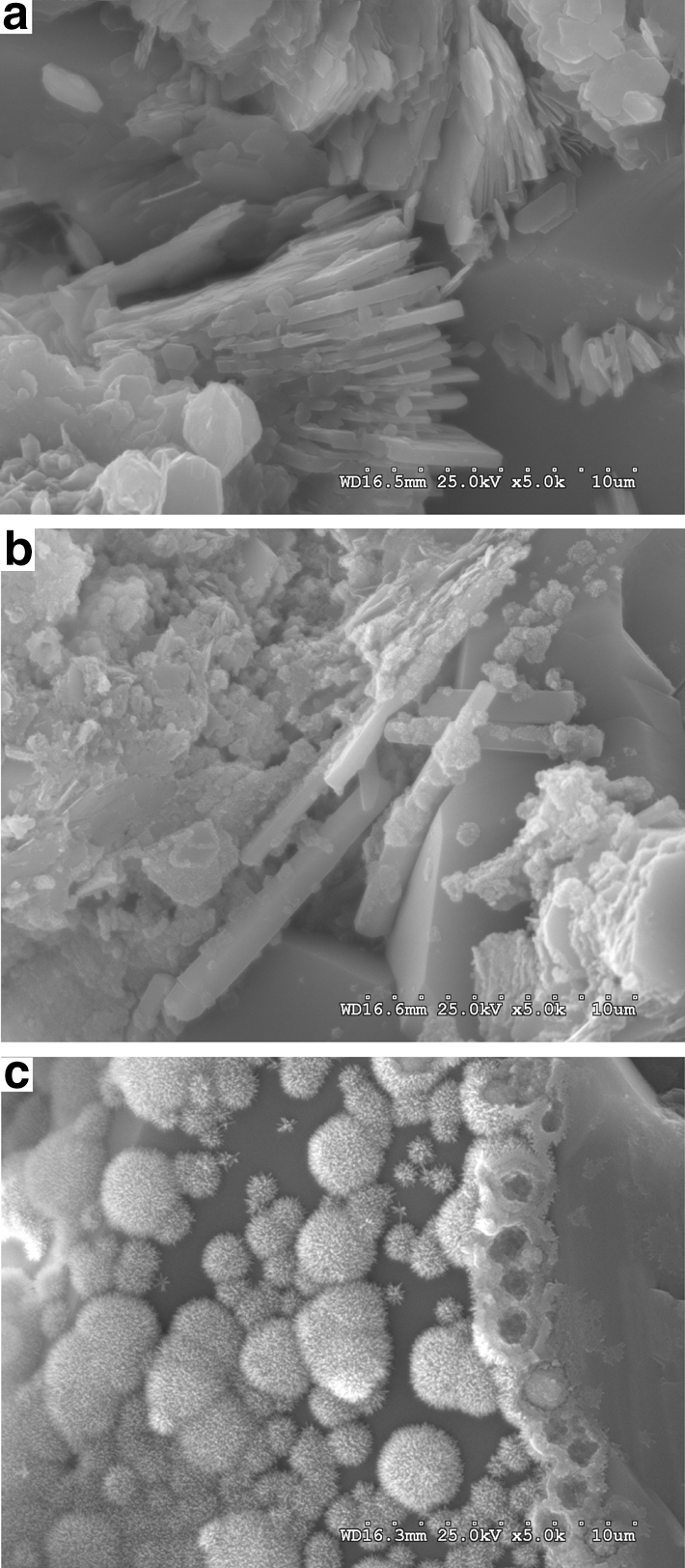
(
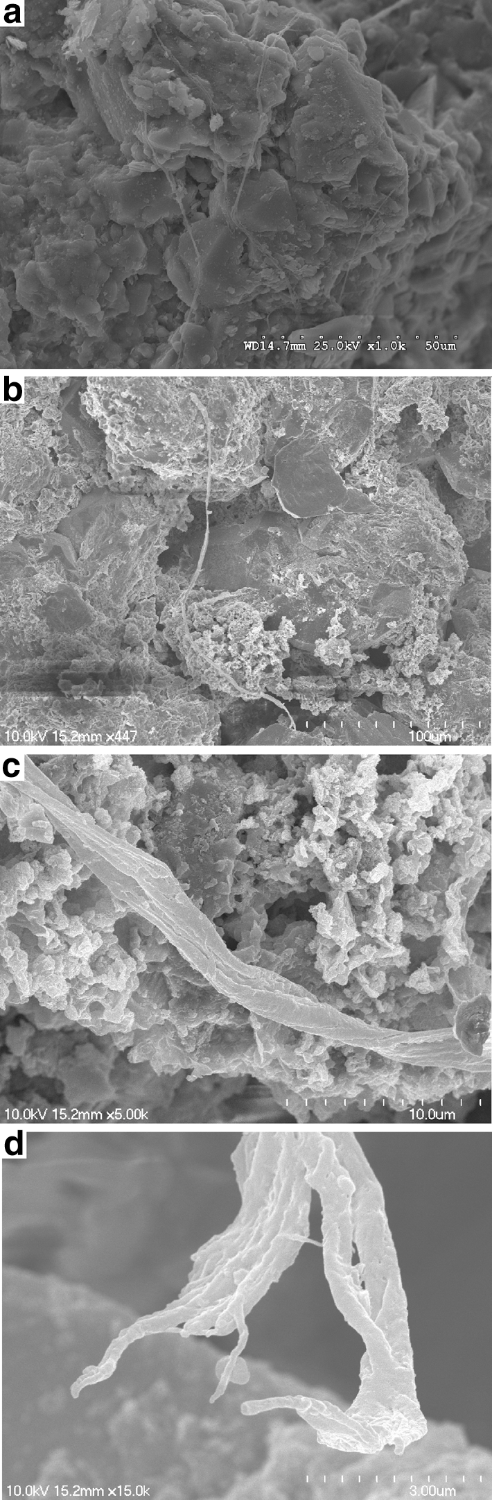
We also observed numerous twisted stalks and filaments (Fig. 7) near the edges of IOC. These stalks are as long as 200 μm and range from 0.7 to 2 μm in diameter. These filaments are locally overgrown with iron-oxide mineralization or trapped within the rock by quartz grains. They typically consist of 20 or fewer individual filaments that are twisted with helical angles ranging from 75° to 85° (Fig. 7c) and are ornamented locally with hexagonal plates. Some of the stalks branch on one end (Fig. 7d). Analysis of the stalks by wavelength-dispersive spectroscopy indicated that they consist of C, Fe, Si, and Al with local traces of Cu (Fig. 8).
(
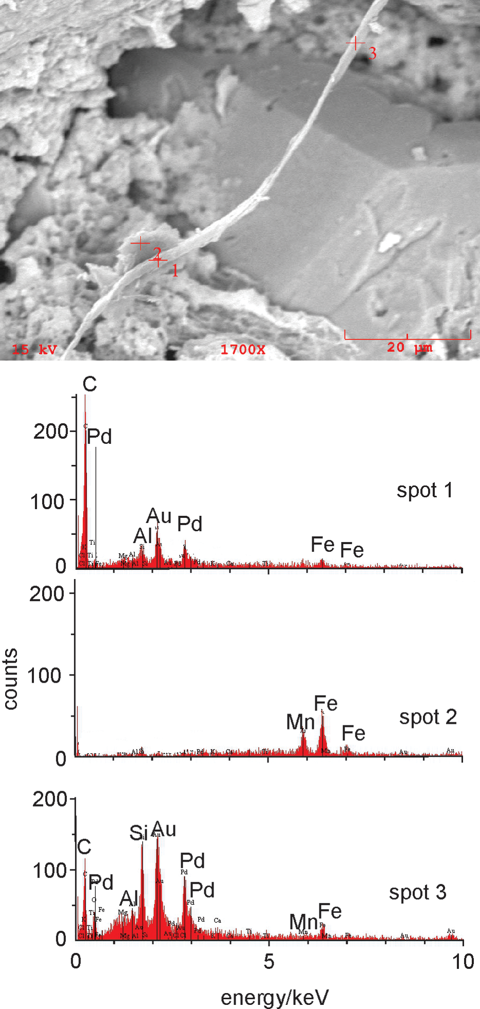
SEM image of portion of stalk shown in Fig. 7b with the energy-dispersive spectra generated by analysis of the spots labeled in the SEM image. Bar scale is 20 μm long. Note large euhedral quartz grain for reference. Numbered crosses indicate positions of electron beam during analyses. Spectra are presented as generated by the instrument after redrafting the axes and relabeling some peaks for clarity. Spot 2 is located off the stalk and detects only Fe and Mn, whereas the analyses located on the stalk (Spots 1 and 3) detect Fe, Mn, C, Si, and Al. The Pd and Au response is from the conductive coating placed on the samples before analysis. (Color graphics available at
In addition to the IOC and IOS, we also observe local disseminated iron-oxide mineralization in the Shinarump Member. Much of this disseminated mineralization comprises equant iron-oxide rhombohedra (Fig. 9) when viewed in thin section.
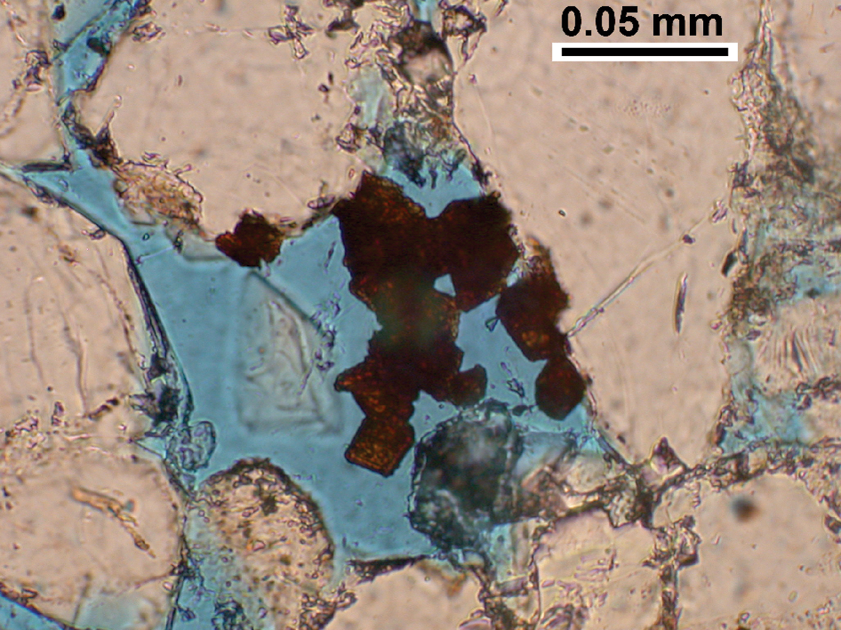
Photomicrograph of sample of sandstone from Shinarump Member. The large, light grains are quartz clasts, whereas the blue areas are pore space that has been filled with dyed epoxy. The reddish-brown rhombohedral grains in the center of the image are interpreted to be iron-oxide pseudomorphs after siderite. Image taken in plane-polarized light. (Color graphics available at
4.2. Spatial analysis of IOC and IOS
Iron-oxide mineralization similar to wonderstone has traditionally been interpreted as a type of periodic precipitate, typically referred to as Liesegang banding. Liesegang bands are distinctive because the spacing of the bands of precipitate is neither random nor even. Band spacing may increase or decrease as the distance from the solution interface increases (Ortoleva, 1984, p 289). However, so-called revert spacing, wherein the distance between bands decreases systematically as distance from the solution interface increases, is rare (Karam et al., 2011). The exact mechanism by which Liesegang bands form remains controversial (Henisch, 1988; Jamtveit and Hammer, 2012). For example, one model involves supersaturation of a pore fluid with the requisite mineral phase, followed by nucleation and growth of precipitate bands. Bands of precipitate then grow at the expense of potential nucleation sites on either side of the bands; solute is driven to the nucleation sites within bands by diffusion (e.g., Antal et al., 1998). In an alternative model, precipitate bands form as larger particles grow at the expense of smaller ones, dissolution and reprecipitation being driven by the lower surface free energy of the larger particles (e.g., Feeney et al., 1983). Experimental work has shown that nanoparticle precipitates will grow, re-dissolve, and form bands of coarser, re-deposited precipitate with the passage of time after two solutes have been introduced at a solution interface (Al-Ghoul et al., 2012).
We use geometric and morphological features of the IOC and IOS to test whether the Liesegang model can be applied to wonderstone. The hypothesis that the entire collection of iron-oxide cementation and staining in wonderstone is the product of the Liesegang phenomenon can be dismissed readily. In particular, the discordant orientation of the bands of IOC and IOS (Fig. 1), the asymmetric distribution of bleached and stained rock, and orientation of these elements with respect to the convex and concave sides of each band of IOC require that cementation proceeded in a specific direction. We therefore tested the hypothesis that the IOC and IOS were separate Liesegang systems.
Liesegang bands typically obey a number of laws with respect to the spacing between bands, band width, and the timing of band formation (e.g., Antal et al., 1998). The spacing law (or Jablczynski law) (Jablczynski, 1923) states that the ratio of the distances of two consecutive bands from the solution interface is given by
where Xn and Xn -1 are the distance of the n and n - 1 bands, respectively, from the interface between the two solutions, and p is a constant between 0.05 and 0.4.
We performed this analysis on IOC in blocks of Shinarump wonderstone (Figs. 3 and 4). No single solution front can be assigned a priori to any series of IOC bands present within any block of Shinarump sandstone. On the other hand, the ratio defined in Eq. 1 should still converge to 1+p if the distance is measured from any arbitrary point in a direction approximately orthogonal to the IOC bands if they are true Liesegang. If the bands are spaced randomly, then the value of p approaches zero and indicates that the Liesegang phenomenon is not a likely explanation for the band geometry and spacing.
Regressions of Xn on Xn -1 yield straight lines with slopes that approach 1 (Figs. 10 and 11). The ANOVA analyses of the regressions (Table 1) test the null hypothesis that the slope of each line is 1 against the alternative hypothesis that the slope is greater than 1. The null hypothesis cannot be rejected for any of the profiles (Fig. 10) obtained from Image P3180044 with an attained significance less than or equal to 0.05. The null hypothesis can be rejected for two (Profile 3 and Profile 6) of the eight profiles (Fig. 11) obtained from Image P3190192. The slopes of these two profiles are still very close to 1, indicating very small values for p. The value of p in Eq. 1 is, therefore, considered to be 0 for the bands of IOC in the wonderstone samples from the Shinarump Member. Because the distance between bands does not increase systematically in any direction, we conclude that the bands of IOC are not Liesegang.

(
(
In contrast to the IOC, coherent sets of IOS do follow the Jablczynski spacing law and are likely true Liesegang. Two examples of bands of IOS that parallel bands of IOC are shown in Fig. 5. The bands of IOC are taken as the origins for profiles orthogonal to the bands. The spacing law can be tested by measuring the distance from the midpoints of each of the bands to the origin. These results are used to test the spacing law in Fig. 12 with the results of the regression of Xn on Xn -1 presented in Table 2. The T statistic presented in Table 2 is calculated for testing the null hypothesis that the slopes of the regression lines are 1 compared to an alternative hypothesis that the slopes are greater than 1. In both cases, the null hypothesis can be rejected in favor of the alternative hypothesis with an attained significance less than 0.01. The calculated slopes for the two regression lines are 1.3 and 1.44, values that are consistent (within assigned error) with the Jablczynski spacing law (Antal et al., 1998).

(
4.3. Test of IOC band thickness versus distance between bands
Inspection of a number of blocks of wonderstone led us to conclude that the thicker bands of IOC were associated with larger intervals between bands of IOC. This relationship is only evident, however, if one moves across the block in the direction in which the IOC bands are convex. We tested this relationship by regressing Wn against Xn - Xn -1, where W is the width of a particular band of IOC (Figs. 13 and 14). The null hypothesis that there is no relationship between band width and distance between bands cannot be rejected (with attained significance <0.05) when distance is measured in the concave direction (Table 3). On the other hand, when the analysis is performed in the convex direction (Wn regressed against Xn +1 - Xn ; Figs. 13 and 14), the null hypothesis can be rejected (with attained significance <0.05) for Profile 3 from Image P3180044 and Profiles 1 and 5 from Image P3190192. Each of the profiles from an individual block should examine the same set of IOC bands. The results, therefore, can be combined to minimize random error in a fashion similar to stacking of seismic data to increase signal-to-noise ratio (Robinson, 1985). After stacking the profiles for both images, the null hypothesis can be rejected with an attained significance less than 0.001 for both images (Table 3). Thus, there is a relationship between width of the IOC bands and distance between bands, but only when measured in the convex direction.

(
(
5. Discussion
Any interpretation of the wonderstone pattern must accommodate the five observations and conclusions as follows: (1) The bands of IOC are not Liesegang. (2) The thickness of any band of IOC is related to the distance to the next succeeding band of IOC, but only when proceeding in the convex direction. (3) The bands of IOC contain petrographic evidence of microbially mediated precipitation of iron oxide. (4) The bands of IOS are Liesegang. (5) One can find local patches of IOC consisting of rhombohedra that we interpret to be pseudomorphs after siderite.
Our interpretation of the Shinarump wonderstone is that the sandstone was cemented locally with siderite during early methanic diagenesis. The sandstone was fractured by joints during Basin and Range tectonism and infiltrated by oxygenated groundwaters during uplift of the Colorado Plateau. Iron-oxidizing bacteria colonized the redox interface between the oxidizing groundwaters and siderite cement. Aqueous Fe2+ that diffused to the stabilized redox interface was oxidized by the microbes and precipitated as iron oxide.
The insolubility of ferric hydroxides requires that iron be transported in aqueous solution either under reducing or strongly acidic conditions. We have found little evidence for strongly acidic conditions in the Shinarump. Although pyrite is ubiquitous in sedimentary systems, the depositional environment of the Shinarump is not favorable for deposition of abundant pyrite. The Shinarump is a non-marine sandstone sandwiched between the continental red beds of the Moenkopi Formation below and those in other members of the Chinle Formation above. The sandstones contain little evidence of acid-sulfate weathering. Minerals such as jarosite or alunite are absent. Some pyrite remains preserved within the Shinarump and can be observed in outcrop to be oxidizing slowly. Cubic or pyritohedral iron oxides that might be produced by oxidation of pyrite are, however, rare. We propose that the iron accumulated locally in the Shinarump as siderite cement. The equant iron-oxide rhombohedra (Fig. 9) are direct evidence for the presence of siderite. Partially oxidized rhombohedral (“diamond-shaped”) siderite grains have been observed in ancient rocks (Lai et al., 2012), and siderite is a typical early diagenetic mineral in fluvial sandstones (Berner, 1981).
The interpretation that iron-oxide cementation proceeded in a specific direction was made based on the discordant orientation of the bands of IOC and IOS (Fig. 1) and the asymmetric distribution of bleached and stained rock relative to the IOC. This interpretation is confirmed by the relationship between IOC band width and distance between bands. This relationship is best explained if the iron that now resides in the bands of IOC was obtained from the region on the convex side of each band of IOC. We conclude, therefore, that the bands of IOC are not produced by solutes diffusing freely throughout the rock or the Ostwald ripening of nanoparticles distributed throughout the rock, but rather result from the oxidation of preexisting Fe-bearing minerals located only on the convex side of each band of IOC.
The iron-oxidizing bacterium Gallionella spp. “can be easily identified because of its distinctive stalk, which appears in the form of bundled fibers arranged in a double helix” (Hallberg and Ferris, 2004). Recent research supports that the distinctive helical stalk is a product of the physiology of microaerophilic iron-oxidizing bacteria and can be considered a biosignature for past iron-oxidizing metabolic activity (Krepski et al., 2013), which can also be preserved in porous medium (Weber et al., 2012). The morphology of the stalks and filaments observed here is identical to that described by Hallberg and Ferris (2004). The stalks have similar lengths, diameters, and numbers of filaments, and the filaments are twisted with similar helical angles. Although the individual filaments are slightly thicker than those observed in modern Gallionella spp., the aggregate diameter of the bundled filaments is comparable to that observed for both modern Gallionella spp. (Hallberg and Ferris, 2004) and documented examples of microfossils of Fe(II)-oxidizing organisms (Weber et al., 2012; Krepski et al., 2013). The branching shown in Fig. 7c and 7d is similar to that observed in stalks formed by Fe(II)-oxidizing bacteria. In those stalks, it is interpreted to result from cell division during stalk growth (Krepski et al., 2013). The composition of the filaments is similar to that reported for samples of modern Gallionella spp. (Ridgway et al., 1981; Kim et al., 2002; Suzuki et al., 2011). More importantly, carbon was detected only in the filaments; adjacent plates consist of only iron, manganese, and apparently oxygen (Fig. 8). The morphological and chemical evidence collectively comprise strong evidence that the filaments were produced biogenically.
The density and abundance of preserved filaments similar to modern Gallionella spp. (Fig. 7) are less than typically observed in modern seeps (cf. Fig. 2 of Emerson and Revsbech, 1994). The relatively low density of filaments may reflect the difference between a fossilized accumulation of iron oxide and a modern setting chosen because of the dense and abundant growth of iron-oxidizing bacteria. Even within a modern setting in which Gallionella stalks are abundant, such stalks are best observed on the top 1–2 mm of the mat. At greater depth within modern microbial mats, Gallionella stalks become progressively encrusted with particulate iron oxides (Emerson and Revsbech, 1994). We had greater success identifying twisted stalks on the periphery of the bands of IOC as opposed to the central portions of the bands. In these latter regions, pore space was filled completely with cement consisting largely of hexagonal plates of iron oxide (Fig. 6a and 6b). Furthermore, community succession during iron oxidation could also comprise a transition from Gallionella spp. dominance to a community dominated by another iron-oxidizing microorganism (Fleming et al., 2014). We have observed iron encrusted bacilli-like structures in IOCs previously (Weber et al., 2012). Given the distinctive morphology and evidence for preservation of Gallionella and similar species (e.g., Weber et al., 2012; Krepski et al., 2013), however, we focused on the twisted stalk morphology as a biosignature indicator for past metabolically active iron-oxidizing microbiota.
The composition, size, and overall morphology of the filaments illustrated in Fig. 7 are evidence that Fe(II)-oxidizing bacteria such as Gallionella spp. were present during oxidation of iron and precipitation of ferric oxyhydroxides. Iron-oxidizing bacteria are chemolithotrophs that oxidize ferrous iron to ferric iron following the reaction
Whereas some microbes can utilize aqueous CO2 as a carbon source (Hallberg and Ferris, 2004; Emerson et al., 2010), Gallionella spp. can utilize FeCO3 as a substrate and precipitate Fe3+ as ferric oxyhydroxide following the net reaction
The production of acid during the latter reaction is important for three reasons, as follows: (1) Production of acid in close proximity to the cell membrane has been proposed to increase the proton motive force and thus the energy-generating potential of the cell (Chan et al., 2004). (2) Excess acid is neutralized by dissolution of siderite tending to drive the reaction from left to right. This mechanism of siderite oxidation is very similar to one we proposed for iron-oxide concretions in the Navajo and Dakota Sandstones (Loope et al., 2010, 2011, 2012; Weber et al., 2012). (3) The iron oxide that is latest paragenetically comprises spherical and hemispherical aggregates of iron-oxide needles (Fig. 6c). This pin cushion habit is characteristic of schwertmannite (Bigham and Nordstrom, 2000), an iron-oxide phase that is stable under acid conditions (Majzlan et al., 2004). The precipitation of schwertmannite apparently reflects local exhaustion of carbonate and, consequently, depletion of the buffer capacity of the rock. Solution pH should decline as iron oxide precipitates, thus stabilizing schwertmannite. Although schwertmannite is associated typically with acid rock drainage, it is stable relative to ferrihydrite “over a wide range of pH when the system contains even a small amount of sulfate” (Majzlan et al., 2004).
Life thrives by exploiting systems that are out of equilibrium to obtain the energy necessary for catabolic and anabolic processes. Although most interpretations of patterned rocks do not explicitly consider the possibility that life was involved in the generation of the pattern (e.g., Singer and Navrot, 1970; Hintze, 1986; Fu et al., 1994; Frankie and Jacobson, 2001; Marko et al., 2003; McBride, 2003; Pickett, 2003; Wells et al., 2003; Serra et al., 2010; Jamtveit et al., 2011), biological systems (e.g., bacteria, Lacasta et al., 1999; concentric demyelination in the human brain, Khonsari and Calvez, 2007) are known to generate patterns that have similarities to Liesegang. How does the formation of the thick bands of microbially oxidized IOC differ from the formation of Liesegang bands? Each band of IOC began precipitation at a steep gradient in oxygen/aqueous Fe2+. This gradient was apparently stabilized by microbial activity. Each band of IOC represents a distinct iron-oxide precipitation event. Precipitation began when microbes began to exploit the gradient and ended when life could no longer preserve that gradient in space. The true Liesegang process, on the other hand, requires that solutes diffuse freely through the region where multiple bands of iron oxide may be precipitating simultaneously. It seems unlikely, therefore, that microbes would generate thick Liesegang bands of IOC.
Our interpretation of the Shinarump wonderstone is that the sandstone was cemented locally with siderite during early methanic diagenesis (Fig. 15). The sandstone was fractured by joints during Basin and Range tectonism and infiltrated by oxygenated groundwaters during uplift of the Colorado Plateau. Iron-oxidizing bacteria colonized the redox interface between the oxidizing groundwaters and siderite cement. The microbes oxidized aqueous Fe2+ that diffused to the stabilized redox interface. The IOS marks reaction finger fronts (Ortoleva, 1994) produced as acid diffused away from newly precipitated ferric oxyhydroxide toward the reduced, siderite-cemented sandstone (Fig. 15). Each band of IOC represents a stabilized redox interface between the oxidizing portion of the aquifer and the more reduced, siderite-bearing portion of the system. Eventually, each stabilized interface in turn became unstable. It is possible that the microbial system lost the capacity to occlude oxidizing waters, or the iron-oxidation reactions might have become less exergonic with local depletion of Fe2+ as the siderite front became more distant, or the accumulation of acid may have driven local pH values below the range in which iron-oxidizing bacteria could thrive. In any event, a new interface was established and colonized by microbes. Finally, the geometry of the interface was such that microbes were able to completely isolate a volume of rock (Fig. 15m–15n) until all the reduced iron within had been oxidized.
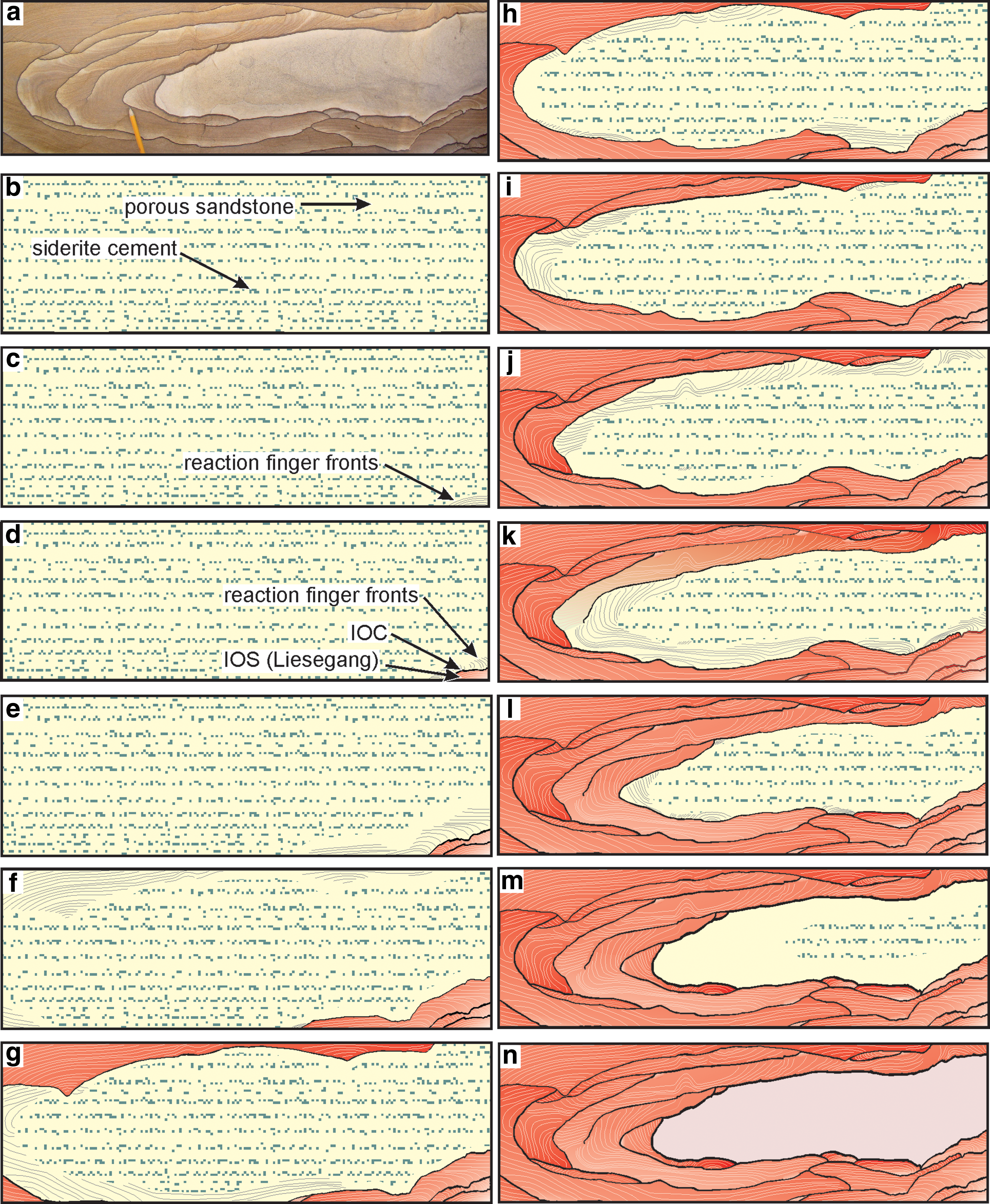
Image of wonderstone and a sequence of diagrams that illustrate development of the wonderstone pattern. Initial porous sandstone (yellow) is cemented locally by siderite [green in (b–m)]. IOC forms out of field of view in (c) from iron obtained by dissolution of siderite in lower right-hand corner. Arcuate lines in (c) represent reaction finger fronts (sensu Ortoleva, 1994). After abiotic oxidation of residual iron material in lower right corner to produce iron-oxide-stained rock (shades of red), another redox interface is established, and IOC (black) begins to accumulate in (d). The iron in this band of IOC was derived from the cement dissolved in the area above and to the left of the band of IOC. Panels (e–l) illustrate the continuing development of the bands of IOC and IOS. Panels (m–n) illustrate the isolation and complete bleaching of the iron in the interior of the diagram as the last band of IOC formed. (Color graphics available at
We interpret the IOS to have been produced during the breakthrough of oxygenated waters as the redox interface advanced within the rock. The interaction between residual Fe2+ absorbed on sand grains and invading oxygenated waters would mimic the geometry of the Liesegang phenomenon. In this interpretation (and in the absence of evidence for microbial activity in the precipitation of the IOS), the IOS is the product of apparently abiotic oxidation of Fe2+ that remains between a band of IOC and the siderite-cemented sandstone. Bleaching of IOS (Figs. 1 and 15) must occur after the IOS is formed and may be the product of iron-reducing bacteria obtaining electrons from the carbon fixed by the iron oxidizers.
6. Conclusions
The iron-oxide cementation and staining produced by the wonderstone phenomenon comprises a microbially induced structure (cf. MISS of Noffke, 2009). The presence of apparent local rhombohedral pseudomorphs of siderite is evidence that a reduced iron phase was present in the rock prior to infiltration by oxidizing groundwaters. Relationships between the width of IOC bands and interband distance are best explained by dissolution of siderite (FeCO3) with subsequent oxidation and accumulation of iron at a series of redox interfaces. The bands of IOC contain abundant iron-oxide-encrusted stalks similar in size and morphology to modern Gallionella spp. Although the spacing of bands of IOC is incompatible with typical examples of the Liesegang phenomenon, the spacing and width of IOS between bands of IOC are fully consistent with true Liesegang. The wonderstone pattern is, therefore, a microbially induced structure comprising microbially mediated iron-oxide cementation and apparently abiotic Liesegang.
These distinctive patterns have exobiological significance for five reasons, as follows: (1) Because reduced carbon is unstable in the presence of excess Fe(III), it has been argued that ferric oxide–bearing rocks are a poor choice for exobiological exploration (e.g., Sumner, 2004). The results obtained here are evidence that oxidized iron need not be considered a risk when exploring for life on other planets. On the contrary, if interpreted properly, patterns of iron-oxide mineralization could be used to guide selection of samples for analysis on site, as well as for sample return missions. (2) The patterns are megascopic evidence of subsurface microbial activity. Many of the wonderstone and so-called Liesegang features observed in the Shinarump and in other formations can be measured in meters. (3) Because rock cemented by iron oxide is more resistant to weathering and erosion than unconsolidated sediment, these features may be relatively easy to observe using remote methods. (4) The evidence that siderite formed on Mars is compelling (Niles et al., 2013; Tomkinson et al., 2013), and siderite would be expected to be the product of the interaction of carbon dioxide–charged fluids and iron-rich rocks (Niles et al., 2013). (5) Although siderite provides an optimal microbial substrate in that it combines a carbon source and electron donor, iron-oxidizing bacteria are known to thrive on other ferrous iron minerals (Emerson et al., 2010). These features may, therefore, occur in a wide variety of rock types.
Footnotes
Acknowledgments
We thank Dr. H. Chen for assistance with electron microscopy and Mr. Kenneth Brown for samples and field access. Support for this research was provided by the NASA Nebraska Space Grant program and the UNL Vice-Chancellor for Research. Paul Niles acknowledges support from the NASA Johnson Space Center and the Mars Fundamental Research Program. Conversations in the field with P. Reiners and B. Toro have improved our interpretation. Reviews of multiple versions of this manuscript by five anonymous reviewers have helped to improve the final product.
Author Disclosure Statement
The authors declare that they have no commercial associations that might create a conflict of interest in connection with this manuscript.