
Abstract
Planetary protection is governed by the Outer Space Treaty and includes the practice of protecting planetary bodies from contamination by Earth life. Although studies are constantly expanding our knowledge about life in extreme environments, it is still unclear what the probability is for terrestrial organisms to survive and grow on Mars. Having this knowledge is paramount to addressing whether microorganisms transported from Earth could negatively impact future space exploration. The objectives of this study were to identify cultivable microorganisms collected from the surface of the Mars Science Laboratory, to distinguish which of the cultivable microorganisms can utilize energy sources potentially available on Mars, and to determine the survival of the cultivable microorganisms upon exposure to physiological stresses present on the martian surface. Approximately 66% (237) of the 358 microorganisms identified are related to members of the Bacillus genus, although surprisingly, 22% of all isolates belong to non-spore-forming genera. A small number could grow by reduction of potential growth substrates found on Mars, such as perchlorate and sulfate, and many were resistant to desiccation and ultraviolet radiation (UVC). While most isolates either grew in media containing ≥10% NaCl or at 4°C, many grew when multiple physiological stresses were applied. The study yields details about the microorganisms that inhabit the surfaces of spacecraft after microbial reduction measures, information that will help gauge whether microorganisms from Earth pose a forward contamination risk that could impact future planetary protection policy. Key Words: Planetary protection—Spore—Bioburden—MSL—Curiosity—Contamination—Mars. Astrobiology 17, 253–265.
1. Introduction
T
One of the primary issues hindering assessment of the likelihood of the forward contamination of Mars is that there is not enough experimental information accumulated about the survival of Earth microorganisms in the martian environment. Over the last decade, a number of studies have reported on the microorganisms that are detectable in spacecraft assembly facilities during prelaunch preparations despite the precautions that were taken to reduce the microbial load (La Duc et al., 2004, 2007b; Kempf et al., 2005; Benardini et al., 2014). Previous studies testing survival of Earth microorganisms in simulated martian conditions have focused on spore-forming microorganisms, particularly bacteria of the Bacillus genus, since spores are hardy forms of terrestrial life that can survive Mars-like conditions (Horneck, 1993; Nicholson et al., 2000; Riesenman and Nicholson, 2000; Schuerger et al., 2003; Link et al., 2004; Newcombe et al., 2005; Tauscher et al., 2006; Zenoff et al., 2006; Fajardo-Cavazos et al., 2010). Several studies have reported on the survival of spore-forming Bacillus species isolated from clean room facilities after exposure of microbes to specific Mars-like environmental conditions (Setlow, 2001, 2006; Nicholson et al., 2002), but recent culture-independent analyses have shown that spore-formers are not the only types of microorganisms present in assembly and clean room facilities (Venkateswaran, 2001; La Duc et al., 2007a, 2009, 2012; Moissl et al., 2007, 2008; Vaishampayan et al., 2010, 2013). Some studies have focused on testing survival of extremophilic microbes that have the metabolic capabilities to inhabit Mars (La Duc et al., 2007a; Morozova et al., 2007; Gómez et al., 2010; Bauermeister et al., 2014), while a lesser number of studies have addressed survival of non-extremophilic, non-spore-forming microbes exposed to Mars-like environmental conditions (La Duc et al., 2007b; Osman et al., 2008; Berry et al., 2010). Few studies have investigated the isolates collected from spacecraft during mission preparations regarding their potential to contaminate Mars.
Protection of Mars is governed by international planetary protection policy, since microorganisms transported on the surface of spacecraft to Mars could hinder the search for past or present life on Mars. This policy restricts the number of microorganisms on a spacecraft's exposed surface areas, mated surface areas, and total encapsulated volume to a bioburden level of less than or equal to 5 × 10⁵ spores (Science Mission Directorate, 2011, NPR 8020.12D). During the preparation stages of a spacecraft such as the Mars Science Laboratory (MSL), a microbial sampling campaign is undertaken to verify the microbial bioburden requirement throughout the mission buildup and testing phases. The microorganisms are collected during the campaign using the NASA Standard Assay (Benardini et al., 2014). Isolates are heat shocked, plated on tryptic soy agar (TSA) at 32°C, enumerated, isolated, and preserved for future study. Although this does not take into account all microbial isolates present on the spacecraft, and leads to the enrichment of mostly spore-forming organisms, the preserved isolates do allow for the study of a selection of organisms residing on the spacecraft.
The goals of this study were to (1) identify microorganisms isolated and preserved from the surfaces of MSL and (2) investigate the potential of the microorganisms to survive specific physiological stresses and utilize energy sources available on Mars. The resulting information will be a step toward recognizing the potential threat of contamination of Mars by microorganisms transported on spacecraft.
2. Materials and Methods
2.1. Sample collection
Samples were collected and processed by the Jet Propulsion Laboratory (JPL) MSL Planetary Protection Implementation Team (Benardini et al., 2014). Cotton swabs and polyester wipes were used to collect samples (∼25 cm2 and 1 m2, respectively) from exterior surfaces of MSL. Swabs were then placed into water and gently sonicated to liberate microorganisms from the swab. Wipes were then submersed in PP rinse solution (NASA, 2010) and gently sonicated to liberate microorganisms from the wipe. Both swab and wipe extracts were heat shocked at 80°C for 15 min in a water bath and a subsample plated onto TSA. Plates were incubated for 3 days at 32°C. Colonies from TSA plates were re-streaked for purity onto the same media, and pure isolates were stored at −80°C in tryptic soy broth (TSB) amended with 50% glycerol. The glycerol stocks (350) were shipped to the University of Idaho for further studies.
2.2. Identification of isolates
A sample from the glycerol stocks was streaked onto TSA and incubated for 24–72 h, until colonies were present, at 30°C. Resulting colonies were re-streaked to ensure purity before proceeding with identification of isolates. Once pure, a colony was inoculated into TSB and grown overnight in a shaking incubator at 30°C at 250 rpm. Cells from grown cultures were collected by centrifugation at 4000g for 7 min. Chromosomal DNA was extracted from cells with a Wizard SV 96 Genomic DNA Purification System (Promega, Madison, WI) or an Ultra Clean Microbial DNA Isolation Kit (MoBio Laboratories, Inc., Carlsbad, CA). This method was only successful with cultures that did not form floating biofilms. For cultures where biofilm formation was an issue for DNA extraction using kits, the following method was used. Cell pellets were suspended in 250 μL of sterile dH2O. Cells were lysed by using three rounds of a freeze-thaw method (15 min at −80°C followed by 15 min at 80°C). Silica beads (1.0 mm) were added to the cells, and the suspension was mixed by vortexing for 30 s. Cell debris was pelleted by centrifugation at 2576g for 3 min, and 10 μL of the resulting cell lysate was used for amplification of 16S rRNA using PCR.
For each isolate, the 16S rRNA gene was amplified with universal bacterial primers 8F and 1525R (Reysenbach et al., 1994; Suzuki and Giovannoni, 1996), and PCR was performed as previously described (Smith et al., 2009). Briefly, reactions were 50 ul, and template DNA was either cell lysate (10 ul) or chromosomal DNA (1 ul) to a final DNA concentration of approximately 200 ng. PCR reagents were added to each tube in the following volumes: 25 μL of Dream Green Taq 2X Master Mix (Fermentas-Thermo Scientific, Glen Burnie, MD), 2.5 μL of 12.5 μmol primer 8F, and 2.5 μL of 12.5 μmol primer 1525R; water was added to bring the final volume up to 50 μL after the addition of cell lysate or chromosomal DNA. PCR conditions were as follows: an initial denaturation step at 94°C for 5 min was followed by 32 cycles of 95°C for 1 min, 51.4°C for 1.5 min, and 72°C for 1.5 min. Completion of the 32 cycles was followed by a final elongation step at 72°C for 5 min. PCR-amplified fragments were purified with Exonuclease I, 10 U, and Antarctic Phosphatase, 2 U, per 5 μL of PCR product. The reaction was heated at 37°C for 15 min followed by heat inactivation of the enzymes at 80°C for 15 min.
Polymerase chain reaction products were sequenced with the primer 27F (Lane, 1991) and a commercial sequencing service (Macrogen, Rockville, MD). The partial 16S rRNA gene sequences were analyzed by the University of Idaho rRNA analysis pipeline, HiSTA, as previously described (De Gelder, 2005). Briefly, sequences of 500 bp or longer were analyzed with BLAST (Altschul et al., 1997) to search for similarity among eubacterial type strains in the Ribosomal Database Project (RDP) (Cole et al., 2003). The RDP sequence for the closest relative of each input sequence was determined based on the highest sequence similarity. From results of the initial identification, isolates with potentially unique 16S rRNA sequences, or one representative of isolates with identical 16S rRNA sequences, were further sequenced to obtain a more complete 16S rRNA gene sequence with primers 27F, 518F, and 1492R (Muyzer et al., 1993; Frank et al., 2008). Contiguous sequences were assembled with Vector NTI Advance 11 (Invitrogen, Carlsbad, CA), and assembled sequences were reanalyzed as described above. Sequences are deposited in the NCBI GenBank under accession numbers KX453857–KX454155.
2.3. Anaerobic growth studies
Pure isolates were inoculated into 1000 μL of TSB in 96 well plates. Plates were incubated at 250 rpm at 30°C for 24 h. Cells were pelleted by centrifugation at 5000g for 12 min, and the supernatant was discarded. Cell pellets were suspended in 260 μL of phosphate-buffered saline (PBS) and inoculated into modified ATCC #2106 medium, without acetate or sodium chlorate, in an anaerobic chamber (Table 1). The media was boiled, autoclaved, and then placed in an anaerobic chamber for a minimum of 24 h prior to use. Sterile electron acceptor and carbon source concentrates were added to the medium to reach the desired concentrations prior to use. All tests were performed in triplicate. Growth with perchlorate, sulfate, or arsenate was determined by measuring turbidity at 600 nm at 0, 7, 14, 21, and 28 days. Growth of cultures with selenite or selenate was determined visually by the formation of a red precipitate (insoluble Se). Growth of cultures by the reduction of insoluble Fe(III) was determined visually by a color change of the solid-phase iron from brown to black.
2.4. Aerobic growth studies (temperature, pH, and NaCl)
Cells were grown overnight and suspended in PBS as described for the anaerobic studies. To study growth of isolates in medium containing NaCl, aliquots of cells were inoculated into TSB containing 0.5, 5, 10, or 20% (w/v) NaCl. To determine growth of isolates at alkaline pH, cells were inoculated into buffered TSB media at pH 7, 8, 9, 10, 11, and 12. Buffers (100 mM) used in the pH range of 7–10 were previously described by Nielsen et al. (1995), and buffers used at pH 11 and 12 were 1 M Na2HPO4 and 1 M KCl, respectively. Cells were grown at 30°C and turbidity monitored at 600 nm at 0, 1, 3, 7, and 14 days. To determine growth of isolates at low temperature, cells were inoculated into TSB, incubated at 4°C, and turbidity monitored at intervals of 0, 1, 3, 7, 14, and 28 days.
2.5. Desiccation studies
Isolates were inoculated into TSB in 96 well plates and incubated at 30°C overnight. Approximately 30 ul of cultures was added to 96-well flat-bottom plates, covered with a gas-permeable film, and placed in a biosafety cabinet overnight to allow evaporation of the medium to occur. The resulting plates were placed into a glass desiccation chamber containing silica gel desiccant for 14 days at room temperature (∼20°C) at Rh <10%. After 14 days of desiccation, plates were removed, and cells were rehydrated with 200 μL of TSB. Initial turbidity readings were recorded immediately after rehydration and again at 24, 48, 72, and 96 h to determine culturability of rehydrated cells.
2.6. Peroxide tolerance studies
Peroxide tolerance assays were performed by using a modified method of that described by Riesenman and Nicholson (2000). Cells were grown overnight in TSB and then transferred into PBS. Unopened bottles of hydrogen peroxide (30%) were purchased from the University of Idaho ChemStore on the day of use. Hydrogen peroxide was added to cell suspensions to a final concentration of 5%, and suspensions were incubated at room temperature with gentle mixing for 1 h. A 100 μL aliquot of the suspension was removed to a new 96-well plate, and 900 μL of bovine catalase (100 μg/mL) was added to the sample. After incubation for 1 h, treated cells (10 μL) were inoculated into 240 μL of TSB, and turbidity was monitored at 0, 24, and 48 h. Catalase activity was confirmed by placing cells on microscope slides, adding 3% and 5% H2O2, and observing for the formation of O2 bubbles (Lim et al., 2004).
2.7. UVC exposure studies
Ultraviolet C exposure studies were performed by using a modified method previously described by Marshall et al. (2003). All strains were grown in TSB and then plated onto TSA for UVC exposure studies. Single colonies of each strain were inoculated into 5 mL TSB and grown for 24 h at 30°C with shaking to a density of 108 to 109 CFU/mL prior to UVC treatment. Cultures were serially diluted in 10-fold steps to 10−5, and 10 μL of each dilution from 10−5 to 100 was spotted onto TSA in square grid plates. The spots were allowed to dry completely prior to UVC exposure, and control plates as well as treated plates were spotted in duplicate.
A UVP multiple-ray lamp (UVP, Upland, CA, P/N 95-0313-01) with a UVC bulb (Sankyo Denki G8T5) was utilized for all UVC treatments, and all treatments were conducted entirely in a dark room in order to minimize the effects of light-dependent repair mechanisms. Prior to treatment, the lamp was turned on and allowed to stabilize for approximately 20 min, after which lamp output was measured with a UVP MS-100 optical radiometer with a 254 nm sensor attached (P/N 97-0100-01 and 97-0099-01, respectively). Exposure time (in seconds) to achieve targeted fluence in J/m2 was calculated by dividing target dose by the meter reading (mW/cm2) multiplied by 10 (T = target fluence/[meter*10]). Plates were uncovered and exposed to UVC for the calculated exposure times to obtain the targeted dose, immediately wrapped with aluminum foil, and incubated at 30°C in the dark for 24 h. Resulting colonies were counted, and the survival fraction was calculated by comparing control and treated plates. Each experiment for all doses was conducted on a minimum of three independent occasions.
3. Results
3.1. Identification
Though ∼1100 isolates were collected from the MSL surfaces during the assembly and testing phases, only the 358 isolates collected from the JPL spacecraft assembly facility prior to May 2010 were used in this study. Of the 358 isolates, 339 were identified based on comparative sequence analyses of 16S rRNA genes. All were identified as bacteria, and the majority of the isolates were representatives of known spore-forming genera (78%), with most (85% of spore-formers) belonging to the Bacillus genus (Figs. 1 and 2). The most commonly identified Bacillus species was B. pumilus (16%), with B. amyloliquefaciens (11%) and B. megaterium (7%) comprising another 18% of the Bacillus population (Fig. 3). Overall, there were 26 different species represented within the Bacillus genus, with an additional 88 isolates that were not identifiable to the species level, due to partial sequence analysis. The remaining spore-forming isolates belonged to five genera: Paenibacillus (5%), Brevibacillus (1.5%), Sporosarcina (2%), Oceanobacillus (0.3%), and Gracilibacillus (0.3%). A number of microorganisms (22%) that were not related to spore-forming genera were identified within the pool of isolates. The most common non-spore-forming isolates comprising 10% of the entire microbial population belonged to the Staphylococcus genus, with seven different species represented (S. aureus, S. capitis, S. epidermidis, S. hominis, S. lugdunensis, S. pasteurii, and S. warneri). Of those identified to the Staphylococcus species level, more than half were identified as close relatives of S. epidermidis (43%) or S. warneri (17%). The non-spore-forming isolates were quite diverse, with over 19 genera represented, and included Acinetobacter (11%), Streptococcus (11%), Moraxella (5%), Leclercia (5%), Pseudomonas (4%), Enhydrobacter (2%), Leuconostoc (1%), and Stenotrophomonas (1%).
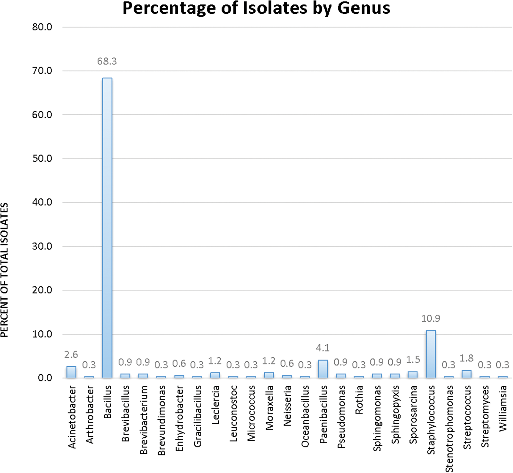
Isolates identified by genus.
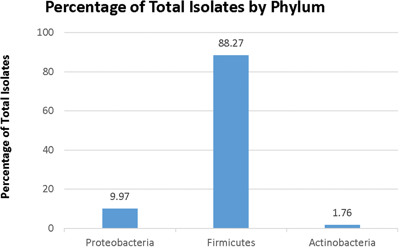
Isolates by phylum.

Bacillus isolates by species.
Four of the isolates identified represented novel species, based on a 16S rRNA sequence identity of <97% to any known microorganism. Two of the four isolates were most closely related to the Bacillus subtilis and Bacillus niabensis (85% and 89% identity, respectively). Two isolates were most closely related to the Brevibacillus borstelensis and Brevibacillus invocatus (87% and 93% identity, respectively), and the remaining isolate was most closely related to Enhydrobacter sp. KB3-12 (95% identity).
3.2. Anaerobic growth studies
All 358 isolates were tested for the ability to grow under anaerobic conditions by using a variety of terminal electron acceptors. Table 1 shows the various combinations of electron donors and acceptors that were tested and the number of isolates that grew under each condition. Of the 358 isolates tested, only 19 isolates grew under anaerobic conditions. Of the 19 isolates, 7 grew using perchlorate coupled with acetate or lactate as the electron donor. Five of the seven perchlorate-respiring isolates grew with lactate only, while the other two grew only with acetate. The isolates that could use perchlorate as a terminal electron acceptor were identified as either Bacillus (6) or Gracilibacillus (1). Two of the 19 isolates grew using sulfate as a terminal electron acceptor, and both isolates were identified as belonging to the Bacillus genus. Both isolates grew on sulfate using lactate only, and neither grew in medium containing acetate. Ten isolates grew with lactate and arsenate, but none of these isolates were able to grow in medium containing acetate in place of lactate. No isolates grew in medium containing selenite, selenate, or insoluble iron as the terminal electron acceptor, and no isolates grew using more than one of the terminal electron acceptors tested.
3.3. Aerobic growth studies (temperature, pH, and NaCl)
All isolates were tested separately for their ability to grow aerobically at 4°C and in the presence of high salt. Growth at 4°C was demonstrated by 132 (37%) of the isolates, which were identified as belonging to the Bacillus, Staphylococcus, Acinetobacter, Stenotrophomonas, Leclercia, Virgibacillus, Williamsia, Arthobacter, Gracilibacillus, or Streptomyces genera. Approximately 35% of the Bacillus species and 14% of the Staphylococcus species grew at the low temperature. There was minimal difference in the number of spore-forming isolates versus non-spore-forming isolates that grew at 4°C (Table 2).
Also shown are results from H2O2, desiccation, and UVC experiments. Ac = Acetate, Lac = lactate, For = formate, NT = not tested.
The majority of isolates (97% of the total) grew in medium containing 5% NaCl. Growth was apparent for 93% (333) of the isolates in media containing 10% NaCl, while only 22% (77) of all isolates grew in media containing 20% NaCl. Of the two most abundant genera, Staphylococcus and Bacillus, 63% of isolates identified as Staphylococcus could grow with elevated salt (20% NaCl), while only 17% of isolates identified as belonging to the Bacillus genus could do the same.
Isolates were tested for growth in media buffered within a pH range of 7–12. As expected, all isolates grew at pH 7, since this is the pH of the medium used in the NASA Standard Assay procedure. Of the 358 isolates, 80% (284) grew at pH 8, and 55% (197) grew at pH 9, while the number of isolates that grew in media buffered higher than pH 9 decreased significantly. Eleven isolates (3%) grew at pH 10, and only one isolate grew in media buffered to pH 11. None of the isolates grew in media adjusted to pH 12. The isolates that grew at > pH 10 were all spore-formers, with one isolate identified as belonging to the Paenibacillus genus and the remaining 11 isolates belonging to the Bacillus genus. Two isolates identified as Bacillus clausii grew in medium at pH 10, but only one of the isolates grew in medium at pH 11.
3.4. Desiccation studies
Isolates showing positive anaerobic or aerobic growth study results, novel isolates (<97% identity based on 16S rRNA gene sequences), or representatives from each clade, were chosen for desiccation studies. A total of 189 isolates were tested for their ability to survive a 2-week desiccation period. Isolates chosen for experiments consisted of representatives from 14 different genera, with 144 isolates representing spore-forming genera and 39 isolates from non-spore-forming genera. The identities of six of the isolates tested have not been determined. Of the isolates subjected to desiccation, 142 (75%) survived the 2-week desiccation period, while 15 isolates did not (Table 3). Desiccation survival on an additional 32 isolates could not be determined due to varying results. Of the spore-forming genera, 80% of the Bacillus isolates and 75% of the Paenibacillus isolates tested survived desiccation. One Gracilibacillus isolate was also tested and survived desiccation. Results obtained from non-spore-forming isolates were similar to those of spore-forming isolates. Of the non-spore-forming isolates, 25 (66%) survived desiccation, while 21% did not (Tables 2 and 4).
Total isolates tested = 189.
3.5. Peroxide tolerance studies
All isolates were tested for their tolerance to hydrogen peroxide by measuring survival after exposure to 5% H2O2 for 1 h. Approximately 19% (70) of isolates grew in TSB after exposure to 5% H2O2, some of which are included in Table 4. Both spore-formers and non-spore-formers representing eight different genera were tolerant to hydrogen peroxide, with 84% of the tolerant strains being spore-formers and 71% of those being Bacillus species. Other isolates tolerant to hydrogen peroxide included representatives from the following genera: Staphylococcus, Acinetobacter, Brevibacillus, Streptococcus, Paenibacillus, Moraxella, and Sporosarcina.
3.6. Isolates surviving exposure to multiple independent stresses
Table 4 shows that a small percentage (11%) of the isolates, 41 of the total, grew after exposure to more than one individually applied stress. For example, Bacillus isolate 236.1.1 grew in medium at pH 10, in media with 20% NaCl, and grew after exposure to hydrogen peroxide. Of the 41 isolates found to grow after exposure to multiple individual stresses, 31 belonged to spore-forming genera. Fourteen isolates survived at least two of the extreme conditions, and 20 isolates survived three of the four conditions shown in Table 4. Six isolates were found to survive all four individual stress conditions, with four identified as Bacillus and the remaining two representing Staphylococcus and Paenibacillus.
3.7. Survival to UVC exposure
Thirteen isolates, listed in Table 5, were chosen for UVC exposure survival, based upon their ability to grow after exposure to the physiological stresses used in this study (e.g., desiccation, hydrogen peroxide, high salt). All strains were initially irradiated at 100 J/m2. Four strains exhibiting >104 log survival were singled out for exposure to higher doses. All four isolates survived irradiation at 500 J/m2. Three of these survived exposure to 2000 J/m2 and exhibited the same level of survival from 500 to 2000 J/m2, the highest dose tested. The isolates were identified as being closely related to Bacillus amyloliquefaciens (isolate #160.2E), Bacillus megaterium (isolate #273.1.3), and an isolate most closely related to Moraxella osloensis (isolate #198.1.1B).
4. Discussion
All Mars-based landers and rovers are subjected to a regiment of microbial reduction protocols to limit the number of microorganisms transported from Earth into space to comply with the Outer Space Treaty. Despite the rigor of the bioburden reduction procedures employed, <5 × 10⁵ microorganisms as enumerated by the NASA Standard Assay (Science Mission Directorate, 2011, NPR 8020.12D) still persist on spacecraft. The purpose of this study was to identify microorganisms collected from MSL prior to launch and to characterize those isolates to determine their potential feasibility for survival in transport to and on Mars. The long-term goal is to obtain knowledge of the hardiness of isolates residing on spacecraft before deployment to space to assess whether a terrestrial-based microbe could potentially compromise a life-detection mission. The study is unique because it is the only set of comprehensive examinations of the survival of isolates, collected from MSL, exposed to stresses that simulate a Mars-like environment.
Until now, it was largely unknown what types of microorganisms populated the external surfaces of MSL. The assumption has been that the isolates were primarily of spore-forming genera, because samples of the MSL surface collected during various phases of assembly were subject to a brief 80°C heat-shock treatment before plating and enumeration, according to the NASA Standard Assay, which should have selected for non-spore-formers. However, our identification of the MSL isolates surviving the heat-shock treatment indicated that almost one-quarter of the isolates were non-spore-forming microbes. In total, the isolates represented 25 genera of bacteria and at least 65 different species, and four of the isolates appear to be novel. Since the isolates were collected after the NASA Standard Assay was performed, they do not represent the total community of microorganisms on the spacecraft but only those that survived the heat-shock treatment and grow under the selective conditions imposed. Thus, it is expected that there was an even greater diversity of microorganisms on MSL when the total bacterial, archaeal, and fungal community is taken into account (La Duc et al., 2014). Previous studies in which culture-independent techniques were used on samples collected from clean rooms of various NASA facilities have shown a wide variety of microorganisms present, but their activity has not been evaluated, so it is not clear whether all the microbes represented are viable (La Duc et al., 2004, 2009, 2012; Moissl et al., 2008; Vaishampayan et al., 2010). At present, international policy is primarily concerned with the number of spore-forming microorganisms/bacteria on board the spacecraft, since spores have a higher probability of surviving the trip to Mars.
Anaerobic growth studies indicate that a portion of the isolates are able to grow using potentially available energy sources on Mars. Tests performed by the Phoenix lander showed that perchlorate was present on the martian surface at concentrations of 2.1–2.6 mM (Hecht et al., 2009), and continued exploration of Mars has confirmed that perchlorate is widespread (Leshin et al., 2013; Ming et al., 2014; Clark and Kounaves, 2016). Seven of the MSL isolates grew using perchlorate as the sole terminal electron acceptor. Two of the isolates grew using sulfate as a terminal electron acceptor. Sulfate has been shown to be a major constituent of martian soils, and because sulfate systems exhibit high water activity, areas with sulfate salts are potentially more favorable to life (Crisler et al., 2012). Although neither selenium nor arsenic has been detected on Mars, various forms of arsenic and selenium are somewhat common on Earth. It is not unreasonable to expect that they may be present on Mars or other planetary bodies, and current thought is that arsenic may have been important to early life on Earth. It is well known that arsenic and selenium are respired by a number of terrestrial microorganisms to conserve energy (Stolz and Oremland, 1999; Zhu et al., 2014; Nancharaiah and Lens, 2015; Rascovan et al., 2016). Interestingly, this study identified 10 microorganisms that grew in media where arsenic was the only terminal electron acceptor available. Although detailed characterization of arsenic respiration was not a focal point of the current project, future work will investigate arsenic utilization by the isolates, as there is still much to be learned about arsenic metabolism in terrestrial microorganisms.
Tolerance to multiple chemical and physical conditions is critical if life is to survive on Mars. Studies have shown that salt tolerance would be required by microorganisms to survive and grow on Mars due to the high salt concentrations found in martian soils (Crisler et al., 2012). The majority of the isolates collected from MSL were able to grow in media containing elevated NaCl (93% of the isolates grew at ≥10% NaCl). It had previously been thought that the martian soil was likely to be acidic, but results by the Phoenix lander show that the soils were mildly basic with a pH of 7.7 ± 0.5 (Hecht et al., 2009). Most of the isolates from MSL (79%, 284 isolates) were able to grow in media at an elevated pH of 8, and 55% (197) of the isolates could grow in media at a pH ≥9, demonstrating that the majority of the isolates tested could survive and thrive in the more alkaline martian soils. Although the average temperature of Mars can range from −10°C to −76°C based on the Mars Pathfinder data, we chose to test all isolates at a baseline of 4°C, with the intent to perform extended experiments at lower temperatures in the future using the isolates that grew at this temperature (Schofield et al., 1997). In this study, 37% (132 isolates) of the isolates grew at 4°C, and future studies will test the resiliency of the 132 isolates to temperatures less than 4°C. Comparing the physical and chemical tolerances exhibited by all of the isolates tested, it is notable that 27% (97) of the isolates were able to thrive under multiple extreme conditions (≥4 of the conditions tested).
Although growth of the isolates in Mars-like conditions (e.g., anaerobic substrates, salt, temperature, and pH) provides a means by which to determine the potential for life to proliferate once on Mars, surviving exposure to harsh environmental stresses is equally important for the organism's survival both in transit to and on Mars. Over 75% (142) of the organisms tested in desiccation studies were able to survive desiccation for 2 weeks. In the future, further studies designed to determine the maximum length of time each of these organisms can survive desiccation will be undertaken, since an organism traveling on spacecraft to Mars would have to endure a 9-month trip without water before even reaching the environment to start growing. Twenty percent of the MSL isolates survived treatment with 5% liquid hydrogen peroxide, indicating that cleaning hardware during spacecraft assembly with a 5% solution of liquid hydrogen peroxide may not be the most effective microbial reduction method. It is noted that liquid hydrogen peroxide was not used during assembly of MSL as a microbial reduction method. Interestingly, it is reported that the surface ice of Europa contains as much as 0.13% hydrogen peroxide generated from the radiolysis of ice (Johnson et al., 2003). Microorganisms traveling to, and surviving on, distant planetary bodies like Europa would need to protect themselves from strong oxidants such as hydrogen peroxide. The ability of some of the isolates to survive oxidizing conditions (e.g., exposure to hydrogen peroxide) and UVC radiation suggests that they may be more likely to survive on a spacecraft destined for Mars or other planetary bodies.
Of the many species belonging to the Bacillus genus, the “gold standard” for testing microbial tolerance to UV exposure is Bacillus pumilus SAFR-032. Bacillus pumilus SAFR-032 was isolated from a spacecraft assembly facility during the time of the assembly of the Viking landers (Puleo et al., 1977). Bacillus pumilus SAFR-032 exhibits only a ∼2 log reduction in viable spores after exposure to 1000 J/m2 UVC (Link et al., 2004; Osman et al., 2008). It was unexpected that some of the MSL isolates from this study that were isolated from spacecraft in the spacecraft assembly facility would show resistance to UVC. Not surprising, three of the four isolates exposed to 500 J/m2 or higher were Bacillus species. The fourth resistant isolate is closely related to Moraxella osloensis, a member of a genus well known for having resistance to ionizing radiation and tolerance to UV at doses comparable to those endured by the extremely resistant Deinococcus radiodurans (Welch and Maxcy, 1975; Keller et al., 1982). Since isolates obtained from clean rooms must, by default, be resistant to numerous assaults resulting from the various cleaning methods applied throughout the assembly phases, it is not surprising that that some isolates survive multiple extreme environmental conditions including resistance to UVC radiation.
The results of the present study augment our current knowledge of the viable microorganisms present on, or associated with, spacecraft surfaces. The study also provides information regarding some of the physiological traits of those microorganisms that are likely required to grow on Mars, and reports on the ability of some of the isolates to survive exposure to extreme environmental conditions analogous to those expected on Mars. Currently, it is not known how the microorganisms residing on spacecraft that are inside clean room facilities adjust to repeated bioburden reduction applications. Nor is it known how the human-controlled environment may influence the overall evolution of the microbial population within this environment. The information collected from these types of studies will allow us to assess the current cleaning procedures of spacecraft components during spacecraft assembly and will provide information on the ability of surviving isolates to withstand extreme conditions similar to those present during space travel and on the martian surface. Future work with the isolates can provide us with new genomic and proteomic information that is invaluable for studies exploring the molecular mechanisms that may account for the stress resistances observed in many of the isolates.
Footnotes
Acknowledgments
This work was funded by the Idaho Space Grant Consortium through the NASA ISGC Research Fellowship Program and the ISGC Research Initiation Grant Program. Additional funding is provided by NASA EPSCoR award # NNX11AQ30A. Computational resources were supported by grants from the National Center for Research Resources (5P20RR016448-10) and the National Institute of General Medical Sciences (8 P20 GM103397-10) from the National Institutes of Health. The research was carried out at the Jet Propulsion Laboratory, California Institute of Technology, under a contract with the National Aeronautics and Space Administration. We wish to thank the non-authored MSL implementation team members, F. Morales, B., Koukol, M. La Duc, and G. Kazarians, for the collection of these isolates. We extended our gratitude toward K. Buxbaum, P. Grunther, J. Spry, and M. Jones for their encouragement and management support for the work conducted at JPL.
Author Disclosure Statement
No competing financial interests exist.