
Abstract
Microbial biofilms can lead to persistent infections and degrade a variety of materials, and they are notorious for their persistence and resistance to eradication. During long-duration space missions, microbial biofilms present a danger to crew health and spacecraft integrity. The use of antimicrobial surfaces provides an alternative strategy for inhibiting microbial growth and biofilm formation to conventional cleaning procedures and the use of disinfectants. Antimicrobial surfaces contain organic or inorganic compounds, such as antimicrobial peptides or copper and silver, that inhibit microbial growth. The efficacy of wetted oxidized copper layers and pure copper surfaces as antimicrobial agents was tested by applying cultures of Escherichia coli and Staphylococcus cohnii to these metallic surfaces. Stainless steel surfaces were used as non-inhibitory control surfaces. The production of reactive oxygen species and membrane damage increased rapidly within 1 h of exposure on pure copper surfaces, but the effect on cell survival was negligible even after 2 h of exposure. However, longer exposure times of up to 4 h led to a rapid decrease in cell survival, whereby the survival of cells was additionally dependent on the exposed cell density. Finally, the release of metal ions was determined to identify a possible correlation between copper ions in suspension and cell survival. These measurements indicated a steady increase of free copper ions, which were released indirectly by cells presumably through excreted complexing agents. These data indicate that the application of antimicrobial surfaces in spaceflight facilities could improve crew health and mitigate material damage caused by microbial contamination and biofilm formation. Furthermore, the results of this study indicate that cuprous oxide layers were superior to pure copper surfaces related to the antimicrobial effect and that cell density is a significant factor that influences the time dependence of antimicrobial activity. Key Words: Contact killing—E. coli—S. cohnii—Antimicrobial copper surfaces—Copper oxide layers—Human health—Planetary protection. Astrobiology 17, 1183–1191.
1. Introduction
T
The objective of the present study was to determine the efficacy of antimicrobial surfaces (pure copper and oxidized copper) as a potential preventive measure against microbial growth and biofilm formation aboard the ISS and for future interplanetary space missions. Therefore, a systematic study of high and low contamination levels (cells applied in a monolayer to multilayer) was conducted. Additionally, possible differences related to Gram-positive and Gram-negative cell wall structures were determined by applying the two microbial model organisms Staphylococcus cohnii (a Gram-positive representative) and Escherichia coli (a Gram-negative representative). Both represent human-associated bacteria frequently isolated from surfaces on the ISS (Novikova, 2004; Novikova et al., 2006; Hans et al., 2013). The relevance of antimicrobial surfaces for spaceflight is shown in this paper for the first time.
2. Materials and Methods
2.1. Bacterial strains and growth conditions
The human-associated microorganisms Escherichia coli K12 (DSM 498) and type strain Staphylococcus cohnii (DSM 20260) were used. Both were grown to stationary phase at 37°C in 10 mL medium. Escherichia coli was cultured in LB (Luria-Bertani: 1% tryptone, 0.5% yeast extract, 1% NaCl, pH 7.0) and S. cohnii in TSY medium (trypticase soy broth with yeast extract: 3% trypticase soy broth, 0.3% yeast extract, pH 7.3). Stationary-phase cells were washed twice by centrifugation for 15 min at 3000g in 10 mL of a 1:10 dilution of a phosphate-buffered saline (PBS) (0.7% Na2HPO4 × 2 H2O, 0.3% KH2PO4, 0.4% NaCl, pH 7.5) and resuspended in 10 mL of a 1:10 dilution of PBS for direct contact killing analysis.
2.2. Material and surface preparation
Square coupons of 2.25 cm2 were prepared from V2A stainless steel (AISI 304: X5CrNi18-10) and 99.99% rolled copper. The coupons were ground, polished (root mean square roughness <50 nm), and sterilized with 70% ethanol (Hans et al., 2013). The fabrication of the copper oxide layers (Cu2O and CuO) was carried out on polished ultrasonically cleaned copper coupons by thermal oxidation in a batch furnace (Carbolite) under atmospheric pressure (Fig. 1). The cuprous oxide layer of approx. 54 nm was generated at 200°C for 20 h and the cupric oxide layer of approx. 240 nm at 350°C for 150 min according to Hans et al. (2013) and Honkanen et al. (2008). After heat treatment, samples were immediately stored at room temperature under nitrogen.
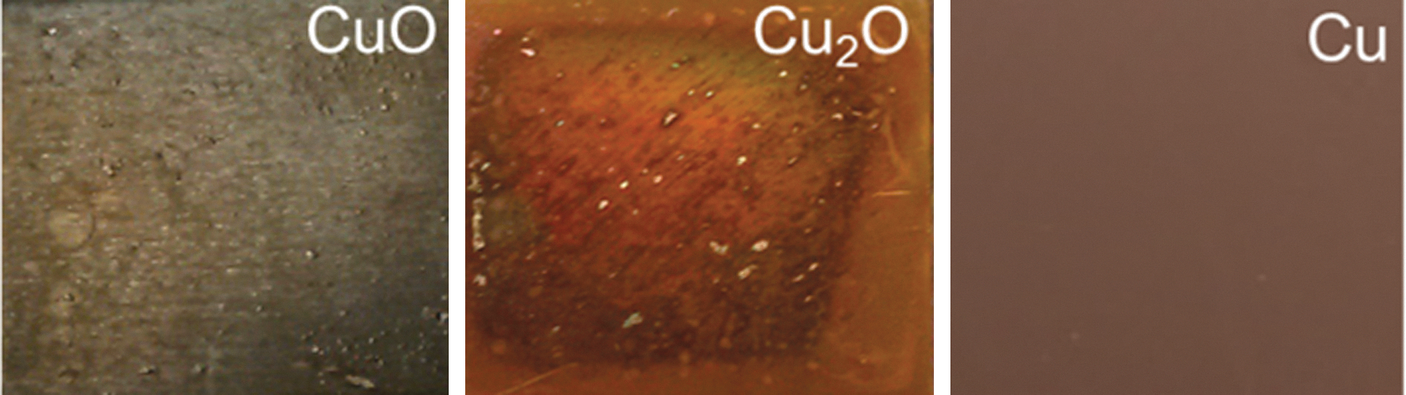
Photograph of a pure copper surface (Cu) and the copper oxide layers (CuO and Cu2O).
2.3. Scanning electron microscopic analysis
Scanning electron microscopic (SEM) analyses were conducted using 50 μL (1:10 dilution of PBS) washed overnight cultures with cell concentrations ranging between 106 and 109 cells/mL. Each of these 50 μL aliquots was pipetted on 0.38 cm2 stainless steel surfaces and immediately dried under laminar flowing air. Accordingly, 105 to 108 cells/cm2 were primarily fixed with 2% glutaraldehyde (Sigma-Aldrich Chemie GmbH, Munich, Germany)/100 mM Na-cacodylate buffer, pH 7.4, followed by fixation with 1% OsO4/100 mM Na-cacodylate buffer. Afterward, samples were dehydrated in an acetone series with subsequent critical-point drying over CO2 (CPD300, Leica Microsystems GmbH, Wetzlar, Germany), essentially as described by Rachel et al. (2010). Samples were finally sputter-coated with 2 nm Pt (Scancoat Six, Edwards, West Sussex, UK) and imaged at 15 kV (TM3000, Hitachi, Munich, Germany).
2.4. Survival after contact killing
Wet contact killing tests were performed as described by Molteni et al. (2010). Briefly, 70 μL of a washed cell suspension was exposed to 0.5 cm2 stainless steel (non-inhibitory control), oxidized copper layers, and pure copper surfaces for 0, 2, and 4 h at 30°C in a water-filled desiccator to ensure the maintenance of a relative humidity of 83%. According to the recommended exposure times, a 1:20 dilution of each cell suspension was prepared, followed by a 1:10 serial dilution. Appropriated dilutions were then spread on corresponding agar plates, followed by a determination of the number of colony-forming units (CFU). Related to the different generation times, the CFU were determined after 1 (E. coli) or 2 (S. cohnii) days of incubation at 37°C.
2.5. Measurement of membrane damage and reactive oxygen species after contact killing
The determination of membrane damage and reactive oxygen species (ROS) production by flow cytometry required greater cell volumes than used previously. In short, overnight cultures of E. coli were washed twice in a 1:10 dilution of PBS and higher volumes; that is, 400 μL of 108 or 106 cells/mL (corresponding to 107 or 105 cells/cm2) was exposed on 2.25 cm2 for 0, 1, 2, and 4 h on stainless steel (control surface) and pure copper surfaces. For LIVE/DEAD analysis, cells were removed after the corresponding times and centrifuged for 10 min at 4000g. The pellet was resuspended in 50 μL sterile filtered double-distilled H2O and stained as proposed by the manufacturer (LIVE/DEAD BacLight Viability kit, Thermo Scientific, Schwerte, Germany). After incubation, 900 μL of sterile filtered 1:10 dilution of PBS was added, and the fluorescence of cells was measured by flow cytometry (FACScan, BD Biosciences). CellROX Green (Thermo Scientific, Schwerte, Germany) was used to determine ROS production. Five hundred microliters of the exposed samples was stained as described by the manufacturer, and subsequently green fluorescence of cells indicating ROS production was measured with the flow cytometer (FACScan, BD Biosciences). The voltage of the FACScan diode/photomultipliers was set as follows: FSC: E01 (log), SSC: 375 V (log), FL1: 600 V (log), FL2: 150 V (log), FL3: 650 V (log), without compensation. For ROS production, FL1 was set to 800 V and FL3 to 150 V. All results were evaluated with Flowing Software 2 (Freeware by Perttu Terho, Turku Center for Biotechnology, University of Turku, Finland,
2.6. Determination of copper ion release
Four hundred microliters of a 1:10 dilution of PBS and 400 μL cell suspensions of 107 cells/mL (106 cells/cm2) were exposed to 2.25 cm2 stainless steel (control surface) and pure copper surfaces for 0, 1, 2, 3, and 4 h. Cu(I) and Cu(II) ions could not be discriminated by the inductively coupled plasma mass spectrometric (ICP-MS) measurements. Therefore, the total release of copper ions after the exposure of the cell suspension was determined. To ensure that only fragmented cells reached the nebulizer and to prevent its clogging, cells were lysed with either 10 μL lysozyme (10 mg/mL) in the case of E. coli or lysostaphin (5 mg/mL) in the case of S. cohnii and 10 μL DNAse (10 mg/mL) for 30 min at 30°C. Prior to the measurement, each sample (350 μL) was diluted 10-fold in double-distilled H2O, containing 150 μL 65% nitric acid and 3.5 μL of an internal standard (10 ppm Ho, 10 ppm Sc). The copper concentration was quantified with a detection limit of 10 ppt by using the ICP-MS 7500 Series, Agilent Technologies; operating parameters and analytical conditions were selected according to the work of Hahn et al. (2015).
2.7. Statistics
All tests were conducted in triplicates that were three individual cultures with three individual tests on each of the three cultures, and presented as a mean with standard deviations. Statistically significant differences were determined with the Student t test where p values <0.05 were considered as significant.
3. Results
3.1. Contact killing of multilayered and monolayered cells
As shown by SEM images, a multilayer of cells was formed after the application of 108 and 107 cells/cm2, whereas most of the cells were in direct contact with the surfaces when 106 cells/cm2 were applied (Fig. 2). Here, only a few cells were detected to be on top of other cells. A complete monolayer of cells was determined to occur when 105 cells/cm2 was applied.
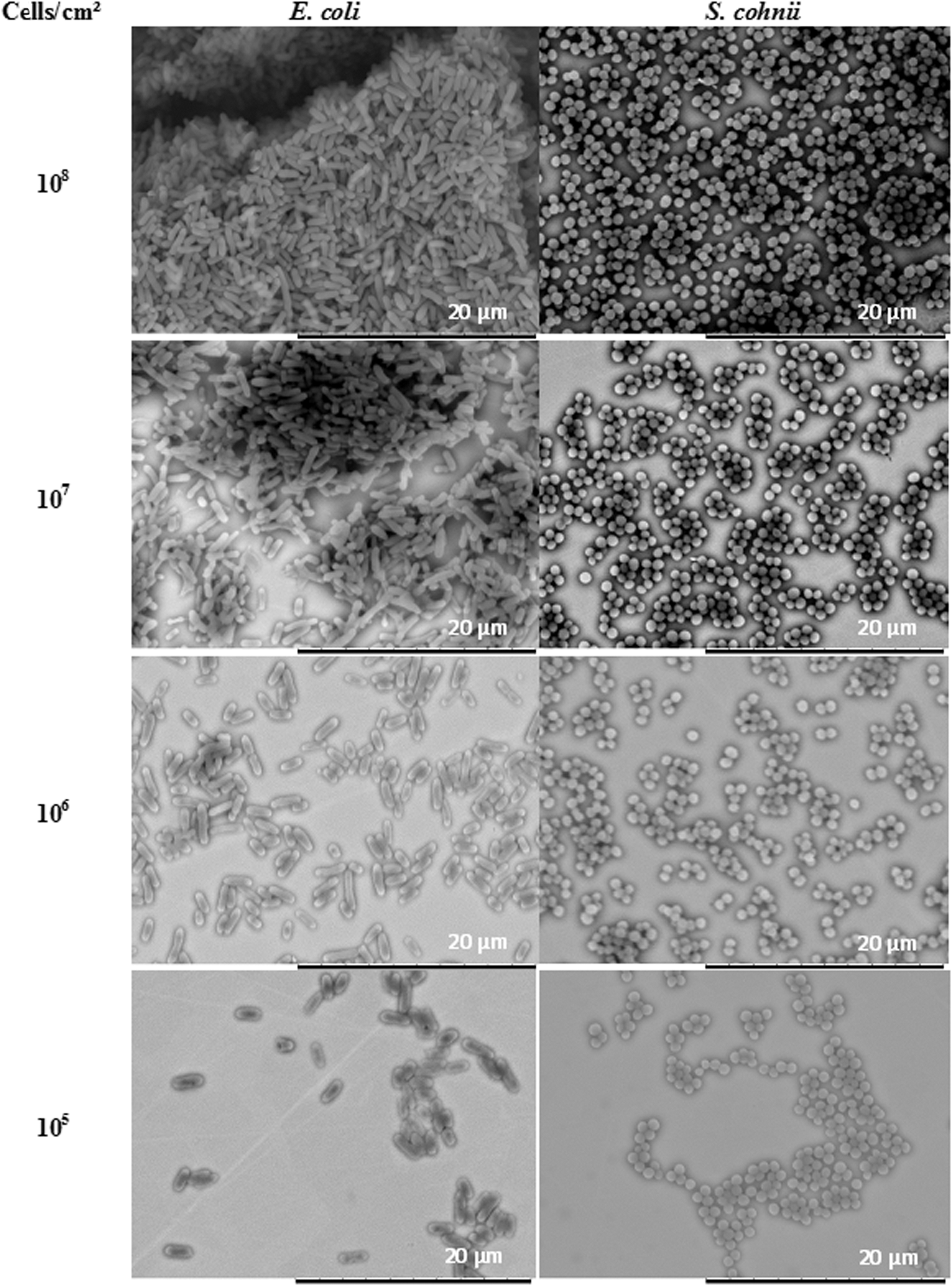
Scanning electron micrographs of 108 to 105 cells/cm2 of E. coli and S. cohnii applied in 1:10 dilution of PBS to stainless steel surfaces.
3.2. Antimicrobial activity of pure copper
The corresponding survival data after exposure of a monolayer or multilayer of cells on pure copper surfaces are presented in Fig. 3. After exposure of 108 cells/cm2 of S. cohnii and E. coli for 2 and 4 h on pure copper surfaces, no significant difference in cell survival was determined. The first significant reduction in cell survival of approximately 1–2 orders of magnitude was detected after exposure of 104 to 107 cells/cm2 for 2 h. Longer exposure of 4 h led to greater variances in cell survival. Exposure of 107 cells/cm2 led to a decrease in cell survival of 1.5 orders of magnitude for E. coli and 2 orders of magnitude for S. cohnii. The cell survival of 106 cells/cm2 of E. coli decreased by 2 and for S. cohnii by 2.6 orders of magnitude. After the exposure of 105 cells/cm2, cell survival in both strains was reduced by about 4.2 orders of magnitude, whereas no survival was detected for either organism after an exposure of 104 cells/cm2.
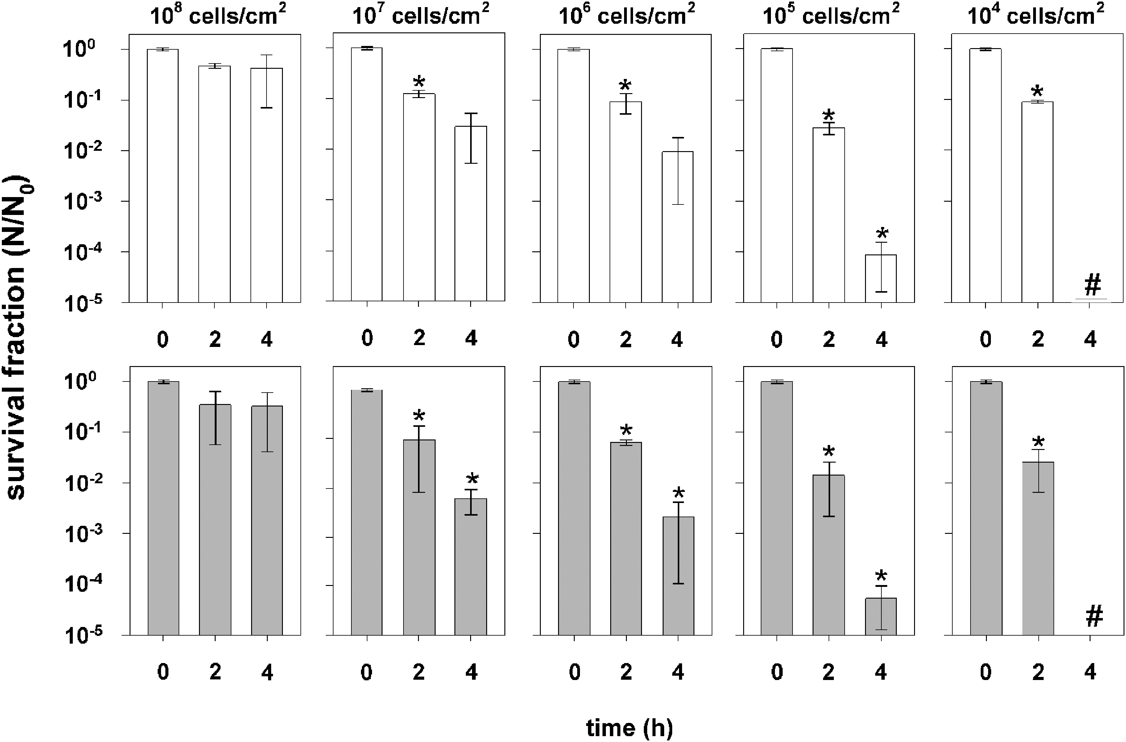
Survival of E. coli (white bars) and S. cohnii (gray bars) suspended in a 1:10 dilution of PBS after the exposure to stainless steel (N 0) and pure copper surfaces (N). Data are expressed as averages ± standard deviations (n = 3) as described in the text. * = Significant decrease in cell survival (p value <0.05); # = below detection threshold.
3.3. Antimicrobial activity of oxidized copper layers
After exposure for 4 h to CuO layers, cell survival decreased by 1 order of magnitude for E. coli and 1.5 for S. cohnii (Fig. 4). In contrast, cell survival decreased after 4 h of exposure on Cu2O layers by 4.5 orders of magnitude for S. cohnii and 5.5 for E. coli. This indicates that the most effective antimicrobial surface was the Cu2O layer, from which certainly Cu+ ions are mainly released, followed by the pure copper surface, whereas the majority of ions released from the CuO layer were Cu2+ ions, which may lead to less efficient contact killing.
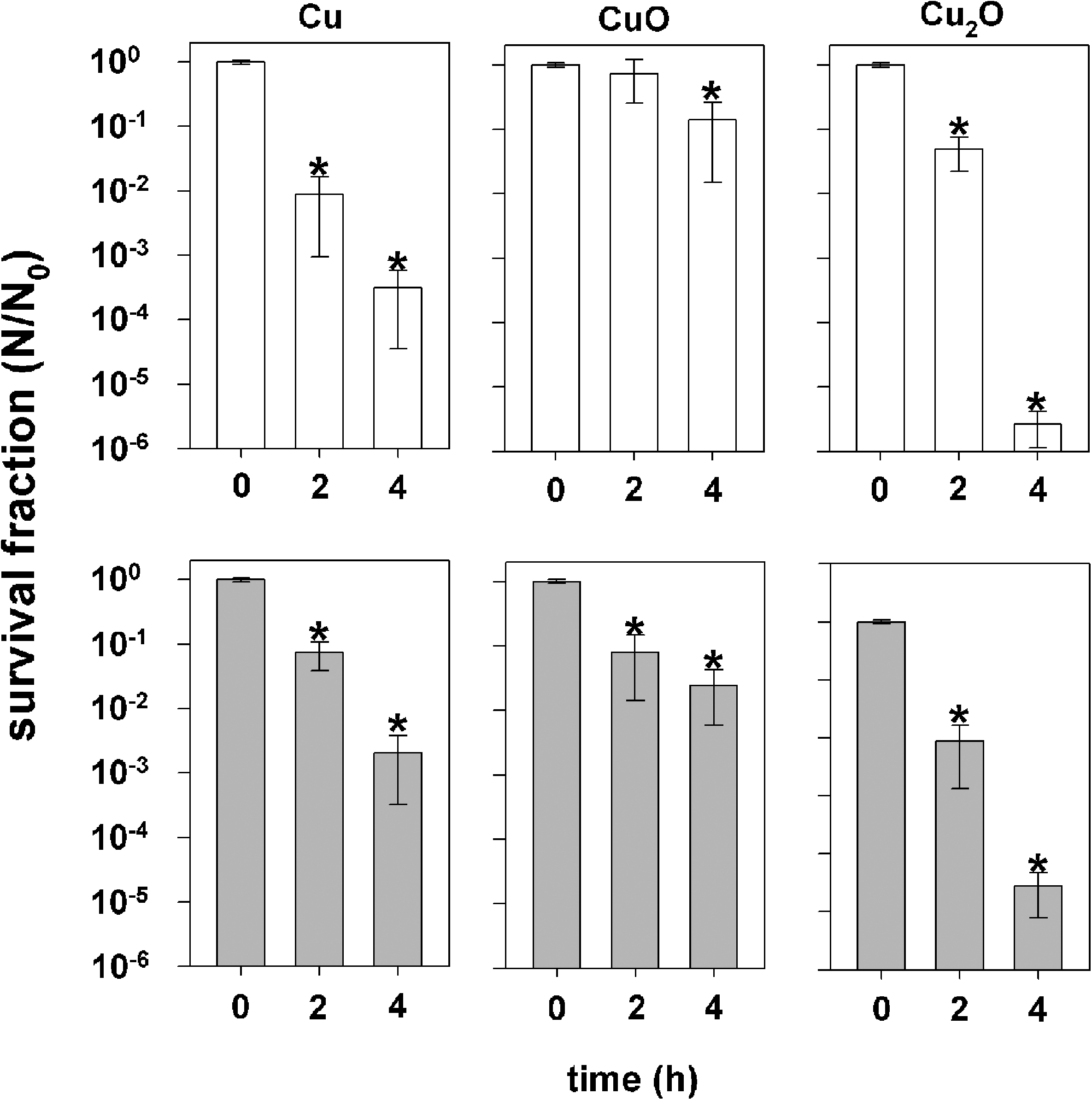
Survival of 106 cells/cm2 of E. coli (white bars) and S. cohnii (gray bars) suspended in a 1:10 dilution of PBS after the exposure to pure copper surfaces and copper oxide layers (N) and stainless steel (N 0). Data are expressed as averages ± standard deviations (n = 3) as described in the text. * = Significant decrease in cell survival (p value < 0.05).
3.4. Membrane damage and ROS production
Exposure of E. coli cells in a multilayer (107 cells/cm2) to stainless steel and pure copper surfaces with a subsequent analysis of membrane damage via the LIVE/DEAD staining assay or ROS production via CellROX green did not result in significant changes in fluorescence. Therefore, results are only presented for monolayers of E. coli cells (105 cells/cm2) after the exposure to stainless steel and pure copper surfaces (Fig. 5).
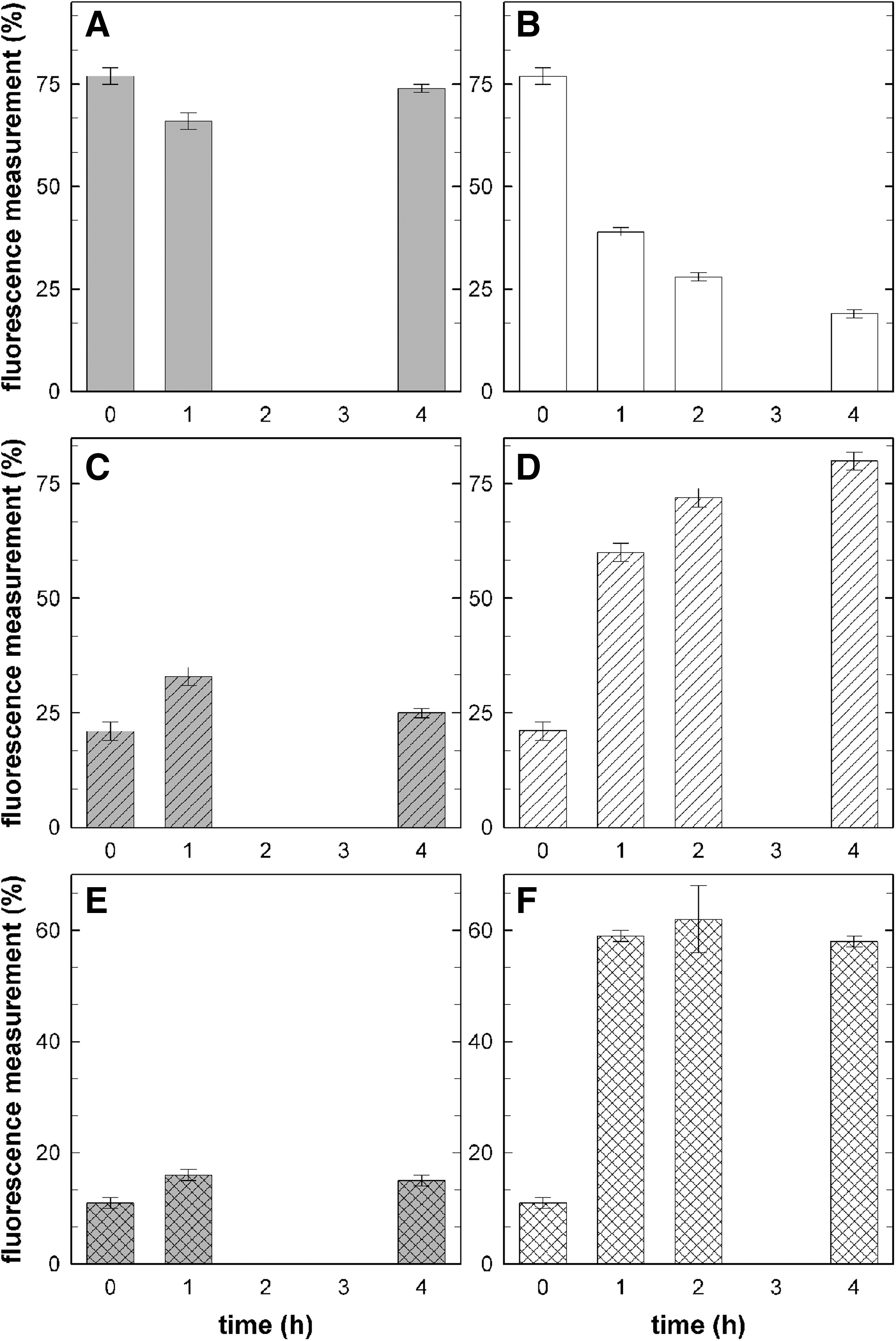
Fluorescence measurement of LIVE (panels
For LIVE/DEAD analyses, approximately 21% of the control sample was stained orange (cell wall/membrane damage) and 77% green (cell wall/membrane intact) essentially for all time points on stainless steel (Fig. 5A–5D). For samples exposed on pure copper surfaces, the viability decreased rapidly as indicated by the loss of green fluorescence. After 1 h on pure copper only 39% of all cells were detected to have an intact membrane. Longer exposure on pure copper led to a further decrease of membrane integrity; after 4 h, only 19% of the cells remained intact (Fig. 5C, 5D).
A general ROS production in cells of about 11% was determined for control samples that were not exposed to any metallic surface. Exposure to stainless steel resulted only in a slight increase of up to 15% compared to the control sample, which was in the range of the measurement accuracy. In contrast, exposure to pure copper surfaces led to a rapid increase in ROS production with a maximum of 59% after 1 h of exposure. Longer exposure did not result in further significant changes in ROS production (Fig. 5E, 5F).
3.5. Copper release from pure copper surfaces
At the beginning of the experiment, the total amount of copper ions in 350 μL cell suspension was 0.01 ± 0.02 nmol (S. cohnii) and 0.03 ± 0.01 nmol (E. coli). After 1 h of exposure on stainless steel, a total of 0.05 ± 0.00 nmol copper was detected in 350 μL cell suspension of E. coli and 0.08 ± 0.04 nmol after 4 h of exposure (Fig. 6). Taking into account the measurement accuracy, no significantly higher concentration was determined.
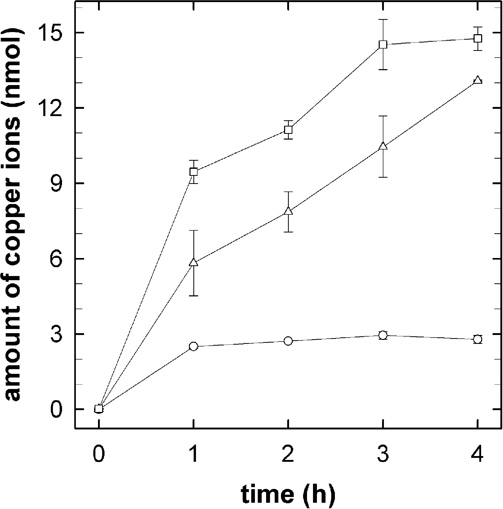
Release of copper ions (in nmol) from pure copper surfaces in the presence of S. cohnii (open triangles; as cells in 0.1 × PBS), E. coli (open squares; as cells in 0.1 × PBS), or buffer without cells (open circles; 0.1 × PBS). Data are expressed as averages ± standard deviations (n = 3) as described in the text.
The exposure of buffer alone on pure copper surfaces led to a quick increase in free copper ions after 1 h of exposure (2.51 ± 0.08 nmol) without a further significant increase after 4 h of exposure (2.79 ± 0.16 nmol). Contrary to the exposure of buffer alone, exposure of cells on pure copper surfaces led to a higher release of copper ions. This is possibly caused by an indirect dissolution of copper ions by cells; that is, no direct contact between the cells and the copper surface occurred. These results indicate that cells dissolve copper ions indirectly from surfaces by excretion of complexing agents. Thereby a complexing agent that contains functional groups like -NH2 (amino), -O- (ether), or -S- (thioether) is known to undergo chemical interaction and bonding, for example, chelate formation with metal ions (Birch and Bachofen, 1990). After 4 h of exposure, the final total amount of released copper ions for E. coli was 14.77 ± 0.47 nmol and for S. cohnii 13.09 ± 0.03 nmol (Fig. 6). Comparing the copper release of E. coli and S. cohnii, the release increased faster for E. coli but resulted in similar amounts of copper ions after 4 h of exposure.
4. Discussion
Previous studies have shown that various microorganisms and fungi occurred on surfaces and in the air aboard the Russian Space Station Mir. The most common airborne microorganism was Staphylococcus sp., whereas the most common fungal species were Aspergillus sp. and Penicillium sp. (Novikova, 2004). Subsequent analysis of samples from the ISS by Novikova et al. (2006) revealed a distribution of the most common species that was similar to that of the isolates from Mir (Novikova et al., 2006). As previously mentioned, under space conditions, the human immune system is affected, and bacteria may become more virulent and resistant to antibiotics (Horneck et al., 2010). To prevent mainly anthropogenic microbial contamination aboard the ISS and in hospitals, copper-based antimicrobial surfaces have been tested in various hospital trials. Under these hospital conditions, single cells adhere on surfaces and can lead to biofilm formation. Therefore, a comparison of hospital and laboratory conditions could be drawn from cells applied in monolayers, which obtain equal stress conditions.
The results of the study reported here indicate that a multilayer was obtained at a density of 107 and 108 cells/cm2. In multilayers, only cells coming in direct contact with the metal surface were affected, whereby cells in the upper layers were protected by shielding from the organisms beneath them. A monolayer of cells was obtained for cell concentrations ranging from 105 to 106 cells/cm2, whereas a significant decrease in cell survival was detected at a cell concentration of 105 cells/cm2. At this concentration no shielding effects occurred, and most of the cells were in direct contact with the metal surface. Additionally, most of the cells were in direct contact with each other. As a result, these cells were able to interact and communicate, which enabled them to better interact with their environment and adapt to it (Miller and Bassler, 2001; Voloshin and Kaprelyants, 2004). This led to a higher survival rate compared to 104 cells/cm2, where apparently the cell concentration was too low for direct interaction of cells. The missing interaction and therefore lack of convection, which is also missing under microgravity conditions (Benoit and Klaus, 2007), could allow a comparison of ground experiments and possible experiments of microbial contaminations on board the ISS.
Contact killing experiments indicated that pure copper surfaces were very effective antimicrobial surfaces for both E. coli and S. cohnii in a monolayer, although both cell types differ in their cell wall and membrane structure; that is, S. cohnii has a thick Gram-positive cell wall, whereas E. coli has a thinner cell wall and an outer membrane that does not occur in Gram-positive cells. Nonetheless, the similar survival rates between the two organisms indicate that these differences were negligible with respect to the killing effect by copper (Rojan, 2013). Certainly, both forms of copper ions were released from pure copper surfaces whereby Cu+ ions may be primarily released from Cu2O layers and Cu2+ ions from CuO layers. Comparing the survival of cells after exposure to these surfaces and layers, the most effective antimicrobial surface layer was Cu2O followed by the pure copper surface and the less efficient CuO layer. However, with respect to versatility, the rough surface structure, and the look of the copper oxide layers, the application of pure copper surfaces is the means of choice. These results are supported by experiments with Enterococcus hirae as recently described by Mathews et al. (2015). Additionally, a comparison of Gram-positive and Gram-negative strains indicates no differences in cell survival after exposure to Cu(I) and Cu(II) oxide layers. This again confirmed that differences of cell walls and membranes were negligible.
The higher toxic effect of Cu2O layers may be due to the release of Cu+ ions, which more easily penetrate intact cytoplasmic membranes than does Cu2+. Once inside the cytoplasm, Cu+ can lead to severe DNA, RNA, and protein damage (Dowjat et al., 1996; Macomber and Imlay, 2009). Additionally, while in the periplasm, Cu+ undergoes Fenton-like reactions producing ROS (Halliwell and Gutteridge, 1984; Macomber et al., 2007; Grass et al., 2011). ROS in turn leads to additional damage of the cell membrane, proteins, DNA, and RNA. Before Cu2+ can cause damaging effects in the cytoplasm, it needs to be taken up by cells either actively via, for example, the Zn(II)-uptake system ZupT (Nies and Herzberg, 2013) or through lesions in the membrane.
The determination of membrane damage and ROS production indicates no increase when multilayers were exposed to pure copper surfaces. The reason for this could be that the number of cells directly in contact with the surface was insufficient compared to the cells of the upper layers so that in the end no differences could be detected. Although no decrease in cell survival was observed after exposure of cells lying in a monolayer for 2 h, experiment results indicate that ROS production and membrane damage occurred within 1 h of exposure. From these results, it can be concluded that ROS production and membrane damage occurred directly after making contact with the antimicrobial surfaces, but effects needed some time to result in cell inactivation. One possible explanation for this could be that free copper ions were bound to the cell membrane, sequestered, or chelated by ligands as previously shown by Borkow and Gabbay (2005) and Lemire et al. (2013). Thereby, membrane damage and ROS production occur, but the intracellular free copper ions were kept at a minimum; therefore further toxic effects were prevented in the beginning of the experiment.
These results indicate that membrane damage and ROS production are major effects caused by contact killing on pure copper surfaces. Similar conclusions were drawn by Espírito Santo et al. (2011), whereas Warnes et al. (2012) postulated that DNA damage is the primary effect of contact killing (Warnes and Keevil, 2011). Due to contrary conclusions reported, further analysis concerning the primary contact killing effect should be conducted.
Previous experiments by Molteni et al. (2010) and Espírito Santo et al. (2011) indicated that the release of copper ions could be of major importance for contact killing. Our results were compared to those obtained by Espírito Santo et al. (2011). The absolute number of copper ions per milliliter could not be directly compared because higher cell and salt concentrations were used by Espírito Santo and coauthors in their experiments. Nevertheless, although the released copper ion concentration indicated in the work of Espírito Santo et al. (2011) was higher than those reported here, the ratio was the same. This means that for the buffer an increase of released copper ions from 1 ppb after 0 h to 10 ppb after 3 h was determined, and for exposed cell suspensions the release increased from 1 ppb after 0 h up to 100 ppb after 3 h of exposure.
By comparing the results of total copper ion release by buffer alone to that of the experiments containing cells, it is clear that the copper ion release was much greater when cells were exposed to pure copper surfaces. By taking into account the results of Mathews et al. (2013), this release may be caused by an indirect dissolution of ions (as described previously). Mathews and coauthors examined cell survival and ion release after exposure on an untreated pure copper surface and a thin perforated plastic layer–coated pure copper surface. For both surfaces, equal release of ions was obtained whereby cells exposed on the coated surface were not affected in cell survival, possibly because cells were not able to adhere directly to the metal surface (Mathews et al., 2013).
Pure copper surfaces were successfully applied and tested in hospital trials where it was shown that the microbial repopulation of copper touch surfaces was decreased when compared to aluminum or plastic materials (Mikolay et al., 2010). Experiments conducted in this study indicated that the release of ions is important for cellular uptake to produce ROS and cause damaging effects to the cell membrane and potentially other intracellular components. Nonetheless, the results presented here illustrate that direct contact with pure copper surfaces is more important than the presence of free copper ions. Under aerobic conditions, copper reacts with oxygen whereby dissolution of copper ions occurs. Oxidized pure copper surfaces still exhibit antimicrobial effects, but oxidization causes changes of the surface roughness, stability, and appearance. Therefore, the application of pure copper surfaces is difficult in spaceflight facilities. To determine better suitable antimicrobial surfaces that will result in lower corrosion and cost and longer durability at constant antimicrobial effects, follow-up experiments should be completed with the use of other materials such as eutectic alloys of copper and/or silver metals such as AlCuAg. Kawakami et al. (2008) tested several metallic elements of the periodic system for their antimicrobial effects and determined antimicrobial properties for aluminum. Aluminum and its alloys have low mass and are used in aerospace and space materials. However, such spaceflight-relevant aluminum alloys have not been tested for their antimicrobial efficiency (Peters and Leyens, 2009; Grilli et al., 2010). Possibly more suitable antimicrobial alloys could be ascertained for the prevention of microbial biofilm formation and the propagation of microorganisms. This would ensure astronaut health and the stability of spacecraft facilities.
Footnotes
Acknowledgments
This work was supported by the Helmholtz Space Life Sciences Research School (SpaceLife) and the German Aerospace Center of the Helmholtz Association (PhD fellowship oc Claudia Hahn; VO-KH-300).
Fixation of the SEM samples was conducted by Christine Maaßen from the Regensburg University, Germany.
C.H., P.R., C.E.H., and R.M. were supported by DLR grant DLR-FuE-Projekt ISS LIFE, Programm RF-FuW, Teilprogramm 475.
R.M.'s contribution is part of the ESA projects: “Testing antimicrobial metal surfaces under spaceflight conditions—an effective strategy to prevent microbial biofilm formation” [No. ESA-HSO-ESR-ILSRA-2014-054; BIOFILMS (Biofilm Inhibition On Flight equipment and on board the ISS using microbiologically Lethal Metal Surfaces)] and “Antimicrobial materials, surfaces and textiles on-board the Concordia Antarctic Research Station as a test-bed approach for the reduction of the microbial contamination on future human space missions (ANTI-BACS)” [No. ESA-AO-11-Concordia-022].
Author Disclosure Statement
No conflict of interest declared.