
Abstract
We propose a model whereby microscopic tunnels form in basalt glass in response to a natural proton flux from seawater into the glass. This flux is generated by the alteration of the glass as protons from water replace cations in the glass. In our proton gradient model, cells are gateways through which protons enter and alter the glass and through which cations leave the glass. In the process, tunnels are formed, and cells derive energy from the proton and ion fluxes. Proton flux from seawater into basalt glass would have occurred on Earth as soon as water accumulated on the surface and would have preceded biological redox catalysis. Tunnels in modern basalts are similar to tunnels in Archean basalts, which may be our earliest physical evidence of life. Proton gradients like those described in this paper certainly exist on other planetary bodies where silicate rocks are exposed to acidic to slightly alkaline water.
1. Introduction
M
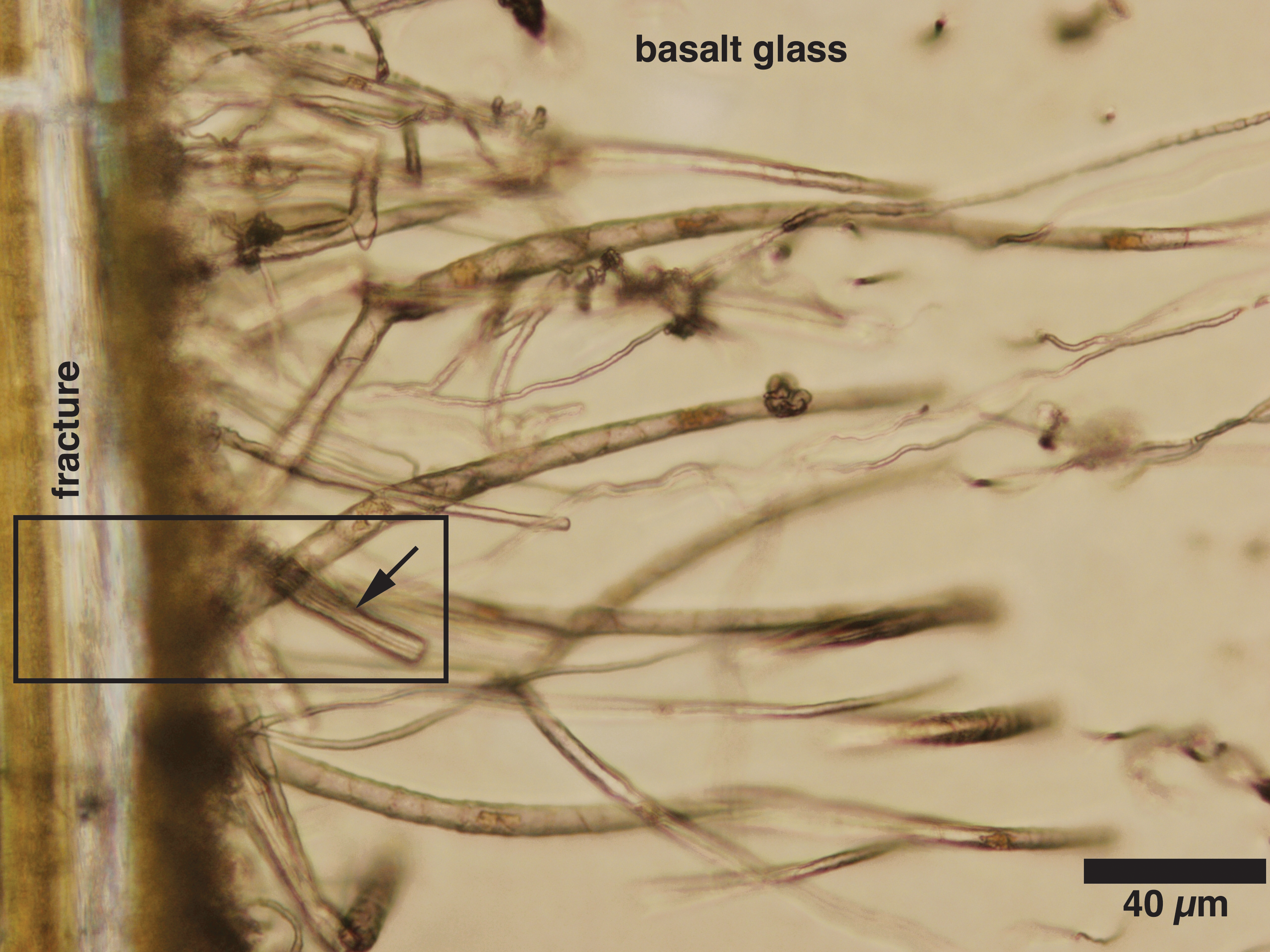
Photomicrograph in plane polarized light of a petrographic thin section of basalt glass from Section 2 of Core 20 of Site 713A on the East Maldive Ridge in the Indian Ocean (Bachman et al., 1988). A fracture is located at the left of the image. Tunnels extend to the right into the glass from the fracture. The box outlines a tunnel with smooth sides and a blunt end, which is used for the model in Fig. 3. Image modified from Fisk and McLoughlin (2013).
Microorganisms and abiotic reactions are known to pit glass, but tunnels matching the size, shape, and distribution of most of the wide variety of the tunnels found in basalt glass have not been made in basalt glass under controlled conditions. Microorganisms, nucleic acids, and biologically important elements have been found associated with tunnels (e.g., Thorseth et al., 1995a; Furnes et al., 1996; Torsvik et al., 1998; Banerjee and Muehlenbachs, 2003; Fisk et al., 2003); and hypotheses have been advanced for tunnel formation involving biological processes (e.g., Thorseth, et al., 1995a; Furnes et al., 1996; Torsvik et al., 1998, Staudigel et al., 2006). Abiotic processes can produce tunnels (e.g., Lepot et al., 2011; French and Blake, 2016), but they match only some of the common chemical and physical characteristics of tunnels in basalt glass. Because abiotic tunnels are not like most tunnels in basalt glass and because microorganisms have not been cultured that make tunnels, how they form remains an open question.
We hypothesize that individual tunnels are created by a natural flux of protons from ambient water through microorganisms and into glass, and this flux is complemented by a counter-flux of Na+ and other cations from the basalt glass to ambient water. Cells blocking the entrances of tunnels use these fluxes for chemiosmotic production of adenosine triphosphate (ATP) (Mitchell, 1961) and other cell functions such as selective membrane transport.
2. Physical and Chemical Characteristics of Tunnels
McLoughlin et al. (2010) summarized physical features of tunnels; and detailed imaging and analysis of cross sections of tunnels by Thorseth et al. (1995b), Alt and Mata (2000), Benzerara et al. (2007), Knowles et al. (2012, 2013), Wacey et al. (2014, 2017), and Pedersen et al. (2015) showed that they have some chemical features in common. In particular, high spatial resolution chemical studies of simple tunnels (Benzerara et al., 2007; Knowles et al., 2012; Wacey et al., 2017, see Appendix Table A1) have shown that (1) tunnels are cylinders filled with phyllosilicates; (2) their interiors are enriched in Fe and K and depleted in Mg, Ca, and Na relative to the surrounding glass; (3) they are encircled by a rim enriched in O and Si and sometimes Al relative to the alteration products in the tunnels and sometimes also relative to the glass; and (4) the rims have a sharp boundary with the unaltered glass outside the tunnel (Fig. 2). In a recent comprehensive elemental microanalysis of simple tunnels (Wacey et al., 2017), the depletion of Si, Al, Mg, Ca, Mn, and P inside the tunnel relative to the basalt glass indicates that these elements were removed from the glass during alteration. In contrast, Fe, K, and sometimes C are enriched inside tunnels relative to the basalt glass, indicating that these elements were either passively enriched due to the removal of other elements or were added to the tunnels. Ti and Na are depleted in most regions inside tunnels but may exhibit patchy enrichment in some tunnels.

High-angle annular dark-field scanning transmission electron microscope image of a cross section of a tunnel in 145 Ma ocean basalt glass from Ocean Drilling Program Hole 765D in the Argo abyssal plain, eastern Indian Ocean (Gradstein et al., 1990). Unaltered basalt glass makes up the background of the image. A homogeneous, dark gray, nearly circular rim composed of Si, Al, and O separates the glass from the lumen, which was once glass and is now fibrous material. The dark area between rim and glass is a void space, which suggests shrinkage of the tunnel contents. Elsewhere the boundary between the rim and the glass is sharp, and fibrous material is absent outside the rim. Image modified from Wacey et al. (2017). Vertical line near the center is TEM beam damage.
3. Hypothesized Origin of Tunnels
When tunnels were first reported in basalt glass, Morgenstein (1969) attributed them to stress fractures in glass due to rapid cooling followed by filling with secondary minerals. Zhou and Fyfe (1989) proposed their formation to be caused by the dissolution of basalt glass. More recently, due to evidence of nucleic acids, carbon, and cells in or near tunnels, biological mediation of glass dissolution has been the favored hypothesis of tunnel formation (e.g., Thorseth et al., 1992; Staudigel et al., 1995; McLoughlin et al., 2010; see French and Blake, 2016, for an additional 28 publications in which biological alteration is the preferred interpretation for tunnel formation). Abiogenic production of tunnels also has advocates (e.g., Lepot et al., 2011; Pedersen et al., 2015; French and Blake, 2016). Whether biotic or abiotic, it is generally accepted that tunnels result from the dissolution of basalt glass and the precipitation of phyllosilicate and amorphous alteration products. One hypothesis is that microorganisms use organic acids to dissolve glass in order to obtain phosphorus or reduced iron, manganese, or sulfur, whose free energy of oxidation can support microbial metabolism (Thorseth et al., 1995a; Bach and Edwards, 2003; Staudigel et al., 2006).
Experimental biotic alteration of basalt glass can produce pits and grooves but not tunnels like those in Fig. 1 (Staudigel et al., 1995; Thorseth et al., 1995b; Buss et al., 2007; Chen et al., 2014), and the same is true for the experimental abiotic alteration of basalt glass (e.g., Crovisier et al., 2003; Fisk et al., 2013, French and Blake, 2016). Based on observations of 121 Ma basalts cored from the flank of the Mid-Atlantic Ridge, the alteration of glass along alpha-recoil tracks that accumulate over time in glass has been hypothesized as one way in which alteration can be localized to form tunnels (French and Blake, 2016), but the distribution, size, and form of alpha-recoil tracks do not match those in Fig. 1 or the forms of many other tunnels in basalt glass (McLoughlin et al., 2010). Tunnels that are superficially similar to those in Fig. 1 and that are attributed to mineral or organic particles being forced through or dissolving their way through rocks have been termed ambient inclusion trails (AITs) (Lepot et al., 2011); however, McLoughlin et al. (2010) rejected the hypothesis that AITs are responsible for the formation of tunnels in basalt glass. Presently, we do not know the conditions needed to dissolve tunnels in basalt glass as in Fig. 1, but as discussed in the next section, we have a good understanding of the mechanism of basalt glass dissolution by water.
4. Alteration of Basalt Glass
Basalt glass is composed primarily of network-forming silicon-oxygen and aluminum-oxygen tetrahedra. The network also includes less abundant ferric iron-oxygen and titanium-oxygen tetrahedra. Shared “bridging” oxygen atoms bind these four network formers to each other. Network modifiers, ferrous iron, Ca, Mg, Na, Mn, P, and K are interspersed in this network and are bound to nonbridging oxygen. In the preferred models of glass dissolution based on laboratory experiments (Berger et al., 1987; Daux et al., 1997; Oelkers and Gislason, 2001; Gislason and Oelkers, 2003), protons first replace the network-modifying metals, and the glass surface becomes a leached layer enriched in Si, Al, Fe3+, and Ti. This layer thickens over a matter of hours until the diffusion of the network modifiers through the leached layer matches the dissolution of Si, Al, Fe3+, and Ti in the leached layer so the dissolution of the glass becomes stoichiometric (Gislason and Eugster, 1987; Crovisier et al., 1992, Oelkers and Gislason, 2001). See Appendix Table A2 for the experimental rate of basalt glass dissolution at conditions relevant to subseafloor aquifers.
Cations of the network modifiers, Mg2+, Ca2+, Na+, K+, and P5+, are soluble, and if the water is anoxic, iron and manganese will be divalent and also soluble. At high pH, silica and alumina will hydrate to Si(OH)4 and Al(OH)3 monomers, which can oligomerize in the presence of salts or organic catalysts into amorphous silica and minerals (Iler, 1979). Also, multivalent metals in solution with soluble silica and alumina will form insoluble metal silicates (e.g., phyllosilicates) and in some experiments a colloidal aluminosilicate (Iler, 1979).
5. Model of Cellular Promotion of Glass Alteration and Tunnel Formation
Everywhere acidic to moderately alkaline water contacts basalt glass, protons will alter the glass by the mechanism just described. For alteration to form a tunnel, there must be a barrier to protons on the glass surface and a point source of protons to the glass through the barrier. It is known from experiments that microorganisms (including those attached to basalt glass) surround themselves with an organic matrix. This matrix can bind with mineral grain surfaces and cations and if composed of exopolysaccharides can either increase or decrease the rate of mineral dissolution depending on the conditions (Welch and Vandevivere, 1994). In some situations, an amorphous Si-Al layer forms between polysaccharide and the mineral (Welch et al., 1999). We hypothesize that cells attach to glass and surround themselves with a barrier composed of organic and/or an inorganic material. A cell or cluster of cells located in these barriers is a point source for protons that enter and alter the glass and initiate tunnels that are about the same diameter as the cell or cell cluster (Fig. 3).

Model of an open fracture in basalt glass containing water from an ocean crust aquifer. A cell attached to the fracture wall has initiated a tunnel. The tunnel contains fibrous reaction products of glass alteration (Fig. 2) and water. The cell remains at the open end of the tunnel, and the tunnel grows from the fracture wall into basalt glass. The aquifer, cell cytoplasm, and tunnel fluid have pH values of 7.5, 9.5, and 9.8, respectively. Orange arrows indicate a proton flux from the aquifer through the cell to the tunnel fluid and from the tunnel fluid into the glass and a Na+ flux in the opposite direction. Not shown are fluxes of Fe2+, Mg2+, Mn2+, Ca2+, K+, and P5+ from the glass into tunnel, which along with Na+ balance the charge of the H+ entering the glass. A ∼100 nm layer of impermeable Si-Al-O oxides lines the tunnel (red). The walls of the fracture near the cell are coated with an H+ impermeable material (green), so for protons to react with the glass they must pass through the cell. The H+ and Na+ fluxes are based on a Si dissolution rate of 10−13 mol/cm2·s at the tip of a 4 μm diameter tunnel. Blue arrow indicates fluid flow through the fracture. Basalt glass and aquifer chemistry are from Appendix Table A3. A single cell is shown at the tunnel entrance, but a cluster of smaller cells is also possible.
Protons replace Na+, K+, and other cations in the glass, which dissolve in the fluid in the newly formed tunnel, and Si and Al precipitate as phyllosilicates or an amorphous phase in the tunnel. Counter-fluxes of protons and cations are established between the aquifer and the basalt glass, and because the charge carried by protons is balanced by the charge of cations, there is no charge buildup in the tunnel. The opposing fluxes of protons and cations pass through the cell or cells at the tunnel opening, which can harvest energy with ATP synthase if there is sufficient potential drop across the cell membrane. ATP may also be produced by the flux of Na+ and K+ to the aquifer using Na+/K+ ATP synthase (Schulz et al., 2013).
6. Proton Motive Force
We use published pH values to model the pH gradient between the aquifer and the tunnel. Gislason and Eugster (1987) reacted basalt glass with water without allowing exchange with the atmosphere, and in 2000 h the pH rose from 5.6 to 9.8, where it was buffered by silicic acid (Appendix A). These conditions are similar to what would be expected for water isolated in a blocked tunnel in basalt glass, and we take a pH of 9.8 for the water in the tunnel in our model (Fig. 3). Jungbluth et al. (2013) measured a pH of 7.4–7.5 in an aquifer in the igneous ocean crust, and we take 7.5 as the pH of water that fills the fracture in our model (Fig. 3). These values give a ΔpH of 2.3 between the aquifer and the tunnel. Cells can maintain a pH of 9.8 in their cytoplasm (Cook et al., 1996; Wiegel, 1998) and pH gradients of 2.0 or more across cell membranes (Booth, 1985; Krulwich, 1995; Slonczewski et al., 2009), and we have chosen a pH of 9.5 for the cell cytoplasm. The ΔpH values for our model are similar to those in the model of Sojo et al. (2014) and Lane (2017) for a primitive protocell at a boundary between high-pH hydrothermal fluids and seawater. In our model, a ΔpH of 2.0 exists between the aquifer and the cell cytoplasm (Fig. 3), but larger pH gradients could exist if the aquifer has a lower pH typical of deep-sea hydrothermal vent water (e.g., Von Damm, 2000). The ΔpH of 2.0 between aquifer and the cell interior is sufficient to drive ATP production (-128 mV at 50°C) as long as there is no electrical charge gradient across the cell wall (see Appendix A).
7. Power from Proton Flux
In this model, ATP production is driven by the flux of protons from water in the aquifer into basalt glass, so the rate of ATP production depends on the magnitude of the proton flux. This proton flux depends on the basalt glass alteration rate and thus the rate at which protons exchange with metals in the glass. We use the rate of alteration of basalt glass and the area of the end of the tunnel to calculate the proton flux into the tunnel and into the glass. The glass dissolution rate is 10−13 mol of Si/cm2·s for a fluid pH of 9.8 and temperature of 50°C (Gislason and Oelkers, 2003). At this dissolution rate and an area at the blunt end of a tunnel of 12.6 μm2, the complete replacement of the network-modifying cations of a typical basalt glass requires about 10,000 protons per second. Using the ΔpH of 2.0 between the aquifer and the cell interior and temperature of 50°C, the potential across the cell membrane is −0.128 V. A proton current of 10,000/s produces about 2 × 10⁻¹⁶ J/s, an amount sufficient to maintain a cell (Hoehler and Jørgensen, 2013). See Appendix A for details.
8. Tunnel Shape
One question not yet addressed by the model is how alteration creates long, nearly straight cylinders (Fig. 1). We point out that tunnels take on other forms as well (Fisk et al., 1998; McLoughlin et al., 2009, 2010; Fisk and McLoughlin, 2013) in addition to the smooth-sided, nearly straight, blunt-ended tunnels as shown in Fig. 1 and modeled in Fig. 3, and we believe the model can be applied to some or many of these other forms. High-resolution transmission electron microscopy images illustrate the 100 nm wide, Si-Al-O rim that forms the outer surface of tunnels (e.g., Fig. 2). It is clear from Fig. 2 that phyllosilicates only form in the lumen and that the glass outside the rim remains pristine. This can be interpreted to be due to the absence of a proton flux through the Si-Al-O rim. This led Wacey et al. (2017) to conclude that a tunnel grows by increasing its length and not its diameter, which is manifested by their great length relative to diameter and nearly constant diameter. For the tunnel to grow in length but not in diameter, glass dissolution must only occur at the closed end of the tunnel, and an impermeable rim must form as the tunnel lengthens.
We speculate that, as the tunnel grows, the tunnel lining composed of Si, Al, and O forms by the aggregation, precipitation, and oligomerization of Si(OH)4 and Al(OH)3 monomers from the dissolved glass. Salts and organic compounds can catalyze this process, resulting in deposits of minerals and amorphous silica (Iler, 1979) and in some conditions producing halloysite, which is similar to the composition of the walls of tunnels (Wacey et al., 2017). Perry and Keeling-Tucker (2000) point out that organisms have an unsurpassed ability to build inorganic structures for specific uses. In the model proposed here, microorganisms control the chemistry of their tunnel aquifer “crystal garden”; thus we hypothesize cells can control the rate of glass dissolution at the end of the tunnel and the composition and location of the impermeable rim by manipulating the pH, and cation and organic content of the tunnel. Microorganisms' control of the sizes and shapes of tunnels in glass and of the composition of amorphous and mineral precipitates in tunnels easily seems within the realm of biological processes.
In our model, protons replace Fe2+, Mn2+, Mg2+, Ca2+, Na+, K+, and P5+ in the glass. Mg2+, Ca2+, Na+, K+, and P5+ are soluble, and if the fluid in the tunnel is anoxic, Fe2+, and Mn2+ are also soluble, so they all can diffuse from the tunnel. If the oxygen level in the tunnel results in the oxidation of iron and manganese to insoluble Fe3+ and Mn4+, they could be incorporated in clays or the Si-Al-O rim of the tunnel or be actively transported by siderophores or other cation shuttles to the aquifer.
9. Rate of Tunnel Growth
Based on the rate of dissolution of basalt glass at conditions expected for the fluid in a tunnel (pH 9.8 and 50°C) and for a subseafloor aquifer (pH 7.5 and 50°C), the rate at which protons enter glass through the end of a 4 μm diameter tunnel is 10,000 per second (Fig. 3 and Appendix Table A3). If these protons are confined to the 4 μm diameter tunnel, then for each 1 μm that the tunnel is extended, a volume of 12.6 μm3 of glass (3.4 × 10⁻¹¹ g) will be altered. For the complete replacement of the network-modifying metals in this volume of glass, 2.3 × 10¹¹ protons are needed (Appendix Table A3). With a flux of 10,000 protons per second, all the metals in this volume can be replaced in about 260 days (0.71 years). At this rate, it would take decades to make tunnels of substantial length as in Fig. 1. At 75°C, the glass alteration rate will be 10 times that at 50°C (Appendix Table A2) and would significantly increase tunnel growth rate.
The proton flux from the aquifer into the tunnel and into the glass is proportional to the area of the exposed glass. If the tunnel walls allowed protons to diffuse into the glass, then the proton and ion fluxes through the cell would increase as the tunnel lengthened. The cell may not be able to survive these increased fluxes. Maintaining a constant surface area at the blunt end of the tunnel and at the same time making the tunnel rim impermeable creates a constant flux of protons for the cell and results in a constant-diameter tunnel.
10. Documentation of Cells at Tunnel Entrances
Our model suggests that cells occupy tunnel entrances, and some studies show nucleic acids, images of cells, and/or concentrations of biologically important elements at tunnel entrances. Torsvik et al. (1998) showed increased C and N at the contact of fresh glass with alteration but not clearly in tunnels' entrances. Nucleic acids were found at the intersection of a tunnel with a fracture in a 6 Ma basalt (Furnes et al., 2001), and Banerjee and Muehlenbachs (2003) found cells and biofilms at the glass-clay boundary but possibly not at tunnel entrances. A ∼1 Ma basalt from 1300 m below the surface of Hawaii was enriched in phosphorus, nucleic acids, and cells where tunnels intersect the glass surface and also inside tunnels (Fisk et al., 2003). These studies indicate that cells might be present in tunnel openings, but their presence has not been confirmed by imaging.
Cells or biogenic organic materials may also occupy the interiors of tunnels. Carbon has been found in tunnels in Archean metabasalts (Banerjee et al., 2006; Fliegel et al., 2010), in Cretaceous basalts (Banerjee and Muehlenbachs, 2003; Benzerara et al., 2007; Knowles et al., 2012; Wacey et al., 2014, 2017), in Miocene basalts (Torsvik et al., 1998; Furnes et al., 2001), and in a 1 Ma Hawaiian basalt (Fisk et al., 2003). Due to the ages of these rocks, it is not known if the carbon in tunnels represents cell remains or if the carbon is contemporaneous with tunnel formation.
11. Energy and Maintenance
Organisms in extreme oligotrophic environments spend most of their energy on cell maintenance rather than cell growth (D'Hondt et al., 2014; Jørgensen and Marshall, 2016). Thauer et al. (2008) calculated that 50 to 55 × 109 ATPs are needed for cell division, so the flux 10,000 protons per second is sufficient to produce two cells per year, which are free to enter the aquifer if cells are located at the tunnel opening. Doubling times on the order of years have been calculated for microorganisms in marine sediment based on cell numbers and substrate consumption rate (Hoehler and Jørgensen, 2013), so the doubling time calculated for cells at the entrance of tunnels is reasonable. Cells that divide less than twice a year can expend energy on cell maintenance, carbon assimilation, organic compound production, facilitating the precipitation of the amorphous Si-Al-O rim of the tunnel, for active ion transport, and for other functions.
Average basalt can supply elements for most of the biological needs of a cell except for N (Appendix Table A3), which can be supplied by the aquifer (Wheat et al., 2010). Solutes will diffuse down their concentration gradients from the dissolving glass at the blunt end of the tunnel to the cell at the entrance to the tunnel. For example, glass can supply 13 fg of phosphorus per 1 μm length of tunnel altered (12.6 μm3). Exponentially growing cells contain between 0.4 and 25 fg of P/cell, so it appears that the glass can supply sufficient P for static or growing cells without any contribution from the aquifer.
We expect cells to also exert some control over the composition of the fluid in the tunnel for their own benefit. This can be accomplished by one or more cells blocking advective exchange between the aquifer and the tunnel and actively, passively, or selectively transporting ions and organic molecules to or from the tunnel. The enrichment of K and depletion of Na in the tunnel contents relative to the surrounding glass (Table A1) are a possible manifestation of such control. Maintaining a constant composition and chemical gradient in the tunnel could be possible because the new volume of tunnel created per year is small compared to the volume of a cell or cells at the tunnel opening.
12. Tests of the Model
The model we propose can be tested by demonstrating that microorganisms produce tunnels in glass, but the difficulty of growing endolithic microorganisms even when they are abundant in their environment remains a problem (e.g., Vartoukian et al., 2010). This difficulty is compounded by the slow growth of low-abundance, oligotrophic organisms, but methods of culturing “the uncultured” have been used with some success. An approach for culturing the organisms from natural proton gradients would be to enrich cells on basalt glass suspended in the confluence of neutral spring and alkaline lake water or where high-pH hydrothermal vent water mixes with seawater. Then inoculate cultures containing basalt glass with this enriched glass.
Our model predicts that basalt glass in a low-to-near-neutral pH medium creates a natural proton motive force (PMF) due to the ΔpH between the medium and the water in contact with the glass (pH 9.8). Matching other conditions of subseafloor aquifer such as being anaerobic, oligotrophic, and 50–70°C could also promote microorganisms that rely on a natural proton gradient for energy. Basalt glass in cultures should be polished and examined before and after incubation and should be of sufficient size to be made into polished thin sections so tunnels can be studied in transmitted light and with high spatial resolution techniques (e.g., Benzerara et al., 2007; Knowles et al., 2012; Wacey et al., 2017). At the hypothesized growth rates, tunnels will be visible in yearlong experiments.
Another approach for testing our hypothesis would be to predict and find genomic characteristics of organisms that rely on chemiosmotic gradients, such as multiple genes for ATP synthase, Na+/K+ATP synthase, Ca2+ or even possibly Mg2+ ATP synthases, carbon fixation, and the absence of genes for importing organic matter and for oxidative phosphorylation. The water in the tunnel will be enriched in Fe2+, Mn2+, Ca2+, Mg2+, Na+, K+, and P5+ from the basalt, so the cell will need to have the capacity of transporting these metals into the aquifer or catalyzing their precipitation in the tunnel. Average basalt glass also contains more than 50 ppm each of these potentially toxic trace metals Co, Cr, Cu, Ni, and Zn (Gale et al., 2013; Appendix Table A3), which will accumulate in the tunnel unless transported to the aquifer. The ability to transport these ions should be detectable in the genome.
13. Origin of Life
Russell (2006), Martin and Russell (2007), Lane et al. (2010), Lane and Martin (2012), Herschy et al. (2014), Sojo et al., (2014, 2016), and Lane (2017) proposed the natural proton gradients where alkaline hydrothermal vent water mixed with seawater promoted the development of life on early Earth. The proton gradient they propose is similar to the one we have described that develops in a basalt-seawater system, which certainly existed on prebiotic Earth as soon as water accumulated on the planet's surface. The flux we describe could have influenced the development of the earliest protocells such as those proposed by Lane (2017) and others referenced immediately above. Koonin and Martin (2005) review evidence for life originating in “geologically formed, inorganic, abiogenic compartments” rather than inside a protocell membrane, and we note that the abundant vesicles in basalt glass are abiogenic compartments. A semipermeable membrane or protocell isolating the fluid in a vesicle from ambient water could lead to the concentration of complex organic compounds in the vesicle even without a temperature gradient mechanism suggested by Mast et al. (2013). Water trapped between a protocell or a membrane and basalt glass could establish a ΔpH between the ambient and trapped water. Even with primitive lipid chemistry, protocells could form selectively permeable membranes that simulate cellular processes (Mansy, 2010). The composition of the water in these abiogenic compartments would have varied with membrane selectivity, composition and mineralogy of the basalt, and composition, temperature, and pressure of seawater. Permutations of these variables and the 1024 m2 of specific surface area of marine basalts exposed to seawater (Nielsen and Fisk, 2010) provide countless opportunities for protocell experimentation, perhaps leading to additional metabolic complexity.
Here, we suggest that on early Earth the natural proton gradient from water into basalt could have been harnessed and evolved to drive the production of ATP. We also propose that microscopic tunnels in basalt glass, which can be found today in the ocean basins and in some other environments, are a modern manifestation of this primitive process. Tunnels in 3.4 Ga Archean basalts, which are similar to those in modern basalts (e.g., Furnes et al., 2004; Fliegel et al., 2010; but see Grosch and McLoughlin, 2014, for an alternative view), may be the earliest physical trace fossil evidence of life; however, even tunnels in Archean basalts would be the products of highly evolved cells compared to the first cells proposed by Lane (2017).
14. Potential for Extraterrestrial Life
Igneous rocks and water are common components of the Solar System, and dissolution of silicate minerals and basalt glass both proceed by proton replacement of cations in the silicate matrix (Oelkers, 2001). Therefore, proton fluxes from water into silicates will certainly exist on planetary bodies where silicate rocks are exposed to surface and subsurface liquid water. On the surface of Mars, igneous rocks were once transported in streams (Williams et al., 2013), and Grotzinger et al. (2014) describe ∼3.7 Ga sedimentary rocks that contain igneous clasts and minerals. These rocks were once submerged in a freshwater lake in Gale Crater. Today in Gale Crater, water vapor cycles from the atmosphere into shallow martian subsurface each sol, and up to 8 wt % water is found in the near-surface rocks and soil, possibly allowing brines to form (Martin-Torres et al., 2015). Shallow ice has been found in the north pole region (Mellon et al., 2009), subsurface water was detected below the south polar ice cap at 81°S (Orosei et al., 2018), and physical models estimate that liquid water is present below the martian cryosphere at depths of 3–10 km (Travis et al., 2003) and <1 to 11 km at the equator and the poles, respectively (Clifford et al., 2010), suggesting that Mars can host an aquifer physically similar to the one in Earth's ocean crust. These measurements and models point to past and present martian environments where natural proton gradients were or are present. If the biological production of tunnels can be demonstrated in cultures on Earth, then retrieving martian pillow lavas or other martian basalts that were once exposed to water could provide physical or chemical evidence of life there.
Some moons of Jupiter and Saturn have an ice crust, and Europa and Enceladus have acid-to-neutral-pH subsurface oceans (Porco et al., 2006; Roth et al., 2014). If the interiors of these and other water-bearing moons contain silicate minerals, then proton fluxes would exist from the water to the silicates. Outside the Solar System, there will be many planets and moons containing water and silicates. A star's habitable zone is defined as the range of distances from the star at which liquid water can exist on a planet's surface, and our solar system's moons confirm that subsurface water is present well beyond this defined range. In the region of sky investigated by the Kepler mission, about 26% of G and K class stars have rocky planets in the habitable zone (Petigura et al., 2013). Assuming 26% of the Sun-like stars have planets in their habitable zones and these stars also have moons with liquid water beneath frozen surfaces, then there are plenty of locations in the Universe for natural proton gradients to promote and sustain life and reason to expect life nearby in the Milky Way.
In this paper, we hypothesize that some microscopic tunnels form in basalt glass by a flux of protons from water into the glass, and additionally the proton flux and sodium ion counter-flux are moderated and harnessed by microorganisms for cell maintenance and growth. The temperature, pressure, and water chemistry in the ocean crust aquifer where most of the microscopic tunnels have been found indicate the hypothesized microorganisms are polyextremophiles—growing in an oligotrophic, aquifer that contains modified seawater at 50–75°C, 100–400 atmospheres pressure, and pH 7.5–10. Perhaps the most extreme feature of the organisms is sustaining a ΔpH of 2.3 across the cell (Fig. 3). The pH is 7.5 on the aquifer side of the cell and 9.8 on the tunnel side of the cell. This is very similar to the ΔpH proposed by Sojo et al. (2014) and Lane (2017) for protocells harnessing the pH gradient of mixing alkaline springwater with seawater. The model (Fig. 3) shows a single 4 μm cell at the tunnel opening, but a cluster of smaller cells is a possible alternative. Wiegel (1998), in a paper on novel extremophiles, expressed his belief that because we know only a small fraction of existing microbes, we can expect the boundaries of life will be extended as extreme environments are explored. The microorganisms hypothesized in this paper may not have yet been isolated nor observed in nature due to the limited collection of viable samples from deep ocean crust aquifers.
15. Conclusions
Microscopic tunnels in basalt glass start at a surface that is exposed to water and extend a few to more than a hundred micrometers into the glass. We hypothesize that tunnels are created by the dissolution of glass by protons that pass through a cell or cells located in an impermeable barrier on the glass surface and the protons replace cations in the glass. Phyllosilicates and fluid fill the tunnel, and the pH and soluble cations Fe2+, Mn2+, Mg2+, Ca2+, Na+, K+, and P5+ increase in the fluid. The proton flux into the glass creates ΔpH of 2.3 between the aquifer and the tunnel, and we assume a ΔpH of 2.0 between the aquifer and the cell interior; this generates −128 mV of PMF and 2.1 × 10⁻¹⁶ J/cell·s of power, which appears sufficient to maintain cells and to allow them to increase their mass and divide. Cells that lodge in the openings of tunnels will be in a position to harvest energy from the natural proton gradient. The rate of tunnel growth (1–2 μm/year) makes the biological production of tunnels in the laboratory a long-term experiment and may explain why no tunnels have been reported in newly erupted seafloor basalts.
Prokaryotes extract energy from proton gradients that they create and maintain. Although ATP synthases are nearly universally present in organisms, there are no known, or clear-cut, examples of microorganisms that extract energy from an abiotically sustained proton flux through the organism. We propose a unique model by which cells use a natural proton gradient that is established by the reaction of water with basalt glass. We hypothesize that microorganisms will be found that maintain but are unable to create a proton gradient and therefore use the energy from natural gradients of protons and sodium and potassium ions as their only means of synthesizing ATP.
Aqueous environments with natural proton gradients have been proposed as locations where life could have originated, and we describe silicate-water reactions that create natural proton gradients. Cells can exert some control over the composition of water in microscopic tunnels in basalt glass, which are enclosures that can sequester and concentrate organic compounds. Organisms capable of using natural proton gradients for energy have a home on or in any body in the Universe where silicate rocks are submerged in acidic to slightly alkaline pH, fresh to moderately salty water. Confirmation that natural proton gradients support prokaryotes will provide impetus for recovering once-submerged igneous rocks from Mars.
Appendix A
A.1. Tunnel definition and tunnel names
Tunnels in basalt glass are cylinders with a closed end inside the glass and an open end at a surface exposed to water such as an open fracture (Fig. 1) or the exterior of a pillow basalt or glass shard. Tunnels have been given a variety of names: microtunnels, microtubes, microtubules, microchannels, microtextures, borings, microborings, microcracks, tubular alteration, tubular textures, tubular microtextures, tubular microcavities, etch tunnels, and elongate alteration features. For simplicity in this paper, we include all these names under the single name of tunnel.
A.2. Tunnel composition
Table A1 compares the compositions of tunnels from three detailed studies (Benzerara et al., 2007; Knowles et al., 2012; Wacey et al., 2017). The tunnels have a fibrous interior (lumen) and a silica-enriched rim. The lumen and rim were analyzed by microbeam techniques. The composition of basalt glass from the work of Wacey et al. (2017) is given for reference.
A.3. Glass alteration
The flux of protons into glass through the closed end of the water-and-phyllosilicate-filled tunnel depends on the dissolution rate of glass, which depends on temperature and pH (Table A2), the area of the closed end of a 4 μm diameter tunnel (12.6 μm2), and the composition of the glass (Table A3). The temperature of the aquifer in the oceanic igneous crust can be assumed to be equal to or greater than that of the temperature of the sediment-basement interface, and this temperature depends on crustal age and sediment thickness. Along a transect from 20 to 100 km from Juan de Fuca Ridge, the temperature at the sediment-basalt interface rises from 16°C to 64°C (Davis and Becker, 2002). For our calculation, we use 50°C as the temperature of the aquifer. In our model, we have chosen a pH of 9.8 for water in the tunnel, which is based on the experimental reaction of basalt with water with the atmosphere excluded (Gislason and Oelkers, 2003). Table A2 gives the negative log10 of the silica dissolution rate in mol Si/cm2·s of basalt glass for temperatures and pH values relevant to our model (Gislason and Oelkers, 2003).
Abundance of Elements in Tunnel Lumen and Rim Relative to Glass
Weight percent oxides in basalt glass determined by electron microprobe and the relative abundance of elements in the tunnel lumen and tunnel rim relative to the glass determined from HAADF-STEM elemental maps [1] and inferred from relative heights of TEM-EDX peaks in references [2] and [3]. (+) enriched, (–) depleted, ( = ) approximately equal. References: [1] Wacey et al. (2017); [2] Benzerara et al. (2007); [3] Knowles et al. (2012).
Although mostly depleted in tunnels, Ti, Na, and C can show localized patchy enrichment in some examples. Blanks indicate element was not analyzed.
Basalt glass rate of dissolution at temperatures and pH values relevant to the model. Rates are the negative log10 in mol Si/cm2·s. Data from Gislason and Oelkers (2003).
For the calculation of the PMF generated by dissolving basalt glass, we use an average basalt (Table A3) from the ocean basins (Gale et al., 2013). During basalt glass alteration, protons first replace Na+ and K+ on the glass surface; then protons exchange for metal ions (Fe2+, Mn2+, Mg2+, Ca2+, and P5+) that are bound to silica, alumina, ferric iron, and Ti4+. At high pH, Si, Al, Fe3+, and Ti are hydrated to form Si(OH)4, Al(OH)3, Fe(OH)3, and Ti(OH)4.
The area of a blunt, 4 μm diameter tunnel as in Fig. 1 is 12.6 μm2, so for a dissolution rate of 1 × 10⁻¹³ mol Si/cm2·s (Table A2), the dissolution at the end of the tunnel is 1.26 × 10⁻²⁰ mol Si/s· or 7600 molecules/s of Si(OH)4. Prior to dissolving the silicate matrix, the metal cations Na+, K+, Fe2+, Mn2+, Mg2+, Ca2+, and P5+ are displaced from the matrix by protons. Table A3 shows that for an average basalt glass about 10,000 protons per second are needed to displace cations at the blunt end of the tunnel if the dissolution rate of Si is 1 × 10⁻¹³ mol/cm2·s.
A.4. Proton motive force and maintenance energy
Water in the aquifer outside the tunnel provides the protons that are consumed by glass alteration at the tip of the tunnel. If a cell is perched at the tunnel opening or lodged in the tunnel, these protons must pass through the cell. The PMF is a combination of charge gradient and proton gradient across the cell membrane. Here we assume that there is no charge gradient due to a balanced counter-flow of charged cations from the tunnel into the aquifer and of protons from the aquifer into the tunnel. In this case, the PMF is given by the equation PMF (mV) = 64 mV × ΔpH. The constant (64) is log10 RT/F, where R is the gas constant, T is the temperature, which for this example is 323 K, and F is the Faraday constant. A ΔpH of 2.0 gives a PMF =−128 mV. Voltage (V) times current in coulombs per second gives the energy per second (power) through a single cell in joules per second, so −0.128 × 10,000 protons·s−1·coulomb−1 gives 2.1 × 10⁻¹⁶ J/cell·s. The maintenance energy requirements of anaerobes in culture have been measured to be 3.8 × 10⁻¹⁵ J/cell·s (Tijhuis et al., 1993), which is 18 times the value calculated here for the proton gradient between the aquifer and the tunnel. However, it is generally taken that culture experiments give maintenance energies that are too high (Hoehler and Jørgensen, 2013), and Morita (2000) states that measurements from cultures overestimate the minimum energy requirements needed for life in Earth's subsurface by 3 to 6 orders of magnitude. This statement is consistent with the work of D'Hondt et al. (2014), who calculated the power from organic carbon oxidation in subseafloor sediment to be 2 × 10⁻¹⁶ kJ/cell·a or 6 × 10⁻²¹ J/cell·s. Given these low calculated minimum energy requirements for microorganisms, 2.1 × 10⁻¹⁶ J/cell·s from the natural proton gradient could be sufficient for maintenance and growth of cells in tunnels. If the cytoplasm pH were 9.0 rather than 9.5, the membrane potential would be −96 mV, and the power available per cell would be 1.6 × 10⁻¹⁶ J/cell·s, orders of magnitude higher than the maintenance energy calculated by D'Hondt et al. (2014). Additional energy is available from oxidation-reduction reactions, such as the oxidation of Fe2+, Mn2+, S2- and organic matter that are present in the fluid in the tunnel and aquifer; however, some subsurface waters are anaerobic. In this case, cells can take advantage of reactions of H2 with CO2 and SO3 (e.g., Smith et al., 2016, 2018). Also, additional energy may be available from a Na+/K+ motive force (Schulz et al., 2013) that results from Na+ and K+ diffusion from the dissolving glass into the tunnel and through the cell into the aquifer.
Average Oceanic Basalt Composition and Alteration Rate
Major elements in oxides wt % and minor and trace elements in ppm in ocean basalts. Major and minor elements, Co, Cr, Cu, Mo, Ni, V, W, and Zn from Gale et al. (2013); C and N are from Marty and Zimmermann (1999); S is from Labidi et al. (2012); Cl is from Michael and Schilling (1989); Se is from Forrest et al. (2017).
Rate of proton replacement of Fe2+, Mn2+, Mg2+, Ca2+, Na+, K+, and P5+ at a tunnel terminus (area 12.6 μm2) for an alteration rate of 10−13 mol Si/cm2·s.
Protons needed to replace Fe2+, Mn2+, Mg2+, Ca2+, Na+, K+, and P5+ in a 1 μm × 4 μm diameter volume of glass (3.4 × 10⁻¹¹ g for a glass density of 2.7 g/cm3). (protons = moles of oxide per 3.4 × 10⁻¹¹ g) × (cations per oxide) × (charge per cation) × (Avogadro's number). Aquifer chemistry converted to ppm from Wheat et al. (2010). Aquifer pH of 7.5 and DOC from Jungbluth et al. (2013). Alk is alkalinity.
Footnotes
Acknowledgments
We thank Stephen Giovannoni and Ryan Mueller for their discussions of the model. Two reviewers provided valuable and insightful feedback. CEOAS Research Publications Office assisted with formatting the figures. D.W. is funded by the Australian Research Council via a Future Fellowship (FT140100321).
Author Disclosure Statement
No competing financial interests exist.