




Abstract
Highly sensitive and rapid detection of airborne fungi in space stations is essential to ensure disease prevention and equipment safety. In this study, quantitative loop-mediated isothermal amplification (qLAMP) was used to detect fungi in the aerosol of the low-biomass environment of China's space station assembly clean room (CSSAC). A qLAMP primer set for detecting a wide range of aerosol fungi was developed by aligning 34 sequences of isolated fungal species and 17 space station aerosol-related fungal species. Optimization of sample pretreatment conditions of the LAMP reaction increased the quantitative results by 1.29–1.96 times. The results showed that our qLAMP system had high amplification specificity for fungi, with a quantifiable detection limit as low as 102. The detected fungal biomass in the aerosol of CSSAC was 9.59 × 10²–2.20 × 10⁵ 28S rRNA gene copy numbers/m3. This qLAMP assay may therefore replace traditional colony-forming unit and quantitative PCR methods as an effective strategy for detecting fungi in space stations.
1. Introduction
A
Culture-dependent methods are used as NASA standard assays for enumeration of spore populations (Gunter et al., 2009); however, this approach is time-consuming and susceptible to biocontamination. Moreover, most of the aerosol pathogens are in a viable, but nonculturable, state and therefore cannot be detected by culture methods (Sui and Cheng, 2014). Polymerase chain reaction (PCR)-based methods were therefore used as an alternative for molecular microbial quantitation without the need for cultivation. Although quantitative PCR (qPCR) is a highly effective method for quantitatively monitoring fungi on Earth, applying this technology in orbit requires further improvements in simplicity, sensitivity, quantitative capability, and cost-effectiveness.
Loop-mediated isothermal amplification (LAMP) has been developed in 2000 to amplify DNA under constant temperature (60–65°C for 1–2 h) with high sensitivity (Notomi et al., 2000). The advantages of this method are that it is time-efficient and easier to perform in orbit than traditional PCR. Furthermore, quantitative LAMP (qLAMP) can be performed with a fluorescent intercalating dye or by measuring the increase in turbidity, and positive results can be observed by the naked eye.
In this study, fungal diversity in the air of China's space station assembly clean room (CSSAC) was analyzed by culturing and compared with isolates from MIR and ISS. A specific primer set was developed for detecting the fungal burden in the aerosol from this low-biomass environment by using a qLAMP assay. The sample pretreatment steps were also optimized. To our knowledge, this study is the first to apply LAMP to microbial detection in a space station. The goal of this study was to develop a rapid and highly sensitive fungal monitoring method that can be easily used in orbit, with the aim of replacing conventional culture-based assays for assessing contamination of spacecraft and associated environments.
2. Materials and Methods
2.1. Sampling methods
Nine samples were collected from the Assembly Integration and Test (AIT) Center (Tianjin, China) from two specific sampling sites: the interior of China's space station during assembly and its encapsulation hall. The sample sites were exposed to a temperature of 22°C (±1°C) and were maintained under ISO 8 (3,520,000 particles >0.5 μm/m3). Two samples were collected from the functional dressing room. Their corresponding IDs and information are shown in Table 1. Sampling took place on April 17, 2017, May 17, 2017, June 15, 2017, and July 4, 2017. Each sample was taken in triplicate.
2.2. Air sampler
Traditionally, the most widely used air impactor is the Andersen sampler. However, the drawback is that it is not portable. In this study, two single-stage inertial impaction samplers commonly used for microbiological sampling were compared side by side using bioaerosols in the space station assembly clean room environment. One of the selected impactors was the DUO SAS SUPER 360 (International PBI, Milan, Italy), which is equipped with a double head that allows the operator to collect two different types of microorganisms at the same time by choosing different culture media. This impaction air sampler was selected for verifying the effectiveness of the filtration air sampler. The other selected impactor was the Sartorius MD8 gelatin filter sampler (Sartorius AG, Göttingen, Germany). Both of these samplers are mentioned in the Spanish standard UNE 171330-2 and they have been used in many studies to monitor air quality control on board the ISS (Sánchez-Munoz et al., 2012; Schiwon et al., 2013). The sampling heads were wiped with 75% ethyl alcohol before each sampling. Sampling conditions are listed in Table 2.
NA, nutrient agar; SDA, Sabouraud's Dextrose Agar.
2.3. Culture conditions
Four types of culture media were used in this study and are commonly employed for fungal detection (Gebala and Sandle, 2013). Standard 90-mm Petri dishes containing Sabouraud's Dextrose Agar (SDA) media were loaded into the sampler head of the DUO SAS SUPER 360. For Sartorius MD8, after air sampling, the filters were removed from the support mesh and completely dissolved in 30 mL of sterile PBS. Aliquots of 5 mL were used for cultivation by spreading the suspensions onto potato dextrose agar (PDA) (P8760; Solarbio, China), SDA, Rose Bengal Agar (RBA), and Czapek–Dox agar (CDA) plates (84086; R1273; 70185; Sigma-Aldrich, Germany) (Novikova et al., 2006; Gunter et al., 2009). All plates were incubated at 28°C until colonies reached the desired size for isolation.
2.4. Culture collection and molecular taxonomic analyses of isolates obtained
Approximately 5 mg (wet weight) of the purified fungal cell was sampled, frozen in liquid nitrogen, and then homogenized in a ball mill before extraction. The Qiagen DNeasy plant mini kit (Qiagen Sciences) was used for genomic DNA extraction, following the manufacturer's instructions. Purified DNA samples were used for subsequent molecular operations. DNA fragments covering the 28S rRNA gene D1/D2 domain were amplified using the following modified primers: 28SF′ (5′-ACCCGCTGAACTTAAGCA-3′)/635 (5′-GGTCCGTGTTTCAAGACGG-3′) (Sugita et al., 2003). PCR products were sequenced by a Sanger ABI 3730 XL 96-capillary sequencer (Applied Biosystems, Foster City, CA). The search for phylogenetic neighbors and calculation of pairwise 28S rRNA gene similarity were performed using the GenBank database. A phylogenetic tree was constructed with MEGA, version 5 (Tamura et al., 2011). The tree was constructed by using the neighbor-joining method and Kimura's two-parameter model. Bootstrap values (obtained with 1000 subsamples) >50% are indicated at the nodes. Scale bar indicates 1% nucleotide substitutions.
2.5. Viability assays
The viability assays in this article refer to biomass of intact cells. It is worth noting that intact cells per se do not have to be viable (e.g., viable but non-culturable [VBNC] states, lack of activity of spores). The propidium monoazide (PMA) assay was used to check the integrity of fungal cell membranes in AIT Center samples. The remaining 25 mL of sample in PBS was concentrated by ultracentrifugation and then split into 5 equal fractions (500 μL for each part), of which 1 fraction was treated with PMA dye (Biotium, Hayward, CA) to a final concentration of 200 μM and the remaining 4 fractions were left untreated and used to determine an effective DNA purification regimen. After addition of PMA, both treated and untreated samples were kept in the dark for 5 min at room temperature (about 25°C) and subsequently exposed to light on ice for 15 min with a 650 W halogen lamp at a distance of 25 cm. PMA-treated samples represent viable microorganisms, whereas non-PMA-treated samples represent the total number of viable and dead microorganisms. After photoactivation, all of the samples were used for DNA extraction.
2.6. Comparison of methods for extracting DNA from filter membranes
To determine an effective DNA purification regimen for assessing microbial contamination in low-biomass samples, four distinct DNA purification methods for extracting nucleic acids were tested in parallel. Method (1) involved the use of a PowerSoil DNA isolation kit (MoBio Laboratories) to extract DNA from filters following the initial processing methods described by Kumari et al. (2014). Method (2) included three additional steps before PowerSoil DNA extraction: Step 1: the sample was frozen in liquid nitrogen and homogenized in a ball mill; Step 2: three freeze–thaw steps were performed; and Step 3: samples were subjected to sonication for 30 min in a water bath at 65°C. Method (3) employed an E.Z.N.A.™ Soil DNA kit (Omega Biotek) following the manufacturer's protocol, with the exception of the last step in which nucleic acid was eluted in 80 μL of molecular-grade water (pH 8.0) instead of elution buffer. Method (4) included the same three steps as described in Method (2) before E.Z.N.A. Soil DNA extraction. The purified DNA samples were used for qPCR analysis and qLAMP.
2.7. Universal fungal qPCR
Traditional qPCR was performed by using real-time PCR equipment (7300ABI; ABI) as a comparison to evaluate qLAMP. In each of the 20-μL reactions, 1 μL of template DNA was mixed with 10 μL of 2× Power SYBR Green PCR Master Mix (Applied Biosystems) and a pair of primers in a final concentration of 10 μM. The copy number of the fungal ITS1 region was quantified with the universal fungal primers: ITS1F (5′-CTTGGTCATTTAGAGGAAGTAA-3′)/ITS2 (5′-GCTGCGTTCTTCATCGATGC-3′). Amplification was initiated with a 15-min denaturation at 95°C, followed by 45 cycles of 15-s dissociation at 95°C and 1-min annealing and extension at 60°C (Kumari et al., 2016). The standard amplicon of the ITS1 region of the isolated strain, TJ-2-12 (Penicillium sp.), was obtained by using plasmid DNA as a template. Plasmid DNAs were serially diluted from 100 to 1010 copies/μL to calibrate the qPCR assay. Each qPCR measurement was performed in triplicate.
2.8. Oligonucleotide primers for the qLAMP assay
Thirty-four fungal nucleotide sequences were obtained from taxonomic analyses in this study. Seventeen additional sequences were obtained from the NCBI (National Center for Biotechnology Information) database, which included common fungal genera that appeared on the ISS and MIR (Novikova, 2004; Novikova et al., 2006). Then, multiple alignments of these 51 fungal DNA sequences were generated with ClustalW (
qLAMP primer sequences were as follows:
F3: 5′-GCGGAGGAAAAGAAACCAAC-3′
B3: 5′-CCCAAACAACTCGACTCGT-3′
FIP: 5′-ACAATGCGGACCCCGAAGGA-GGATTGCCTCAGTAACGGC-3′
BIP: 5′-ATTTGCAGAGGATGCTTCGGGA-CAGACGGGATTTTCACCCTC-3′
qLAMP was performed in a 25-μL reaction volume containing 1.0 μM of FIP and BIP and 0.25 μM of F3 and B3, respectively, 1.0 mM dNTPs, 1 M betaine (Sigma), 20 mM Tris-HCl (pH 8.8), 10 mM KCl, 10 mM (NH4)2SO4, 4 mM MgSO4, 0.1% Triton X-100, 8 U of the Bst DNA polymerase large fragment (New England Biolabs), 1 μL of 20× SYBR Green I (Tiangen, Beijing, China), and 2 μL of extracted DNA (∼100 ng/μL) as the template. The reactions were run on a real-time PCR detection system (BioRad, CA). The reaction was accomplished by incubating the reaction mixture at 63°C for 60 min. Each qLAMP measurement was performed in triplicate.
2.9. LAMP primer evaluation
To assess minimum detection limits with these LAMP primers, plasmids carrying the D1/D2 region of the 28S rRNA gene of strain TJ-2-12 (Penicillium sp.) were constructed as the template DNA for the LAMP assay. Plasmid DNAs at 100 to 1010 copies/μL were used in qLAMP reactions. The same procedures were followed to generate the qLAMP standard line.
To assess the specificity of the new set of LAMP primers for amplification of fungal species, extracted total DNAs (1–2 ng) from each of the 34 isolated fungi and 40 isolated bacteria and human blood samples were added to individual LAMP assays as templates. Positive results were assessed by the naked eye through turbidity changes. The 40 isolated bacteria comprised the following species: Bacillus, Paenibacillus, Staphylococcus, Lactococcus, Sphingomonas, Pseudomonas, Cronobacter, Janthinobacterium, Streptomyces, Parapusillimonas, Acinetobacter, Arcobacter, and Enterococcus.
3. Results
3.1. Culturable fungal diversity in the CSSAC aerosol and qLAMP primer design
More than 1000 fungal colonies were obtained by culturing. Colonies were initially divided into 34 types according to morphological characteristics, and about 600 bp of the 28S rRNA gene was sequenced (GenBank accession numbers MH021120–MH021153). Based on this result, the design of a universal primer for detection of fungi by qLAMP was possible. In addition, the diversity of culturable fungal species in the CSSAC aerosol was investigated. A total of 34 colony types were isolated, which belonged to 12 genera: Aspergillus, Candida, Chaetomium, Exophiala, Fusarium, Histoplasma, Paecilomyces, Penicillium, Scopulariopsis, Trichophyton, Malassezia, and Rhodotorula. A neighbor-joining phylogenetic tree (Fig. 1) based on partial 28S rRNA gene sequences was constructed. The 12 genera belonged to seven orders: Eurotiales, Saccharomycetales, Capnodiales, Chaetothyriales, Hypocreales, Onygenales, and Microascales. Notably, Aspergillus sp. and Penicillium sp. were the dominant fungal genera isolated from the AIT Center aerosol by culture-based methods.

Phylogenetic tree of 28S rRNA genes showing the position of 34 isolated fungi.
3.2. Minimum detection limit and specificity of the qLAMP assay
Amplification curves for the LAMP reaction were evaluated by using 100 to 1010 copies of template DNA for each reaction (Supplementary Fig. S1A; Supplementary Data are available at
When examining specificity of the assay, genomic DNAs from each of the isolated fungi and bacteria, along with human blood, were used as templates. The results showed that amplification of DNA from all isolates did occur, whereas DNA from nonfungal sources did not amplify. Therefore, the designed LAMP primer set can be used for fungal detection in the spacecraft assembly room.
3.3. DNA extraction efficiency assessment and detection of fungal burden in the CSSAC aerosol using the qLAMP method
Despite advances in specificity and sensitivity of molecular biological technologies, the ability to efficiently extract DNA from low-biomass environments remains a challenge (La Duc et al., 2007). The effectiveness of DNA extraction from low-biomass AIT Center samples was tested with two commercial DNA extraction kits, the MoBio PowerSoil DNA isolation kit and the Omega E.Z.N.A. Soil DNA kit, by traditional qPCR. The results in Figure 2 show that there were no significant extraction efficiency differences between these two kits. The ITS1 gene copy numbers were 6.77 × 10⁴ (PowerSoil kit) and 7.01 × 10⁴ (E.Z.N.A. kit) for EHa-4/17-2, 9.17 × 10⁴ (PowerSoil kit) and 1.07 × 10⁵ (E.Z.N.A. kit) for EHa-5/17-2, and 1.86 × 10⁵ (PowerSoil kit) and 2.02 × 10⁵ (E.Z.N.A. kit) for CIa-5/17-2. Notably, when the preprocessing step was omitted before extraction, significant differences were observed. Specifically, the number of fungi detected by qPCR decreased by 1.29–1.96 times for all samples compared with those subjected to pretreatment. These data indicate that the two kits selected in the experiment did not differ in extraction efficiency when tested with low-biomass samples. The addition of ultrasound, ball milling, and three rounds of freeze–thawing pretreatment significantly increased the efficiency of DNA extraction. For sample EHa-4/17-4, the number of fungi was lower than the detection limit of the qPCR method, so no data were obtained with this sample.

Fungal concentration in the aerosol of the AIT Center detected by qPCR and qLAMP based on four different genome DNA extraction methods. In the two bars on the right, the gray background represents the number of viable fungi. All error bars represent the standard error of the mean. AIT, Assembly Integration and Test; qLAMP, quantitative loop-mediated isothermal amplification; qPCR, quantitative polymerase chain reaction.
Based on the findings above, DNA that had been pretreated and extracted by the E.Z.N.A. kit was used as the template for qLAMP. The amplification curves of samples without PMA treatment are shown in Supplementary Figure S1B, black lines, indicating the total fungal burden (Fig. 2). The 28S rRNA gene copy numbers detected by qLAMP assessment were comparable with traditional qPCR results. Notably, due to its high sensitivity, 9.59 × 10² copy numbers/m3 were detected in EHa-4/17-4 by qLAMP.
The amplification curves of samples with PMA treatment are shown in Supplementary Figure S1B, gray lines. The number of viable fungi in samples were calculated and shown in Figure 2, gray bars. The results of viability assays using qPCR and qLAMP show no significant differences for samples EHa-4/17-2, EHa-5/17-2, and CIa-5/17-2, with rates between 72.43% and 86.98%. For EHa-4/17-4, a viability ratio of 89.93% was detected by qLAMP only.
3.4. Detection of fungal burden in the CSSAC aerosol using the traditional culture method
The numbers of culturable fungi in each sample are shown in Figure 3. As illustrated, there was no significant difference in the number of fungi in aerosols from the encapsulation hall and interior of the cabin between April and July, with values ranging between 31 and 126 CFU/m3. Neither changes in sampling means nor changes in media caused significant differences in the concentration of culturable fungi. Compared with the encapsulation hall and interior of the cabin, the number of culturable fungi in the dressing room was significantly lower, with only 2–42 CFU/m3, which was due to the performance of a strict sterilization prevention and control system in this environment.
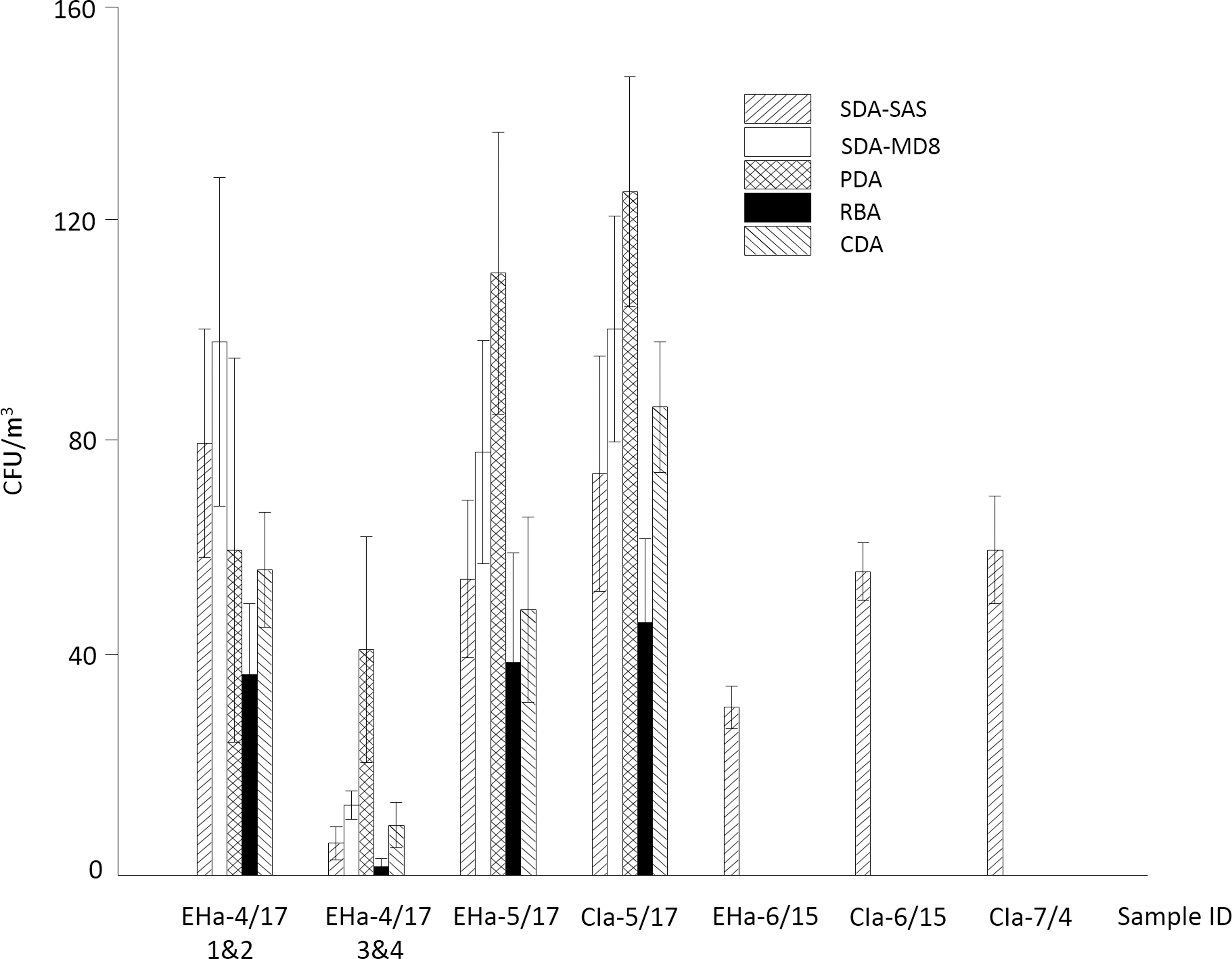
Culturable fungal concentration in the aerosol of the AIT Center as determined by the use of various culture media. Bars are pattern-coded to represent different culture media (as indicated in the figure). SDA-SAS represents the result of direct sampling of the sampler, and the remaining four represent results of the Sartorius MD8 sampler. CDA, Czapek–Dox agar; PDA, potato dextrose agar; RBA, Rose Bengal Agar; SAS, DUO SAS SUPER 360 sampler; SDA, Sabouraud's Dextrose Agar.
4. Discussion
In this study, an effective LAMP primer set was designed for detection of fungi in aerosols from low-biomass spacecraft assembly clean room environments. The positive detection rate of the LAMP assay was the same as that obtained with traditional culture methods and higher than that obtained with qPCR methods. The qLAMP assay designed in this study has the advantage of providing results within 1.5 h. This is not only remarkably quicker than the time required for culture-based and qPCR methods but there is also no risk of secondary pollution, which is a requirement of microbial detection on a space station. The LAMP reaction time would be expected to be shortened in the future by adding loop primers (Nagamine et al., 2002). Moreover, when compared with traditional qPCR, the qLAMP reaction can be carried out at 60–65°C under isothermal conditions. In fact, the LAMP assay has already been applied to a microfluidic multiplex electrochemical chip for differentiating bacteria (Luo et al., 2014). All of these characteristics are promising for the design and implementation of future in-orbit equipment.
Twelve genera of fungi were isolated from the aerosol sampled from the spacecraft assembly clean room. These microbes are likely to be taken in orbit in the future along with the launch of China's space station. Eight of the genera (Chaetomium, Exophiala, Fusarium, Histoplasma, Paecilomyces, Scopulariopsis, Trichophyton, and Malassezia) were not found in the environment of the ISS, and four genera (Exophiala, Histoplasma, Trichophyton, and Malassezia) were not found in the environment of the MIR (Novikova, 2004). Aspergillus sp. and Penicillium sp. dominated the fungal population, which was consistent with biocontamination on board the ISS (Novikova et al., 2006). The concentrations of airborne fungi in the ISS were lower than 44 CFU/m3, which was similar to contamination levels in dressing room samples in this study. According to preliminary data obtained during this research, theoretically, this concentration range should be detectable by our qLAMP assay. However, the detection efficiency of qLAMP in orbit requires further verification.
The number of fungi detected by qPCR and qLAMP were higher than the number detected by plate counts. There are at least two reasons for this. (1) The number of gene copies per genome can potentially influence qPCR and qLAMP quantitation (Manter and Vivanco, 2007). It has been reported that the number of rRNA copies per genome ranges from 50 to 100 in filamentous fungi (Rooney and Ward, 2005), therefore the ITS1 and 28S rRNA gene copy numbers in isolates in this study may be relatively high because Aspergillus and Penicillium were the predominant genera. (2) Estimates in some reports predict that in some environments, the majority of microorganisms are, as yet, unculturable. Therefore, it is undoubtedly true that there are a variety of fungal genera in spacecraft assembly clean rooms that cannot be isolated, but may be detected by qPCR and qLAMP because of the universality of the ITS1 primer and the designed 28S rRNA LAMP primer. In general, both molecular and culture-dependent methods showed similar results. At present, standards and limits of microbial burden in orbit and preflight are based on culture-dependent methods (Pierson et al., 2013). With continuous improvement and perfection, we expect that molecular methods will be used as new standards in the future.
The two DNA extraction kits selected for this study have been commonly used in the analysis of environmental microorganisms (Kumari et al., 2016; Yu et al., 2016) and our data indicate that they have the same extraction efficiencies. A series of preliminary experiments were performed in this study and the results indicated that yields were significantly higher when a combination of ball milling in liquid nitrogen, freeze–thawing, and sonication-based methods was employed. These three methods aid cell disruption (Richard et al., 2002), but may not be appropriate when applying qLAMP reactions to a microfluidic chip. Incorporation of a qLAMP assay onto a microfluidic chip would be potentially convenient for microbial detection in orbit in a space station because of its ease of manipulation, rapid results, and protection against biological contamination.
Multiple studies have shown that the use of PMA, a dye that can differentiate between total and the biomass of intact cells, enables more accurate analysis of viable microorganisms to be achieved (Nocker et al., 2007; Vaishampayan et al., 2013). It was also shown that PMA treatment might be successfully used to determine dead and viable counts for many fungal species (Vesper et al., 2008; Crespo-Sempere et al., 2013). In this study, PMA-treated and PMA-untreated samples varied significantly in biomass. Since viable microorganisms are usually most critical in determining whether the presence of a microorganism poses a risk to human and equipment safety, our result confirmed that PMA treatment is important when accurately determining the molecular fungal biomass in space station environment samples.
SYBR Green I was used as the dye for both qPCR and qLAMP in this study. This reduced the uncertainty factor caused by dye difference when comparing the two methods. Cross contamination of samples did not occur. SYBR Green I is nearly the most common DNA dye used in previous reports on the study of spacecraft microbes (Mayer et al., 2016; Koskinen et al., 2017) and also showed good performance on qLAMP (Huang et al., 2015). However, some studies show that when some certain species are used as research objects, Syto-82 was demonstrated to be the best dye for both qPCR (Gudnason et al., 2007) and qLAMP (Oscorbin et al., 2016). Therefore, it is worth comparing the properties of different DNA dyes on space station samples in the future.
It is well known that humans and microorganisms in space are exposed to both space radiation and microgravity. It is possible that a variety of microorganisms, especially fungi, can cause unexpected infections or allergic diseases in astronauts whose immune systems are disturbed in an uncertain degree and way in extreme space environments (Makimura et al., 2001). At the same time, microbial mutations could occur more frequently in the space environment (Elena and Lenski, 2003; Nickerson et al., 2004). At present, fungal strains that may cause harm are not clear enough. With more and more comprehensive understanding of species of harmful fungi, it would be important to monitor for specific fungal species by use of specific primer systems instead of a universal primer in the future.
5. Conclusions
A specific real-time qLAMP method was developed for detecting low-biomass aerosol fungi with a minimum detection limit of 102 28S rRNA gene copy numbers/m3. Three simple pretreatment steps were designed that ensured high-efficiency DNA extraction. The effectiveness of this method was successfully verified by testing aerosols from the CSSAC. Findings revealed the culturable concentration and diversity of the airborne fungal pollution in China's space station during assembly and in its assembly clean room. This qLAMP method is a promising candidate for microbial detection in orbit on manned spacecraft.
Footnotes
Acknowledgment
This work was financed by the National Natural Science Foundation of China (Project No. 31600404).
Author Disclosure Statement
No competing financial interests exist.
Associate Editor: Petra Rettberg
Abbreviations Used
References
Supplementary Material
Please find the following supplemental material available below.
For Open Access articles published under a Creative Commons License, all supplemental material carries the same license as the article it is associated with.
For non-Open Access articles published, all supplemental material carries a non-exclusive license, and permission requests for re-use of supplemental material or any part of supplemental material shall be sent directly to the copyright owner as specified in the copyright notice associated with the article.
