
Abstract
Antibody-based analytical instruments are under development to detect signatures of life on planetary bodies. Antibodies are molecular recognition reagents able to detect their target at sub-nanomolar concentrations, with high affinity and specificity. Studying antibody binding performances under space conditions is mandatory to convince space agencies of the adequacy of this promising tool for planetary exploration.
To complement previous ground-based experiments on antibody resistance to simulated irradiation, we evaluate in this paper the effects of antibody exposure to real space conditions during the EXPOSE-R2 mission outside the International Space Station. The absorbed dose of ionizing radiation recorded during the 588 days of this mission (220 mGy) corresponded to the absorbed dose expected during a mission to Mars. Moreover, samples faced, at the same time as irradiation, thermal cycles, launch constraints, and long-term storage. A model biochip was used in this study with antibodies in freeze-dried form and under two formats: free or covalently grafted to a solid surface.
We found that antibody-binding performances were not significantly affected by cosmic radiation, and more than 40% of the exposed antibody, independent of its format, was still functional during all this experiment. We conclude that antibody-based instruments are well suited for in situ analysis on planetary bodies.
1. Introduction
In the context of planetary exploration, the use of miniaturized instruments based on target-binding reagent with high specificity and binding affinity is clearly relevant. Indeed, such instruments can, in principle, detect unambiguously thousands of different targets from small molecules like amino acids to complex molecules and microorganisms in a single assay. The basic principle can be summarized as follows: the target-binding reagents, like antibodies (Ab), are fixed on a solid surface to specifically capture their target. In direct methods, target interaction with the binding site of Ab and the use of appropriate reagents generate a visible signal capable of being measured, for instance, by a CCD camera. In the field of astrobiology, several Ab-based biochips have been proposed as miniaturized high-throughput detection systems, to detect biomarkers, especially organic ones, in the search for extraterrestrial life (Parro et al., 2005, 2008, 2011a, 2011b; Le Postollec et al., 2007; Martins, 2011; Sims et al., 2012; McKay et al., 2013; Smith and Parro, 2014).
However, space is a hazardous environment in particular due to strong irradiation from primary and secondary particles produced by galactic cosmic rays and solar energetic particles, temperature variations, and long-duration storage (during the cruise phase especially). As a consequence, one main concern relies on the resistance of biochips to these cumulative space conditions. In recent years, studies have been performed to evaluate the resistance of Ab to specific space conditions, especially regarding the effect of some ionizing particles on Ab binding performances (Le Postollec et al., 2009a, 2009b; Baqué et al., 2011, 2017; de Diego-Castilla et al., 2011; Coussot et al., 2017). However, these ground-based simulations were limited due to the use of one single type and energy particle at a time and the experimental characteristics of the beam (high fluxes in a short exposure time, unidirectional irradiation). In the space environment, biochips would be exposed to continuous and sporadic fluxes of various particles in a large range of energies and all directions, concomitantly with other parameters inherent to a space mission including thermal variations, vibrations, and storage conditions.
To complement ground-based studies, Derveni et al. conducted in 2007 a 12-day mission on the BIOPAN-6 low Earth orbit platform to demonstrate the effects of cumulative irradiation on two antibodies' ability to bind to their respective antigens (Derveni et al., 2012, 2013). The absorbed dose recorded at the samples' position by the dosimeters was 2.4 mGy during the experiment. Both freeze-dried Ab, in free format and absorbed into laser-cut glass fiber pads, were reported as unaffected with regard to their target-binding abilities after their low Earth orbit platform exposure. Analyses were done with conventional indirect enzyme-linked immunosorbent assays (ELISA). However, major loss of Ab activities (>70%) was pointed out and explained by the Ab preparation procedure, in particular the freeze-drying step and storage time period. These issues complicated the interpretation of the results. This study highlighted that using Ab as target-binding reagent necessitates controlling its ability to bind to its target (referred to also as its functionality) after each step of the antibody-based device development. Consequently, for biochips, a control is required after the Ab immobilization onto the solid surface (to determine the initial rate of functional Ab), but also after washing, freeze-drying, storage, and rehydration steps. These controls permit to precisely quantify the number of Ab that remains functional after the preparation procedure and before undergoing some putative deleterious experiments.
In the frame of the BiOMAS project (Biochip for Organic Matter Analysis in Space), we developed a model biochip, in which a specific Ab (anti-horseradish peroxidase Ab, hereafter anti-HRP Ab) was used in free format or covalently immobilized onto a solid surface. A direct detection method called “antibody anti-horseradish peroxidase (A2HRP)” was developed and fulfilled the validation criteria requested by the Food and Drug Administration and the European Medicines Agency for the validation of bioanalytical methods (Coussot et al., 2018a, 2018b). This gold standard method permits to evaluate accurately and precisely the number of free or immobilized functional Ab (interday variation <12.1%). The lower limit of quantification corresponds to 1.40 ± 0.18% of the initial rate of active Ab. The A2HRP method was successfully used in optimizing freeze-drying conditions to better preserve the Ab functionality, notwithstanding its format (Coussot et al., 2018c), and in evaluating the Ab resistance under various stressful environments (Coussot et al., 2017, 2018c).
In this work, we first summarize our experiment (see also Vigier et al., 2013), which was part of the Photochemistry on the Space Station (PSS) project (Cottin et al., 2015), and the analytical developments we conducted prior to the final analyses performed on the samples a few weeks after their return to Earth (Coussot et al. 2018a, 2018b, 2018c). Both formats, free anti-HRP Ab and anti-HRP Ab immobilized onto a surface, have been conditioned into a homemade sample holder unit and installed outside the International Space Station (ISS) on the EXPOSE-R2 platform. We then present the results on the resistance of our model biochip against a long-time exposure (more than 18 months) to real space conditions. Finally, the relevance of antibody-based biochips for space exploration is discussed.
2. Materials and Methods
2.1. Chemicals, reagents, and materials
Mouse monoclonal anti-horseradish peroxidase antibodies (anti-HRP Ab) were obtained from MyBioSource (clone number B215M, batches 2F17811 & 2F15911, USA). Bovine serum albumin (BSA, fraction V, 96–100% protein, batch SLB38588V), horseradish peroxidase (HRP, EC 1.11.1.7, batch SLBF8268V, with a purity index of 1.9), Tween 20 (impurities ≤3% in water), o-phenylenediamine dihydrochloride (OPD-2HCl), hydrogen peroxide (H2O2, 30% (v/v) solution equivalent to a titer of 120V of oxygen (10.73 M) upon opening [Coussot et al., 2018c]), Dulbecco's phosphate-buffered saline (DPBS, 10 × solution, pH 7.4, used in a final concentration of 1 × in water solvent), sulfuric acid (H2SO4, ≥97.5%), citric acid (≥99.5%), sodium hydroxide solution (10 M solution for molecular biology), sodium phosphate dibasic dodecahydrate (Na2HPO4·12H2O, ≥99.0%), sodium bicarbonate (NaHCO3, ≥99.5%), sodium carbonate (Na2CO3, ≥99.5%), D(+)-saccharose (sucrose, ≥99.0%), L-histidine (His, ≥99.0%), D-arginine (Arg, ≥99.0%), ethanol (EtOH, ≥99.5%), hydrochloric acid (HCl, 37%) were purchased from Sigma-Aldrich (Saint-Quentin Fallavier, France). Corning DNA Bind 8-well strip plate with N-hydroxysuccinimide modified surface (NHS-wells) was provided by Sigma-Aldrich (Saint-Quentin Fallavier, France). NHS-wells were manufactured by Air Liquide (Sassenage, France) to fit perfectly with the shape and size of EXPOSE-R2 closed cells provided by the French Space Agency (CNES, Toulouse) (Vigier et al., 2013), called simply “cells” in the following. These home-designed NHS-wells had a diameter of 8.4 mm with a 7.1 mm height. Teflon caps were specially manufactured by Air Liquide to close these custom-designed NHS-wells (Coussot et al., 2019). The ultrapure water was obtained from a Millipore purification system. Other chemicals are analytical grade and used as received. During the assays, the relative humidity and air temperature were controlled with a thermohygrometer Testo 605-H1 (Type 05600610, identification number 39227197/205), equipment whose calibration is traceable to national standards (certificate number 1306125). During all assays, room temperature (RT) was 23.2 ± 1.3°C, and recorded relative humidity (RH) was 25.7 ± 0.9%.
2.2. Sample preparation
Long-time exposure to real space conditions during the EXPOSE-R2 mission was evaluated on freeze-dried Ab in both covalently immobilized format (labeled “G” for grafted) and in-solution Ab format (free anti-HRP Ab labeled “F” for free). The immobilization strategy, the freeze-drying optimized procedure, and the A2HRP validated protocol are detailed elsewhere (Coussot et al., 2017, 2018a, 2018b, 2018c). The freeze-drying stock solution was freshly prepared and was composed of 1.05 g of citric acid (0.1 M), 38.75 mg of His (5 mM), 52.5 mg of Arg (5 mM) in a final volume of 50 mL of water and adjusted to pH 6.5 with NaOH (10 M) before adding 10 μL of Tween 20.
2.2.1. Preparation of anti-HRP Ab in free format
The home-designed NHS-wells were filled up with 250 μL of BSA (3% (w/v) in DPBS (1 × )). After overnight reaction time at RT in the dark, the wells were emptied and rinsed three times with DPBST solution (DPBS (1 × ) with 0.05% (v/v) Tween 20) and three times with DPBS. An anti-HRP Ab solution was directly prepared at 200 μg·mL−1 in the above freeze-drying solution immediately before deposit into the BSA-saturated home-designed NHS-wells (noted also as inactivated BSA wells). In each inactivated BSA well, a volume of 100 μL of the anti-HRP Ab in the freeze-drying solution was mixed with 20 μL of a freshly prepared sucrose solution (5g·L−1 in water). The wells were placed within the homemade prefrozen aluminum block designed by our group and kept frozen by adding liquid nitrogen to follow the optimized freeze-drying procedure (Coussot et al., 2018c) as described in Section 2.3.
2.2.2. Preparation of anti-HRP Ab in covalently immobilized format
The home-designed NHS-wells were filled up with 200 μg·mL−1 of anti-HRP Ab solution in DPBS (1 × ). After a minimum of 4 h under gentle agitation at RT, a saturation step with 250 μL of BSA solution (3% w/v in DPBS) per well was carried out to prevent nonspecific bindings (Baqué et al. 2011), followed by three washings with DPBST solution and three washings with PBS. In each covalently immobilized and saturated Ab well was added 100 μL of the freeze-drying solution and 20 μL of a freshly prepared sucrose solution (5g·L−1 in water). Samples were then freeze-dried by using the homemade aluminum holder as described in Section 2.3.
2.3. Freeze-drying procedure
The aluminum holder had the advantage of running multiple samples at the same time, avoiding freeze-drying batch effects. As described elsewhere, this device was prefrozen and transferred into the central part of a freeze-dryer (Christ Alpha 2–4 from Martin Christ GmbH, Germany) (Coussot et al., 2018c). Freeze-drying was performed overnight (condenser temperature −85°C, vacuum 0.05 mbar). When the freeze-drying process was achieved, the chamber of the freeze-dryer was filled with nitrogen gas before closing the sample-containing aluminum holder, which kept the samples hermetically sealed and sheltered them from the moisture and light until its opening. The opening of the aluminum holder was done in a glove box under a controlled atmosphere of helium (10% He) in argon (Ar) provided by Air Liquide (Sassenage, France) to maintain a RH level of 10–15%. During our experiments in the glove box, the RH and air temperature were controlled with a thermohygrometer Testo 605-H1 (Testo, France). RH was 12 ± 3%, and air temperature was 22.8 ± 2.3°C. After opening the aluminum holder, freeze-dried samples were capped and directly transferred into the CNES closed cells with a tool vacuum suction pen (FFQ939 from Mayf's Online Shop, China). Into each cell, two thermoluminescent dosimeters were placed at the bottom of the cell, below the samples. These passive dosimeters were analyzed at the end of the mission. The absorbed dose measured into the cells was 220 mGy that corresponds to the dose that can be expected during a mission to Mars including 8 months travel and 18 months at Mars' surface. (Hassler et al., 2014; Coussot et al., 2019). The cells were then screwed with a final tightening of 0.7 Nm with a torque screwdriver TorqueVario-S (Wiha, Germany) (Vigier et al., 2013; Coussot et al., 2019).
2.4. Experiment outside the ISS
To study the impact of all constraints encountered by a biochip during an entire space mission (long-duration storage, transportation, take-off/landing shocks, thermal constraints, and cumulative effects of cosmic ray particles), 41 anti-HRP Ab samples (21 anti-HRP Ab in the free format “F” and 20 in the covalently immobilized format “G”) took part in the “Biochip in PSS experiment” (Vigier et al., 2013; Cottin et al., 2015) during the EXPOSE-R2 mission. An overall view of the EXPOSE-R2 mission is illustrated in Fig. 1. All samples (14 exposed on the ISS and 27 kept on Earth) were simultaneously prepared, within the same freeze-drying batch, as described in the experimental section (2.3). The 14 exposed cells were spread on the two exposure levels of Tray 3 of the EXPOSE carrier dedicated to the PSS experiment (4F and 3G anti-HRP Ab in each level). Disposition of the exposed cells on the sample carrier was the same for the upper and the lower level of Tray 3 (denoted as upper tray and lower tray, respectively). Launch to the ISS (July 23, 2014) and storage inside the ISS was at ambient temperature (22–25°C on average) (Rabbow et al., 2017). Then, the EXPOSE-R2 platform was placed outside the ISS on Universal platform D on August 18 with opening of the valves for venting the inner part of EXPOSE-R2 on August 20 and removal of the UV shield on October 22, 2014. On February 3, 2016, trays were covered and brought back inside the ISS. Thus, the exposed samples spent 566 days outside the ISS over the 588 days of the mission (1 year, 8 months).
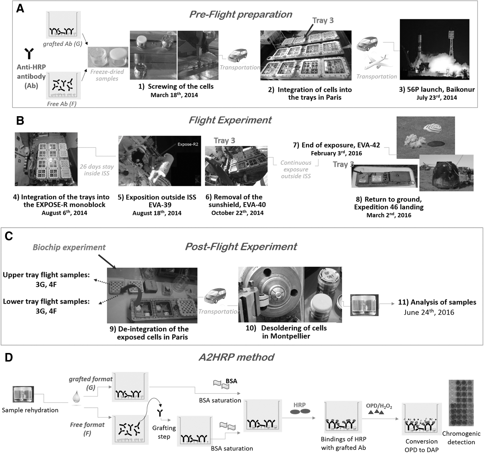
An overall view of the EXPOSE-R2 mission with two formats of freeze-dried anti-HRP antibody (Ab): first is grafted format (G) with Ab covalently immobilized on the well-surface, and second is free format (F) with Ab prepared in solution. (
During their extravehicular exposition, the samples on Tray 3 were submitted to radiation and temperatures varying between −20.9°C and 57.98°C (Rabbow et al., 2017). On March 2, 2016, EXPOSE-R2 Tray 3 landed on Earth. During transit from the Baikonur cosmodrome to Moscow, and from Moscow to the German Aerospace Center (DLR, Germany), a recording of temperatures was done; the temperatures oscillated between 20°C and 24°C. Then, from DLR to LISA (Laboratoire Interuniversitaire des Systèmes Atmosphériques, Créteil, France) (Rabbow et al., 2017) and from Créteil to Montpellier a controlled chamber at 4°C was used during transportation in the summer period. De-integration of the exposed cells from the sample carriers was done according to MUSC/DLR/ESA/CNES internal procedures (Rabbow et al., 2017). On June 24, 2016, the soldered joint of all the cells (including ground references) was removed, by using a mechanical lathe and a manufactured CNES tool, in the mechanics department of the Montpellier University (Coussot et al., 2019). Samples were immediately sealed in a FoodSaver bag (Fischer Scientific, France) and stored in the dark at 4°C until analysis with the A2HRP method (Section 2.7).
2.5. EXPOSE-R2 mission ground references
During the same period, experiments were performed on ground to control the long-term behavior of anti-HRP Ab upon storage but not exposed to space conditions. A total of 27 mission ground controls were studied. Six ground cells (3F and 3G anti-HRP Ab) were stored at the French Space Agency (CNES, Toulouse) by maintaining an accurate temperature control at 3.9 ± 0.8°C over that period. Twenty-one ground cells were kept in DLR (Cologne, Germany): six ground cells (3F and 3G anti-HRP Ab) were stored at 5°C; eight ground cells (4F and 4G anti-HRP Ab) underwent the same thermal history as the ISS samples (DLR ΔT); and seven ground samples (4F and 3G anti-HRP Ab) were stored in conditions combining long-time storage, varying thermal environment as the ISS ones, and UV radiation (DLR ΔT+UV) (Rabbow et al., 2017). All of these 27 ground cells were brought back to Montpellier to be analyzed simultaneously with the 14 exposed cells during all the desoldering process and 4°C storage before rehydration. For the free format, rehydration was done with 100 μL of water and 20 μL of carbonate-bicarbonate buffer (0.1 M, pH 9.2) per well in order to fix the pH of the sucrose-containing freeze-drying Ab solution to 7.4 to preserve Ab functionality. Free Ab were then covalently coupled to NHS-wells following the procedure described in Section 2.2.2. For the grafted format, rehydration was carried out with 120 μL of DPBS. The rate of functional Ab in both formats was determined with the A2HRP method (Section 2.7, Fig. 1D).
2.6. Additional ground thermal cycling experiments
The anti-HRP Ab were freeze-dried in both formats as described in Section 2.3. Thermal cycling experiments were carried out on freeze-dried samples in their sealed bags in a temperature test chamber (Vötsch VT 4004). Two independent thermal variation experiments were done: one with a sharp rise in temperature (5°C/min) reaching a 1 h plateau at 80°C (referred to as “80°C peak” in the text), and a long cycling period (178.8 h, or 7.45 days) mimicking thermal variations of exposed cells (Coussot et al., 2019) with an amplitude of about 70°C (referred to as “long cycle” in the text). Samples were rehydrated as described above and analyzed with the A2HRP method.
2.7. Validated A2HRP method and data interpretation
For all ground-based experiments and that of the overall ISS mission, the A2HRP method (Fig. 1D) was used to evaluate the functionality of anti-HRP Ab toward its antigen HRP. Briefly, the principles are as follows: once the anti-HRP Ab were immobilized onto the NHS-wells to reach a density of 3.9 × 10¹¹ antibody per mm2 (Section 2.2.2) (Moreau et al., 2011), a HRP stock solution (1 g·L−1) was prepared by dissolving the enzyme in a stabilizing solution composed of 0.1 M Na2HPO4 and 0.05 M citric acid, pH 5.2. A HRP working solution at 200 μg·mL−1 was prepared immediately before use from the HRP stock solution by dilution in DPBS. A volume of 110 μL of the HRP working solution was pipetted into wells containing the previously grafted anti-HRP antibody to saturate all of its binding sites (Moreau et al., 2011). At least 2 h at RT or overnight incubations were considered for maximal binding of HRP to anti-HRP Ab surfaces. Unbound HRP was removed by rinsing three times with DPBST and three more times with DPBS. The wells were then incubated with a freshly prepared o-phenylenediamine dihydrochloride/hydrogen peroxide (OPD/H2O2) solution. The OPD/H2O2 reaction solution was composed of 1 mL OPD stock solution at 5 g·L−1 in deionized water, 8.6 mL of stabilizing solution described above, and 400 μL of 30% H2O2. Mixed into each well were 50 μL of this OPD/H2O2 reaction solution and 50 μL of stabilizing solution. The HRP/OPD/H2O2 reaction that converts OPD to 2,3-diaminophenazine (DAP) was then stopped after 4 min by adding 30 μL of STOP solution (H2SO4, 4 N, pH <1). The HRP-catalyzed OPD oxidation into DAP was recorded at 490 nm with an Infinite 200 absorbance microplate reader from Tecan (Lyon, France). The activity of the Ab surfaces is expressed as a percentage of the sample/reference ratio of its net absorbance values. The net absorbance values correspond to the measured assay values minus the mean absorbance of the blank obtained with inactivated BSA wells (Section 2.2.1).
All data were reported as the mean ± the standard deviation from at least three replicate experiments. Statistical significance of the assays was determined with Student's t test (p = 0.05).
3. Results
Stress affects Ab performances in different ways depending on the type and duration of the event (Coussot et al., 2018b, 2018c). Irradiation effects on anti-HRP Ab were assessed here by evaluating the functionality of the anti-HRP Ab before and after the stress event or by comparing the activity of exposed Ab samples to that of non-exposed ones (named ground references [GR] or controls; see above). Analyses were done according to the validated A2HRP method (Fig. 1D). The A2HRP method was demonstrated to have the potential for analyzing the binding ability of the anti-HRP Ab in both formats even if deleterious events occurred on the anti-HRP Ab before its coupling to the surface (Coussot et al., 2018c). This direct assay permits a precise and reliable quantitation of HRP bound on the immobilized Ab surface, and consequently permits an evaluation of the anti-HRP Ab binding capacities. It has also demonstrated its suitability to evaluate discrepancies in Ab bindings after their exposure to short-term irradiation events or forced degradation studies (Baqué et al. 2017; Coussot et al., 2017, 2018a, 2018b, 2018c). In the present paper, we evaluate the Ab resistance after their exposure to real space conditions, and thus for the first time the effect of cumulative and long-term stress events on the performance of a grafted antibody. In order to broadly represent any future Ab-based biochip instruments, we evaluated in a second part free Ab format since both free and grafted Ab are considered for future applications in planetary exploration.
3.1. Resistance of anti-HRP Ab in its grafted format after exposure outside the ISS
Since radiation gradients were observed on the EXPOSE platform in previous ISS missions (Berger et al., 2012), the 14 exposed Ab “flight samples” were spread on the two exposure levels of Tray 3 of the EXPOSE carrier dedicated to the PSS experiment, but with the same disposition on the upper and the lower level of the carrier. All the experiments performed on grafted Ab are reported in Fig. 2. To only take into account the effects encountered during the ISS mission (flights, extravehicular exposure, and all transportation during the mission), the percentage of functional Ab is calculated by using the freeze-dried ground samples stored at 5°C in the DLR facility (n = 2 replicates) during all the duration of the mission. At first view, the results reveal a possible degradation of several samples, including flight samples. Higher variability in the estimated recognition capabilities of our Ab has been observed when using custom-designed NHS-wells with a within-assay precision equal to 10.3% for that custom-designed wells batch, in comparison with conventional ones (within-assay precision is less than 7.1%; Coussot et al., 2018b). The effects of transportation are evaluated by confronting activity of ground controls stored in DLR with that of CNES ground samples. Remaining activity of both CNES and DLR ground controls is not statistically different (Student's t test). No significant effect is also observed with the ground sample named DLR ΔT+UV, which combines long-time storage, varying thermal environment mimicking the ISS ones, and UV radiation. To complement these data, we run here additional ground thermal cycling experiments to evaluate whether an effect due to a long-term exposure to temperature variations during the mission can be invoked. Ground thermal cycling experiments were carried out on the anti-HRP Ab over a 80°C short peak of temperature, a long cycle mimicking thermal variations of flight samples (see Section 2.6, in the experimental part), and compared with an identical storage time at 4°C. All assays were performed with eight replicates. Ab activities are presented in Fig. 3. A degradation of one-third of initially active grafted Ab (corresponding to 66% of surface active Ab) is shown during the freeze-drying process by comparing data obtained with fresh G Ab that did not undergo freeze-drying with that of rehydrated sample upon opening of the aluminum holder (Fig. 3, first two shaded bars with gray lines). In addition, comparing results from the overall process, cumulating in the freeze-drying step and storage, shaded gray bars indicate a slight but significant difference in activity between the three stored freeze-dried samples (55.5 ± 4.1 for the 4°C storage; 48.5 ± 3.2 for the 80°C peak; 46.7 ± 4.1 during the long cycle) and the freeze-dried sample analyzed with no storage (66.8 ± 6.7), whereas the percent Ab activity values after the sole freeze-drying process are 83.1 ± 14.5, 69.8 ± 13.1, and 72.5 ± 12.1 for the freeze-dried 4°C storage, the 80°C peak, and long cycle, respectively, indicating that there were not significant differences (p > 0.05) between the reference sample with no storage and the three stored ones (Fig. 3, filled gray bars).

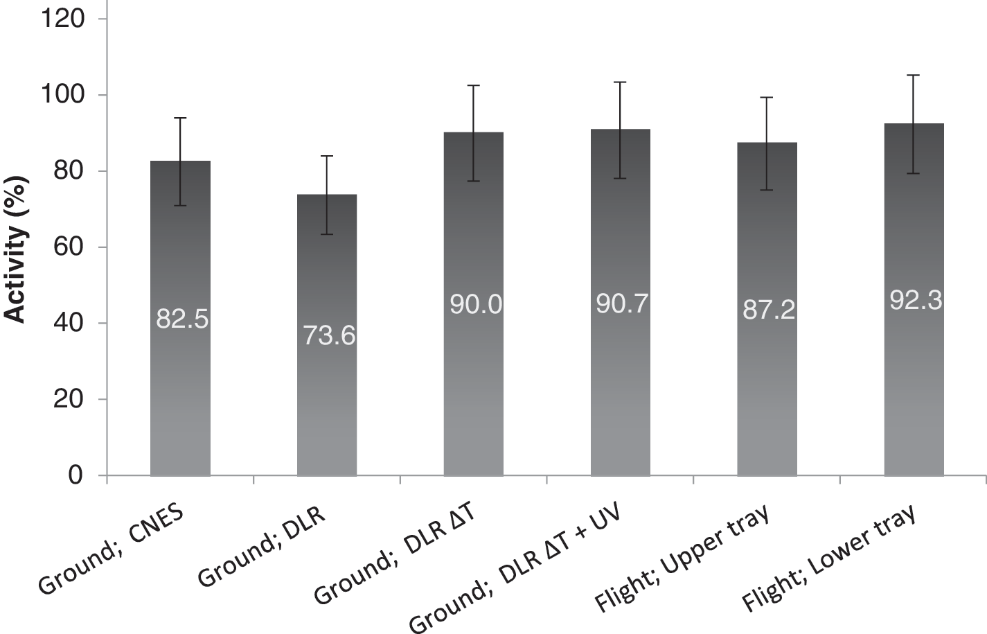
Binding activity of the grafted anti-HRP Ab obtained for ground controls (number of replicates: CNES n = 3; DLR n = 2; DLR ΔT n = 4; DLR ΔT+UV n = 3) and flight samples (n = 3 replicates for both lower and upper tray) by the A2HRP method. Each filled gray bar represents the mean and standard deviation of the Ab activity (normalized value in %, calculated against freeze-dried ground controls stored in DLR at 5°C [n = 2 replicates]).
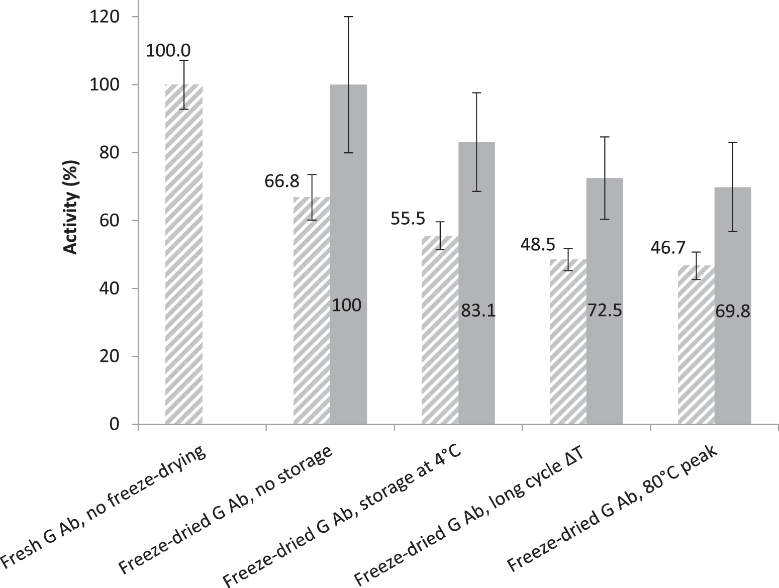
Binding activity of the grafted anti-HRP Ab (G Ab) obtained without and after freeze-drying, without storage (instantaneous rehydration after freeze-drying), with storage at 4°C for freeze-dried control, with sharp rise in temperature (80°C peak), and a thermal cycling experiment mimicking thermal cells' exposure outside the ISS (long cycle ΔT) but in accelerated time: 1 h of EXPOSE-R2 mission was converted into 1 min to carry out this experiment in the lab with similar temperature amplitude (Coussot et al., unpublished data). Each bar represents the mean and standard deviation of the Ab activity (n = 8 replicates, normalized value in %) calculated against freshly prepared Ab surface (shaded bars with gray lines) to evaluate cumulative effects or calculated against freeze-dried Ab without storage (filled gray bars) to evaluate the effects of storage and temperature.
3.2. Resistance of anti-HRP Ab in free format after exposure outside the ISS
Since a 30% functional activity difference in freeze-dried Ab, between the free format and the covalently immobilized one, has been reported in literature depending on the freeze-drying formulation components (Coussot et al., 2018c), and only free Ab format has been studied in previous experiments on the BIOPAN-6 low Earth orbit platform (Derveni et al., 2012, 2013), the resistance of free anti-HRP Ab after exposure to real space conditions is detailed hereafter.
Previous data obtained by using the same freeze-drying solution adjusted to pH 7.4 for Ab immobilization to the NHS-wells showed that free Ab activity losses under 25 ± 2% could not be quantified with the A2HRP method due to sample preparation, multistep procedure, and handlings (Coussot et al., 2018c). However, the A2HRP method has the advantage of providing reliable and precise data for stability studies of the free anti-HRP Ab, by comparing with conventional competitive inhibition assays. Indeed, changes in Ab properties upon degrading conditions were proved to dramatically affect the data in conventional immunoassays with high variability of the results. It is also restricted to competitive mechanism of inhibition; if other mechanisms occurred at the same time in the sample, it would generate uninterpretable data. All the results from the EXPOSE-R2 mission samples illustrated in Fig. 4 were obtained with the A2HRP method. No significant alteration of the free anti-HRP Ab is observed after both 80°C peak and long cycle exposures (data from ground experiments not shown). As shown by Coussot et al. (2018c), a possible additional air moisture degrading effect might have occurred during the storage of the freeze-dried F anti-HRP Ab at 4°C (sealed bag not perfectly hermetic). Owing to the inherent conditions of the rehydration step, this result is not surprising and correlates well with the above observations on the freeze-dried G Ab, and that of free GR stored in DLR (73.7 ± 10.3%). Consequently, considering all the results presented in Fig. 4, no significant differences (p > 0.05) are observed in the binding activity of the anti-HRP Ab, under its free format, with the same order of activity for the flight samples as for the ground ones. As before, a 37% drop in remaining free Ab activity is necessary to detect alterations in Ab binding efficiency from cosmic radiation during flight samples' exposure outside the ISS.
Binding activity of the free anti-HRP Ab obtained for ground controls (number of replicates: CNES n = 3; DLR n = 3; DLR ΔT n = 4; DLR ΔT+UV n = 4) and flight samples (n = 3 replicates for both lower and upper tray) by the A2HRP method. Each dark bar represents the mean and standard deviation of the activity (normalized value in %, calculated against a freshly prepared freeze-dried ground reference).
4. Discussion
In this study, we evaluated the effect due to a long-term exposure to temperature variations on the activity of anti-HRP Ab in its grafted format but independently from flight samples (Fig. 3). Indeed, the effect of short exposure of covalently grafted Ab at elevated temperatures has been published previously (Coussot et al., 2018c). It revealed that about 60% of the initial activity of covalently immobilized Ab was preserved during short temperature stress. We reported also that freeze-dried samples suffered less from stress due to temperature exposure than from the rehydration step, probably due to changes in composition of the solid form upon contact with air moisture, which was supposed to lead to only a partial refolding of the freeze-dried Ab. Thus, after exposure of grafted Ab to thermal variations, activity changes, if they occurred, might not be due to an effect of temperature but rather from storage time. Indeed, the results of remaining Ab activity after the freeze-drying process show no discrepancy (p > 0.05) between the reference sample with no storage and the three stored ones (Fig. 3, filled gray bars). This means that an alteration of Ab bindings other than those occurring during the freeze-drying process/rehydration or thermal variations might be evoked only if there is more than a 40% drop in remaining Ab activity (from freeze-dried control) after a long-duration experiment. This is consistent with tests done on GR samples illustrated in Fig. 2.
Measurements performed with passive dosimeters during the EXPOSE-R2 mission revealed, a posteriori, that the absorbed dose difference between cells at upper and lower levels was low (about 3 mGy for the whole mission, corresponding to 1.4% of the total). We explained this difference due to an estimated shielding of thermoluminescent dosimeters of 0.82 g/cm2 on the upper level (Coussot et al., 2019), so the absorbed dose remains quasi uniform between the two levels. This can explain why the results we obtained for the “upper tray” and the “lower tray” samples have no statistical differences. Indeed, we obtained a remaining activity of 50.6 ± 9.0% for the flight “upper tray” samples and 62.9 ± 11.0% for the flight “lower tray” for grafted Ab (Fig. 2). For free format, our results showed that a 37% drop in remaining free Ab activity is necessary to detect alterations in Ab binding efficiency. In other words, this shows that much more than 63% of the free anti-HRP Ab are still functional after their long-duration exposure outside the ISS (Fig. 4). Consequently, based on all the above considerations, our results show that grafted and free Ab partially keep their recognition capabilities during the overall flight mission, and that the degradations from cosmic radiation, if any, are too small to be detected by our analysis protocols.
Nevertheless, due to the high number of potentially hazardous factors encountered during a space mission (McKenna-Lawlor et al., 2012), our experiment demonstrates with confidence, for the first time, that much more than 40% of Ab (whatever the format) survived the long-duration exposure outside the ISS and remained functional.
Antibody-based biochips have not been used yet for planetary exploration missions despite their high potential for searching tracers of extinct or extant life (Parro et al., 2011c). One major concern for an instrument based on this technology is radiation effect issues on the antibodies. Many laboratory experiments have been performed so far at different energies with different particles and high fluences (much more important than suspected for a mission to Mars, for instance) to test the ability of antibodies to recognize their target after radiation exposure (Le Postollec et al., 2009a, 2009b; Baqué et al., 2011, 2017; de Diego-Castilla et al., 2011; Coussot et al., 2017). A 12-day mission on the BIOPAN-6 low Earth orbit platform has been performed to study the effects of cumulative irradiation on two antibodies' ability to bind to their respective antigens (Derveni et al., 2012, 2013). In the present paper, we improved previous studies to better test the effects of real space constraints on antibodies during an EXPOSE-R2 mission outside the ISS. In terms of radiation, the total accumulated radiation dose recorded by the dosimeters during the EXPOSE-R2 is 220 mGy, which is much higher than those measured during the BIOPAN-6 low Earth orbit experiment (2.4 mGy) (Derveni et al., 2012) and is in agreement with the absorbed dose expected during a mission to Mars (Le Postollec et al., 2009a; Hassler et al., 2014). All these studies show two major results: (1) the preparation procedure and analytical steps have to be controlled and validated with care to obtain reliable results; (2) no clear deleterious effects have been reported so far on the antibody and antigen recognition step, in the limit of detection of the analysis protocols.
As a consequence, in our opinion, radiation effects on the antibodies should not be considered anymore as an issue for antibody-based instruments dedicated to a planetary mission (in particular for the exploration of Mars).
5. Conclusions
The aim of our study was to test whether spaceflight conditions might have influences on the performance of Ab-based biochips. A direct, precise, and reliable assay was used to evaluate the remaining activity of ground controls and flight samples that underwent long-term storage, temperature variations, and shocks all along the EXPOSE-R2 mission. The results presented in this paper show that cosmic radiation has no significant effect on the antibody recognition ability, independent of the exposed format, free or immobilized onto a solid surface. These results are in agreement with all the previous ground-based experiments performed on irradiation facilities with different particles at various energies.
In that experiment, since we precisely managed all the steps of the analytical protocol and of the sample conditioning under controlled atmosphere, and with all the necessary steps to consider during the overall mission (Fig. 1), we clearly demonstrated the relevance and adequacy of antibody-based instruments to be used for future planetary exploration experiments.
Footnotes
Acknowledgments
The authors would like to thank the French national space agency (CNES) for financial support (05/2182/00-DCT094). The authors thank Jean-Louis Kergueme from the Mechanics Department of the Montpellier University for his assistance during the desoldering of the cells and Drs. Sonia Khier and Yann Ladner for their assistance that has led to good progress during the analysis of samples.
Associate Editor: Victor Parro