
Abstract
Most adsorption and radiolysis experiments related to prebiotic chemistry studies are performed in distilled water or sodium chloride solutions. However, distilled water and sodium chloride solutions do not represent the composition of the primitive seas of Earth. In this work, an artificial seawater with ion abundances Mg2+ > Ca2+ >> Na+ ≈ K+ and SO4 2− >> Cl− was used, one that is different from the average composition of seawater today. This artificial seawater is named seawater 4.0 Ga, since it better represents the composition of the major constituents of seawater of primitive Earth. The radiolysis of adenine adsorbed onto montmorillonite was studied. The most important result is that adenine is adsorbed onto montmorillonite, when it is dissolved in artificial seawater 4.0 Ga, and the clay protects adenine against gamma radiation decomposition. However, desorption of adenine from montmorillonite was possible only with 0.10 mol L−1 of KOH. This result indicates that adenine was strongly bonded to montmorillonite. Fourier transform infrared spectroscopy showed that NH2 group and electrostatic interactions, between negatively charged montmorillonite and positively charged adenine, are responsible for adsorption of adenine onto montmorillonite. In addition, X-ray diffractograms showed that adenine enters in the interlayer space of montmorillonite.
1. Introduction
John Desmond Bernal (1951) suggested that minerals were relevant for chemical evolution; they could have concentrated biomolecules from dilute solutions, protected biomolecules against degradation from radiation and hydrolysis, catalyzed organic reactions, and worked as a primitive genetic code. There are several works showing that at least the first three of Bernal's suggestions are possible (see reviews from Lahav and Chang, 1976; Mosqueira et al., 1996; Zaia, 2004, 2012; Lambert, 2008).
It is well known that montmorillonite (2:1 clay), a phyllosilicate from the smectite group, is a good organic-molecule-adsorbent material. Montmorillonite emerged on primitive Earth about 4.55–4.00 billion years ago (Hazen et al., 2008) and was also found on the surface of Mars (Poulet et al., 2005). Montmorillonite is able to easily adsorb adenine, an important organic molecule (Ferris et al., 1989a; Benetoli et al., 2008; Carneiro et al., 2011).
Adenine is a purine, one of the DNA/RNA nucleic acid bases. In primitive Earth, there were two possible sources of adenine: (1) the exogenous sources, adenine was delivered to Earth by meteorites, comets, and interplanetary dust particles, and (2) the endogenous sources, adenine was synthesized on Earth (Hayatsu et al., 1975; Basile et al., 1984; Ferris and Hagan, 1984; LaRowe and Regnier, 2008; Martins et al., 2008). Since both montmorillonite and adenine existed on primitive Earth, they are excellent candidates for prebiotic chemistry essays. The adsorption of adenine and adenine nucleotides onto clays depends on the pH and the exchanged cations (Lailach et al., 1968a, 1968b; Lailach and Brindley, 1969; Winter and Zubay, 1995; Benetoli et al., 2008; Carneiro et al., 2011; Feuillie et al., 2013, 2015; Pedreira-Segade et al., 2016).
Ultraviolet radiation from the Sun was the major energy source for chemical reactions on prebiotic Earth; however, other energy sources should not be ignored. Ionizing radiation exists on Earth since its formation, and the major sources were the radioisotopes 40K, 232Th, 235U, and 244Po. The formation of free radicals in aqueous solution by ionizing radiation favored biomolecule reactivity, and they had probably an important role in chemical evolution on Earth (Negrón-Mendoza et al., 2016). The radiolysis of adenine in solution, promoted by ionizing radiation, produces a complex array of chemical species. The adenine decomposition is driven by the free radicals of water produced by ionizing radiation and is dependent on pH and dissolved O2 (Conlay, 1963; Van Hemmen and Bleichrodt, 1971; Yamamoto and Fuji, 1986; Su et al., 2011).
The survival of purines and pyrimidines adsorbed onto montmorillonite exposed to gamma radiation has been reported by several authors (Guzmán-Marmolejo et al., 2000, 2009; Guzmán-Marmolejo, 2003; Meléndez-López et al., 2014). However, most adsorption and radiolysis experiments were performed in distilled water or sodium chloride solutions and far from the estimated composition of primitive Earth's seawater. Izawa et al. (2010) performed sequential leaching of the meteorite samples from Tagish Lake. They obtained the following order for the concentration of cations: Mg2+ > Ca2+ >> Na+ ≈ K+ and for the anions SO4 2− >> Cl−. It should be noted that Tagish lake meteorite samples are probably one of the oldest materials in the Solar System (Brown et al., 2000). Thus, useful information about the soluble ions in the oceans of primitive Earth could be obtained from the experiment by Izawa et al. (2010). Zaia (2012) suggested the use of an artificial seawater model that was based on the work of Izawa et al. (2010). Today's seawater has a high concentration of Na+ and Cl−, and the seawater suggested by Zaia (2012) has a high concentration of Mg2+ and SO4 2−. Thus, this artificial seawater resembles more closely the saline concentration of the primitive seas at 4.0 billion years ago (4.0 Ga).
In a letter to his friend Joseph Dalton Hooker on March 29, 1863, Darwin suggested that life could have arisen in a small warm pond. According to him, if the ponds contained ammonia and phosphoric salts plus energy (heat, light, and electricity), proteins as well as other complex molecules could be synthesized (Peretó et al., 2009). Nowadays, several environments have been suggested for the origin of life. However, two environments must be highlighted as probable for the origin of life on Earth. Hydrothermal vents could be one of these environments where life arose on Earth (Corliss et al., 1979, 1981). Hydrothermal vents may have had several advantages as a prebiotic environment for synthesis of biomolecules and biopolymers: protection from UV rays of the Sun, energy from the heat, gradient of temperature for different reactions, water behavior like an organic solvent, and, due to hydrothermal vents, seawater composition (salts, transition metals, and clay minerals) could catalyze the reactions for the formation of biomolecules and biopolymers (Yanagawa and Kobayshi, 1989, 1992; Holm, 1992; Colín-García et al., 2016). The other environment is a combination of two different environments named as wet/dry cycles: a solid/liquid phase (wet) and a solid phase (dry). Unlike the seawater of primitive Earth that could have a high concentration of dissolved salts, the warm small ponds could have a very low concentration of dissolved salts. This system could be very advantageous for the formation of vesicles. It should be noted that low Mg2+ and Ca2+ concentrations also disrupted vesicles (Monnard et al., 2002; Black et al., 2013). Usually, the wet/dry cycles are important in the formation of polymers (Lahav and Nir, 1997; Bujdák and Rode, 1999; Cheng et al., 2002, Pearce et al., 2017). Biomolecule syntheses are also reported in the literature (Nelson et al., 2001). By using a wet/dry cycles model, Damer and Deamer (2015) suggested that the cycles of hydration and dehydration played a more important role than just synthesizing polymers. According to them, monomers (amino acids and nucleotides) as well as amphiphilic molecules would be concentrated in the dry phase, and also polymers would be synthesized in this dry phase. Upon rehydration, the amphiphilic molecules could capture the polymers, forming several different molecular systems. These systems could be named “protocells.” After each wet/dry cycle, those protocells that are stable in wet phase could, over time, acquire several competencies, such as capturing nutrients and energy, polymer syntheses, and replication of polymers. According to Pearce et al. (2017), protocells could reduce the loss of nucleobases by seepage.
Therefore, the aim of this work is to compare the adenine sorption onto montmorillonite in distilled water and in artificial seawater solutions. Additionally, this paper aims at evaluating the endurance of adenine in distilled water-clay and artificial seawater-clay suspensions exposed to ionizing radiation. The interactions of the adenine-clay systems were investigated by using Fourier transform infrared (FT-IR) spectroscopy and X-ray diffractometry (XRD) techniques.
2. Materials and Methods
2.1. Materials
2.1.1. Adenine and montmorillonite
Adenine (Fig. 1) and montmorillonite KSF were purchased from Sigma-Aldrich and Acros Organic, respectively, and were used as received.

Molecular structure of adenine.
2.1.2. Glassware
All the glassware for irradiation was cleaned, according to the chemical radiation procedures, with a hot mixture of HNO3 and H2SO4 for 4 h, followed by rinsing with bi-distilled water and MilliQ purified water (ultrapure water). The glassware was heated at 300°C overnight in order to eliminate organic matter.
2.1.3. Artificial seawater 4.0 Ga
The 4.0 Ga artificial seawater was prepared by dissolving in 1.0 L of ultrapure water (MilliQ) the following salts: Na2SO4 (0.271 g), MgCl2·6H2O (0.500 g), CaCl2·2H2O (2.50 g), KBr (0.050 g), K2SO4 (0.400 g), and MgSO4 (15.00 g) (Zaia, 2012).
2.2. Methods
2.2.1. Irradiation experiments
Two sets of experiments were performed, as follows.
Solutions of adenine (500 μg mL−1) in ultrapure water (MilliQ) and artificial seawater were prepared. Argon was bubbled into the samples to eliminate dissolved oxygen before irradiation. Aliquots of 5 mL of the adenine solutions were added to 100 mg of montmorillonite, and the suspensions were stirred for 3 h. The samples were then centrifuged for 10 min at 20,000 rpm at 15°C in a Beckman Coulter Allegra 64R centrifuge. The amount of adenine in the supernatant was measured by UV spectrophotometry or by high-performance liquid chromatography (HPLC). Separation conditions are explained below.
For the irradiation procedure, adenine was dissolved in distilled water or artificial seawater (500 μg mL−1) and was adsorbed onto montmorillonite (100 mg). The samples were stirred for 3 h and then exposed to radiation. The samples were irradiated in a gamma ray source (Gammabean 651-PT) at room temperature (298 K). The irradiation dose was determined by using a ferrous sulfate copper sulfate dosimeter. The dose rate was 197 Gy min−1. Irradiation doses were from 0.0 to 94.52 kGy. After irradiation, the samples were centrifuged for 10 min at 20,000 rpm at 15°C, and the supernatant was removed for analysis. Thus, adenine was desorbed from montmorillonite by stirring the samples with 5 mL of KOH solution (0.1 mol L−1) for 3 h. The amount of desorbed adenine was measured by UV spectrophotometry or HPLC.
2.2.2. UV/vis spectrophotometry
The absorbance was determined in a UV/vis spectrophotometer, Varian model Cary 100 Scan. The amount of adenine was determined by reading the absorbance at 260 or 266 nm. Equation 1 was used for the determination of the amount of adenine adsorbed onto montmorillonite. Equation 2 was used for the determination of the amount of desorbed adenine from montmorillonite. The terms C and Abs are the concentration and absorbance, respectively, from the amount of adenine at standard solution, sample supernatant, adsorbed and desorbed.
where
2.2.3. High-performance liquid chromatography (HPLC) analysis
Adenine was quantified with a Varian 9005 chromatograph equipped with a UV/vis detector and a C-18 column. The mobile phase employed was 77% A (ammonium acetate 0.1 mol L−1 at pH = 4.5) and 23% B (250 mL of acetonitrile, 250 mL of methanol, and 4 mL of tretrahidrofurane). The flow rate was 0.3 mL min−1, and the detection was made at 260 nm.
2.2.4. Fourier transform infrared (FT-IR) spectroscopy
The FT-IR spectra were obtained with a reflectance accessory (PerkinElmer Spectrum 400, USA) in a spectrometer (PerkinElmer Spectrum 400, USA). The spectra were recorded at transmittance mode from 4000 to 650 cm−1 and a resolution of 4 cm−1 over 10 scans.
2.2.5. X-ray diffractometry (XRD)
Samples were analyzed in non-oriented samples and powder form by powder X-ray diffraction. Measurements were made in an Empyrean diffractometer operating with an accelerating voltage of 45 V and a filament current of 30 mA, using CuKα radiation, nickel filter, and PixCELL 3D detector. All the samples were measured over a 2θ angle range of 2 to 80° with a step size of 0.04° (2theta) and 40 s of scan step time. The interplanar distance was measured by Bragg's law (Eq. 3). The terms refer to X-ray wavenumber, λ, interplanar distance, d, trigonometrical function sine, sin, and incident angle, θ.
2.2.6. Statistical analysis
The Tukey test was performed to analyze the adsorption differences with a significance level of p < 0.05.
3. Results
3.1. Quantitative essays
For distilled water and artificial seawater, the amount of adenine adsorbed onto montmorillonite was 99.8% and 98.4%, respectively; these values are not statistically different from each other (Table 1, p > 0.05). The pH of the suspensions after sorption ranged from 2.20 to 2.74 (Table 1). At this pH range, due to protonation of nitrogen 1 (pKa1 = 4.2), adenine is positively charged (Christensen et al., 1970). Montmorillonite has an isoelectric point, pHiep = 2.04 (Carneiro et al., 2013); thus the edge groups are negatively charged. However, the substitutional charge of the layers is negative and pH independent.
Adsorption of Adenine onto Montmorillonite
The results are presented as mean ± standard error of mean. The number of samples was eight. Five milliliters of adenine solution (500 μg mL−1) was added to 100 mg of mineral. Q represents the amount of adenine adsorbed in mg g−1 of mineral or %. Tukey test (p > 0.05) for the Q (mg g−1). Artificial seawater was prepared as described by Zaia (2012).
The samples of adenine adsorbed onto montmorillonite in distilled water and artificial seawater were irradiated at doses of 23.71, 47.26, 71.12, and 94.52 kGy. To test the endurance of adenine adsorbed onto montmorillonite under gamma radiation exposure, it is necessary to desorb it from the mineral. The desorption was performed with a 0.1 mol L−1 KOH solution. Under these reaction conditions, adenine decomposition did not occur. For adenine, decomposition occurs if it is heated at 95°C with KOH 1.0 mol L−1 for 45 min (Stevens et al., 1959). Adenine in alkaline solutions (pH >9.87) is negatively charged due to deprotonation (Christensen et al., 1970). Therefore, the maximum absorption peak of adenine in the UV spectrum shifts from 260 to 266 nm. For distilled water and artificial seawater samples, the recovery of adenine after desorption with KOH solutions was 78.35 ± 1.72% and 88.05 ± 3.50%, respectively (Fig. 2). These values are not statistically different from each other (p > 0.05). For adenine samples adsorbed onto montmorillonite in distilled water or artificial seawater, the amount of adenine desorbed from the samples irradiated or not irradiated was not statistically different from each other (Fig. 2, p > 0.05). These results show that when adenine is adsorbed onto montmorillonite, it is protected against gamma radiation. No other degradation products from adenine were observed when the samples were analyzed by HPLC (figure not shown). Adenine and the other nucleic acid bases are easily decomposed upon irradiation in solution (Scholes et al., 1960; Hartmann et al., 2007), but in the presence of clay the decomposition decreases (Guzmán-Marmolejo et al., 2000, 2009; Guzmán-Marmolejo, 2003; Meléndez-López et al., 2014).
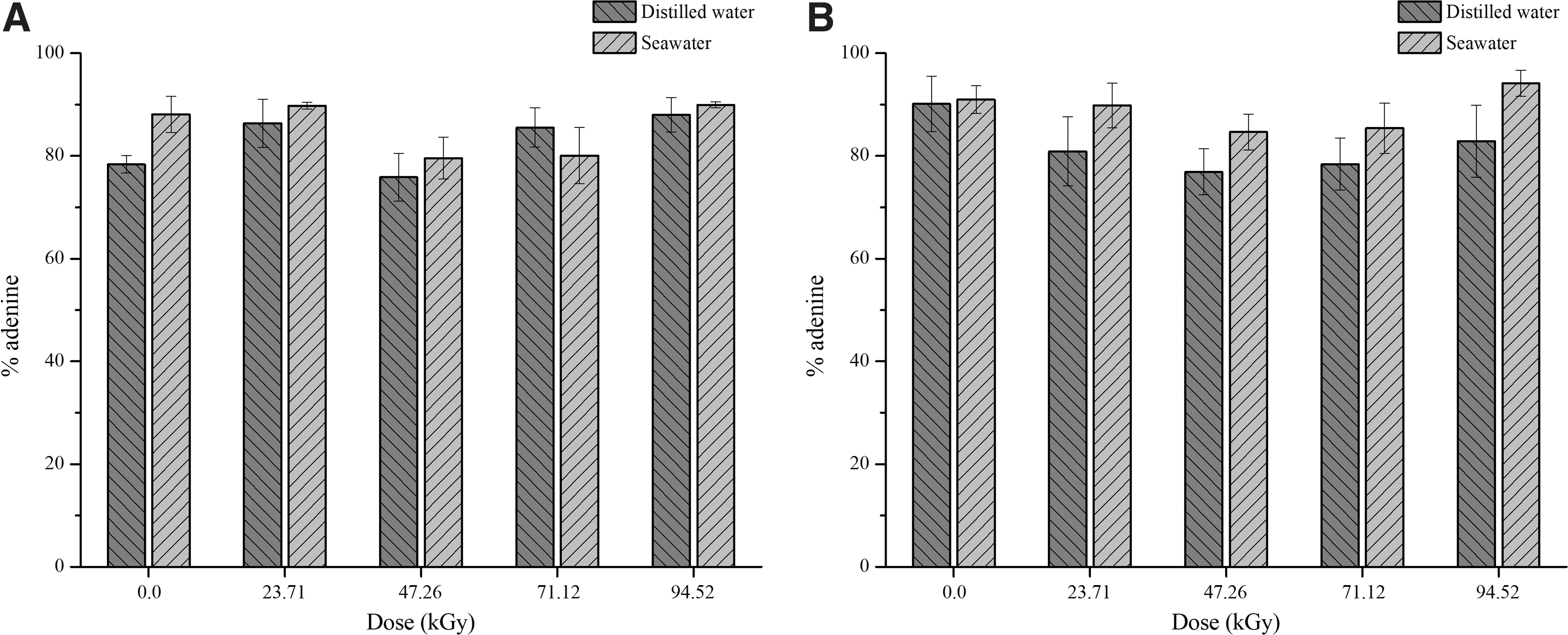
Recovery of adsorbed adenine onto montmorillonite as a function of dose. (
3.2. Qualitative essays
To better understand the sorption process of adenine onto montmorillonite, FT-IR spectra and X-ray diffractograms were obtained. FT-IR spectrum of montmorillonite without any previous treatment showed three major bands at 1010, 1635, and 3072 cm−1, and a shoulder at 3618 cm−1 (Fig. 3, sample 1). The broad band at 1010 cm−1 is a sum of many frequencies attributed to Si-O deformation, -OH deformation of hydroxyl linked to Fe3+ and Al3+, and Si-O-Si and Si-O stretching (Bukka et al., 1992). The band at 1635 cm−1 is attributed to hydration of the clay or H-O-H bending (Bukka et al., 1992; Tyagi et al., 2006). The broad band at 3072 cm−1 and the shoulder at 3618 cm−1 could be attributed to hydration water and O-H deformation of OH structural groups (Madejová, 2003). After stirring montmorillonite samples with distilled water or artificial seawater and lyophilizing them, FT-IR spectra showed that the absorption of the bands in the region at 1635 and 3072 cm−1 decreased due to the withdrawal of water molecules (Fig. 3). In addition, for samples treated with distilled water and artificial seawater, the bands at 1635 and 3072 cm−1 shifted to 1631 cm−1/1622 cm−1 and 3399 cm−1, respectively (Fig. 3), indicating a dehydration of the clay (Madejová, 2003). The sample of montmorillonite treated with artificial seawater (Fig. 3, sample 3) showed a band at 667 cm−1 that could be attributed to SO bending from sulfate (SO4 2−) ion (Iishi, 1979; Anbalagan et al., 2009). The bands attributed to SO stretching in the 1130 cm−1 region (Anbalagan et al., 2009) were not observed, since montmorillonite spectrum showed a large band in this region (Fig. 3). However, it should be noted that X-ray diffraction patterns showed gypsum precipitation onto montmorillonite (Fig. 5).

FT-IR spectra: (
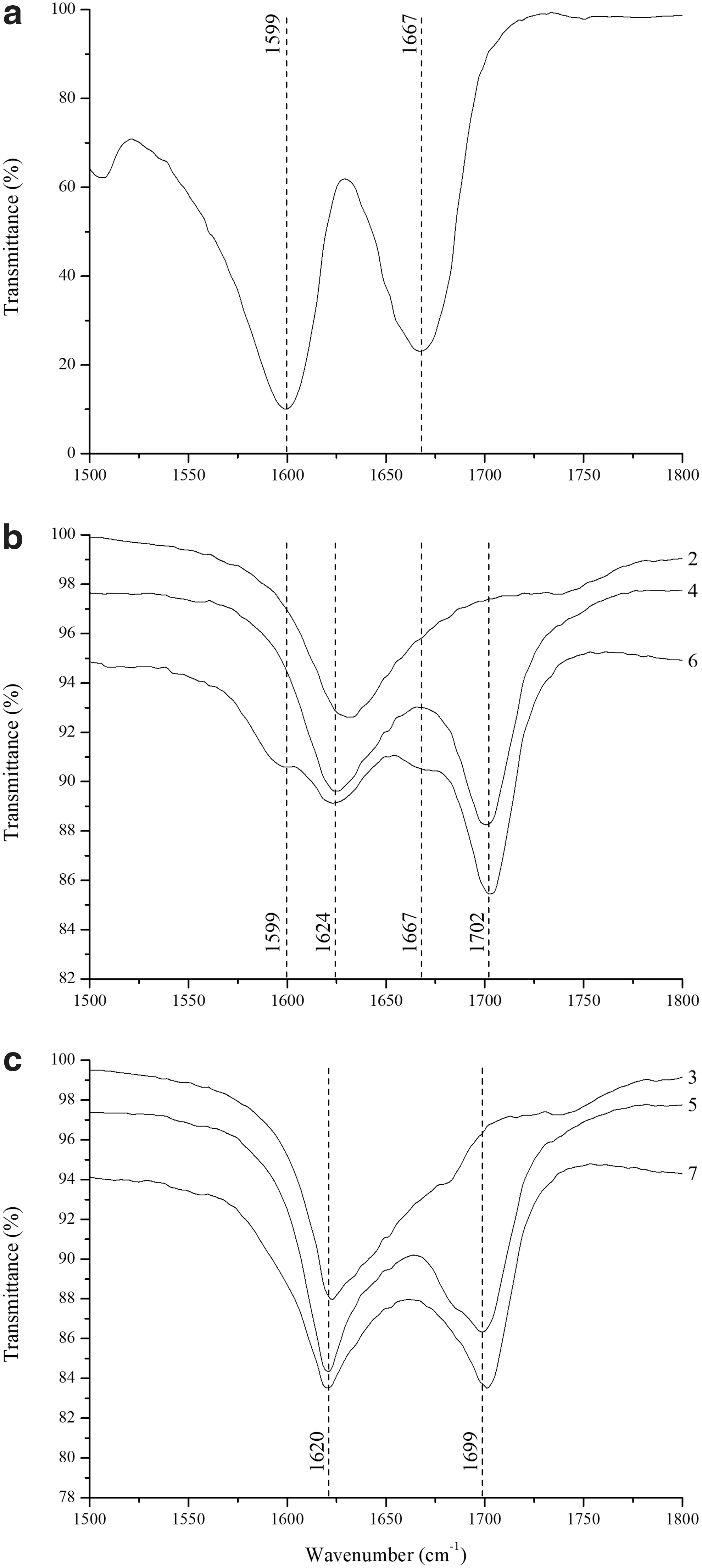
Adenine has two bands at 1599 and 1667 cm−1 (Fig. 4a) which could be attributed to C = C stretching and NH2 in-plane bending, respectively (Anizelli et al., 2014). After the sorption of adenine onto montmorillonite in distilled water and artificial seawater, the band at 1599 cm−1 shifted to 1624 and 1620 cm−1, while the band at 1667 cm−1 shifted to 1702 and 1699 cm−1, respectively (Fig. 4b, 4c, samples 4 and 5). This shift was due to adenine protonation (Anizelli et al., 2014). It could be highlighted that at 1620 cm−1 montmorillonite exhibits a band due to hydration water (Fig. 4, samples 2 and 3). The FT-IR spectrum of adenine adsorbed onto montmorillonite when using distilled water showed two shoulders at 1599 and 1667 cm−1 when exposed to gamma rays (94.52 kGy) (Fig. 4b, sample 6). These two frequencies are the same found at solid adenine (Fig. 4a). Nevertheless, in the experiment in artificial seawater, these shoulders were not observed (Fig. 4c, sample 7). Perhaps the salts provide some stabilization to adenine structure, as previously reported by Anizelli et al. (2014).
FT-IR spectra. (
X-ray diffractograms. (
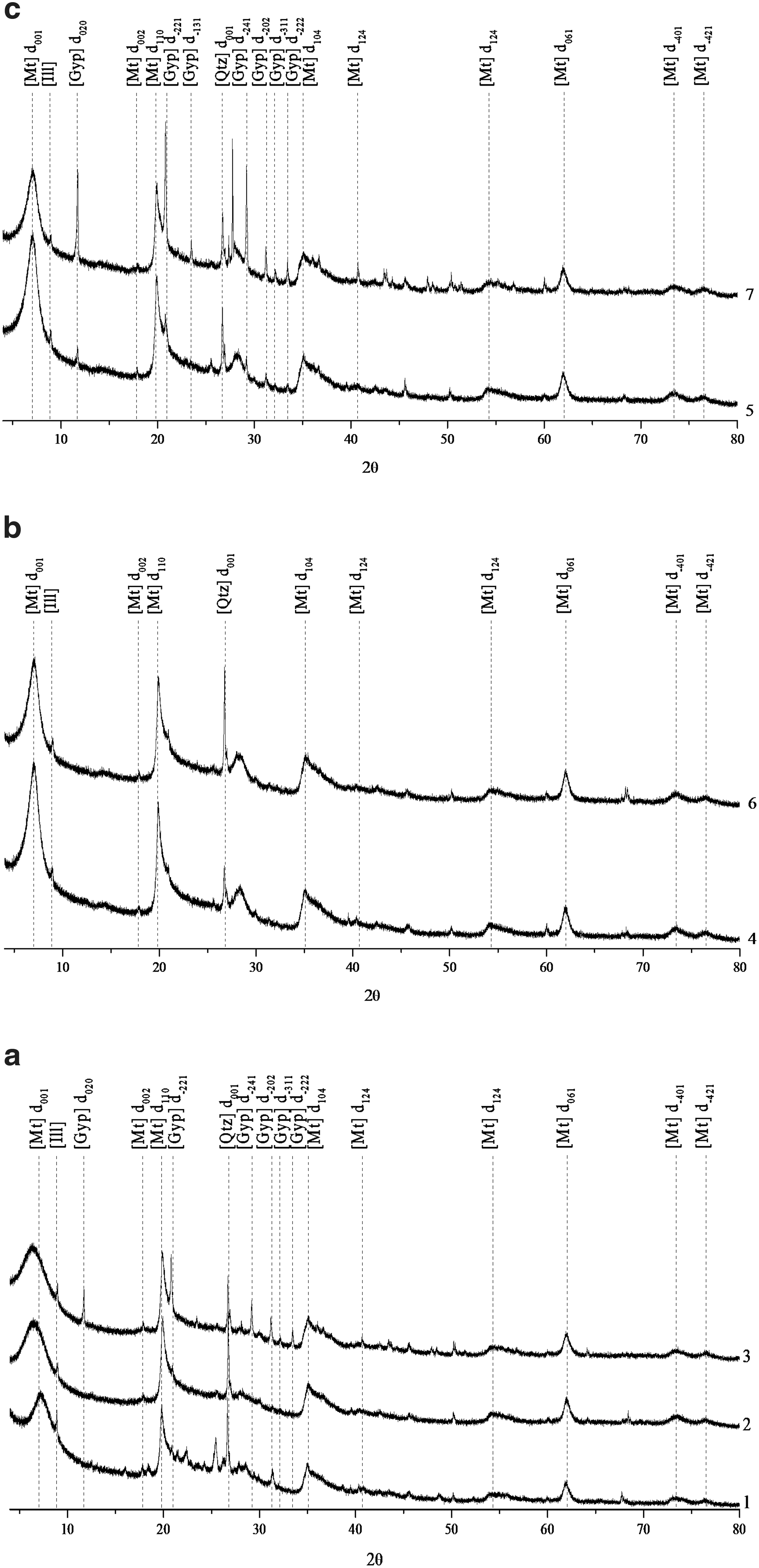
For XRD data, montmorillonite without any previous treatment showed peaks attributed to montmorillonite, illite, quartz, and two small peaks attributed to gypsum (Fig. 5a, sample 1). However, when montmorillonite was mixed with distilled water, the peaks attributed to gypsum vanished, and the other peaks remained (Fig. 5a, sample 2). In addition, when montmorillonite was mixed with artificial seawater, besides the peaks attributed to montmorillonite, quartz, and illite, six peaks attributed to gypsum were observed (Fig. 5a, sample 3). This result was confirmed by FT-IR spectrum (Fig. 3, sample 3) and indicates that the mineral gypsum was formed from Ca2+ and SO4 2− ions present in the artificial seawater solution. For the samples of montmorillonite mixed with distilled water (Fig. 5, sample 2) and the montmorillonite mixed with artificial seawater (Fig. 5, sample 3) when compared to montmorillonite without previous treatment (Fig. 5, sample 1), the peak related to the basal plane 001 shifted to smaller degrees and appeared more well defined. The full width at half maximum (FWHM) was higher for samples 2 and 3 than for sample 1 (Table 2). The d001 basal plane from samples 1, 2, and 3 was 12.3, 13.7, and 13.9 Å, respectively (Table 2). Nguyen-Thanh et al. (2005) also observed a similar interplanar distance for Na-montmorillonite without previous treatment. After the sorption of adenine onto montmorillonite when using distilled water or artificial seawater (Fig. 5b, 5c, samples 4 and 5), the X-ray diffraction patterns of these samples showed the same peaks of the samples of montmorillonite without adenine adsorbed on it (Fig. 5a, samples 2 and 3). It could be emphasized that adenine did not prevent the formation of gypsum onto montmorillonite (Fig. 5c, sample 5). The increase in the crystallinity samples was verified by the estimation of the FWHM value of the peak at d001 (Fig. 5, Table 2). Samples 4 to 7 have FWHM values close to 1.3 for random and oriented samples (Table 2). This implies that the addition of adenine to the system increases the crystallinity of the clay. This is because adenine enters into the interlayer space, improving the regularity of the clay layers' stacking and consequently increasing clay's crystallinity. However, this cannot be due to the same effect since zeolites are not layered minerals.
Interplanar Distance Values and FWHM
Samples:
Interplanar distance.
Full width at half maximum.
To better understand the variations in interlayer spacing, the samples were subjected to oriented deposition and to ethylene glycol swelling. The peak relative to the basal plane 001 changes, depending on the treatment of the samples (Fig. 6). The main difference between the FWHM values from random to oriented samples was observed for sample 1, in which the FWHM changes from 1.6 to 2.3, while the interplanar distance changes from 12.3 to 12.8 Å (Table 2). It should be noted that the orientation process consists of adding distilled water, creating a suspension that was air-dried. After samples 1, 2, and 3 were treated with ethylene glycol, the interplanar distance increased up to 17.1 Å, suggesting that ethylene glycol entered the interlayers of montmorillonite (Table 2). On the other hand, when adenine was adsorbed onto montmorillonite (samples 4–7), a slight increase up to 13.2 Å (Table 2) was observed.

X-ray diffractograms of the samples: (
4. Discussion
It is well known that ionizing radiation has some effects on clay minerals. Gournis et al. (2000) and Plötze et al. (2003) observed that Fe3+ was partially reduced to Fe2+. This reaction was attributed to the formation of the H˙ radical from intercalated water of montmorillonite; due to the small size of hydrogen radicals, they diffuse through hexagonal Si-O rings of the tetrahedral layer to the octahedral layer, thus reducing Fe3+. Moreover, H˙ radical, OH˙ radical, and H2O2 were also formed (Gournis et al., 2000). However, they did not observe an effect of gamma irradiation on several other physicochemical parameters of montmorillonite as well as other clays. Using EPR spectroscopy, Gournis et al. (2001) observed the formation of paramagnetic defects in laponite and SWy-1 after irradiation. They also observed the migration of Li+ toward the empty octahedral sites of SWy-1 and Zenith. H2 could be produced from H˙ radicals from interlayer water or from cleavage of O-H bonds inside the mineral layer (Fourdrin et al., 2013). Using FT-IR spectroscopy and 27Al and 29Si NMR, Negrón et al. (2002) did not observe structural changes of montmorillonite with up to 2 MGy of gamma irradiation, although they observed a small variation in the water content. Therefore, in the present work, a much lower irradiation dose (94.52 kGy) was used, and montmorillonite apparently did not experience any changes.
The adsorption of adenine onto montmorillonite could be explained by two different mechanisms: (1) electrostatic interactions between the surface of montmorillonite (negatively charged) and adenine (positively charged), probably accompanied by intercalation of adenine cations (positively charged) into the interlayers of montmorillonite negatively charged, and (2) interaction of the –NH2 group of adenine with montmorillonite. The shift of the band at 1667 cm−1 (NH2 in-plane bending) to 1700 cm−1 (Fig. 4b, 4c, samples 4 and 5) suggests that the NH2 group of adenine interacts with montmorillonite. However, Anizelli et al. (2014) observed that the band at 1667 cm−1 shifted to 1699 cm−1 and a new band appeared at 1573 cm−1 when the N1 of adenine was protonated. This band (at 1573 cm−1) was not observed in our spectra (Fig. 4b, 4c, samples 4 and 5). It should be noted that the pH of the suspensions indicates an acid medium (Table 1), suggesting that the N1 of adenine was protonated (Christensen et al., 1970). Therefore, it may be inferred that the NH2 group may interact with the clay or that electrostatic interactions might occur. Since ethylene glycol did not enter the interlayers of montmorillonite after sorption of adenine (Table 2), the intercalation of adenine in montmorillonite also occurred. This could mean that adenine (positively charged) interacts with the negatively charged surface in the interlayer of montmorillonite. This probably prevented the entry of ethylene glycol into that space (Lailach and Brindley, 1969; Weckhuysen et al., 1999; Carneiro et al., 2011). Several authors observed a decrease of sorption of adenine when the pH increases (Lailach et al., 1968a, 1968b; Lailach and Brindley, 1969; Winter and Zubay, 1995; Benetoli et al., 2008; Carneiro et al., 2011). Thus, it could be suggested that electrostatic interaction and the entrance to the interlayer of montmorillonite play the main role in the sorption of adenine on montmorillonite. Since an increase of pH implies that adenine first became neutral and then negatively charged, electrostatic interactions diminish (Christensen et al., 1970). In the case of nucleotides, at low pH, the N1 of adenine is involved in the adsorption, while, at high pH, the phosphate group is involved in the adsorption. (Feuillie et al., 2013). There are several studies on the adsorption of nucleic acid bases onto clays, and two mechanisms stand out: electrostatic interaction and the entrance to the interlayer of clay. However, to date, there is no evidence of which mechanism is the most important.
For the ethylene glycol treatment, samples without adenine, the interplanar distance (Table 2) had a higher value than for samples with adenine. This indicates that the ethylene glycol did not enter the montmorillonite interlayer because adenine occupied the interlayer site of montmorillonite.
The adenine that was desorbed from montmorillonite with KOH solution ranged from 78% to 88% (Fig. 2). Guzmán-Marmolejo (2003) also obtained the same recovery of adenine after one extraction with a KOH solution at pH 14. According to the author, after four extractions, about 97% of adenine was recovered from montmorillonite. Desorption of adenine from montmorillonite was also carried out with distilled water, CaCl2 (0.010 mol L−1), and Mehlich-1 (0.050 mol L−1 of HCl plus 0.0125 mol L−1 of H2SO4) extractors. When using distilled water/CaCl2 and Mehlich-1 as extractors, up to 4.1% and 6.6%, respectively, of adenine was desorbed from montmorillonite. These results were confirmed by FT-IR spectroscopy. FT-IR spectra of the samples after desorption with KOH did not show a characteristic band of adenine in the region of 1700 cm−1. On the other hand, when desorption was carried out with CaCl2 and Mehlich-1 extractors, FT-IR spectra showed a characteristic band of adenine in the region of 1700 cm−1 (figure not shown). In soil chemistry, CaCl2 and Mehlich-1 are used to verify if a substance is adsorbed on a solid as an outer-sphere complex or an inner-sphere complex, respectively (Sposito, 1989).
In this work, the recovery of adenine by desorption from the clay was statistically the same for non-irradiated and irradiated samples (Fig. 2). However, using 125 kGy, Guzmán-Marmolejo (2003) observed a decomposition of adenine close to 20%, when 1.0·10−3 mol L−1 of it was adsorbed on 100 mg of montmorillonite. On the other hand, when 1.0·10−2 mol L−1 of adenine was adsorbed on 100 mg of montmorillonite, even when using 180 kGy of irradiation, the decomposition was even lower. It should be noted that in the present work 3.7·10−3 mol L−1 of adenine was used, and the highest irradiation dose was 94.52 kGy. In addition, no product of decomposition was observed in this work. However, for adenine dissolved in distilled water and adsorbed onto montmorillonite, after irradiation, the FT-IR spectrum of the sample showed two shoulders with the same peaks of solid adenine (Fig. 4b, sample 6). It could be highlighted that the chromatograms did not show any peak indicating that adenine was decomposed (figure not shown). The appearance of the bands at 1599 and 1667 cm−1 (shoulders), after irradiation, could be due to a deprotonation of adenine by irradiation. It could also be commented that not all adenine was deprotonated, because the intensity of the bands is low. In addition, the X-irradiation of adenine crystals (Nelson et al., 1992) and later theoretical investigations by density function theory (DFT) suggest that adenine dehydrogenated radicals might be formed upon ionizing radiation (Wetmore et al., 1998). Nevertheless, excited-state intramolecular proton transfer (ESIPT), a mechanism of protonation deprotonation through hydrogen bond, happens. ESIPT is a tunneling phenomenon, where the proton transfer is induced by ionizing radiation which promotes the formation of tautomers (Tang et al., 2011), or also the shuttle of a proton from an initial oxidized molecule to a stronger proton acceptor (Nelson et al., 1998). At this scenario, adenine is protonated at N1 and adsorbed into the interlayers of montmorillonite; thus the loss of the N1-proton, induced by ionizing radiation, might be plausible. Nelson et al. (1992, 1998) demonstrate the deprotonation of N1-protonated adenine species at excited-state occurs more easily at acid conditions (pKa = 4.2), by the subtraction of the N1-prtoton (H˙).
When adenine was dissolved in artificial seawater and adsorbed onto montmorillonite, after being exposed to gamma irradiation, the FT-IR spectrum of the sample did not show any of those bands (shoulders) (Fig. 4c, sample 7). Thus, the deprotonation of adenine occurred in distilled water, but it did not occur in artificial seawater. The salts that are present in artificial seawater are, most likely, consuming the free radicals formed, since anions are considered OH˙ scavengers (Kumagai et al., 2013; Hata et al., 2016). In addition, the divalent cations from artificial seawater are able to coordinate in bidentate position with adenine, resulting in a lower energy of the HOMO orbital, thus making it a less reactive specie (Anizelli et al., 2014). Finally, seawater salts can protect adenine because they decreased the reactivity of it.
5. Implications
It is shown in this work that montmorillonite could concentrate adenine from dilute solution and protect it against ionizing radiation. However, several other works showed that minerals concentrate and protect biomolecules. The main difference from this research relies on the use of an artificial seawater model, which could better represent the seawater of primitive Earth. Most other similar studies have used distilled water or sodium chloride solutions (Zaia, 2012). Thus, by applying conditions that are more closely related to those that probably existed on primitive Earth, we show that Bernal's hypothesis still stands. However, an issue that is rarely discussed in the literature is the strong adsorption of biomolecules on minerals (Lambert, 2008). Desorption of adenine from montmorillonite was only possible when using a high concentration of potassium hydroxide, meaning that it was strongly bonded to the clay. This suggests that adenine will be not easily desorbed, being protected against hydrolysis and radiation.
The fact that adenine is strongly bound to the montmorillonite leads to some implications. First, it could be argued that adenine would not be available for any further reaction. Lambert et al. (2013) showed that when glycine was sorbed into the interlayer of clay, it was weakly reactive for the formation of peptide. Nevertheless, it could also be argued that sometimes immobilization is required for oligomerization reactions to take place. Indeed, it has been demonstrated that nucleotides and polynucleotides are sorbed onto montmorillonite and formation of a nucleotide dimer by a phosphodiester bond occurred (Ferris et al., 1989a, 1989b; Ferris, 2002, 2005). However, there are several critical remarks that could be made about Ferris's work. The most important one is that the experiments were performed with condensing agents or activated monomers that did not exist on primitive Earth. Montmorillonite itself needs to be activated; usually this activation is carried out with saturated solutions of alkali and alkaline earth metal ions. Naturally, salt composition of the seas of prebiotic Earth was a mixture of several cations that could be adsorbed onto clays. Thus, montmorillonite should be activated with different artificial seawaters to verify if it is still a catalyst of the nucleotide polymerization reactions. It should be noted that the highest cation concentration in seawater of prebiotic Earth was probably Mg2+ (Izawa et al., 2010; Zaia, 2012). However, according to Ferris, Mg2+ has no effect on the catalytic activity of montmorillonite (Ferris, 2005). The procedure used by Ferris to obtain the polymers is similar to the one used by Merrifield for solid phase peptide synthesis (Merrifield, 1963). In the procedure used by Merrifield and Ferris, all monomers that did not react or by-products formed were washed out with water or other solvent. In a real system, this washout did not occur; and, after a few cycles, the system will contain a large mixture of monomers, polymers, and by-products. Thus, it will be impossible to obtain any useful polymer in high concentration. In the experiments using activated monomers, the elongation of the polymer was possible because a polymer with 10 monomers was previously adsorbed onto montmorillonite. Thus, these experiments could not be considered prebiotic ones. In addition, nucleotides are different molecules from nucleic acid bases. Nucleotides are made up of a nucleic acid base (such as adenine), a pentose, and phosphate. This different configuration leads to other interactions with clays. This work showed that adenine interacts by electrostatic charges, by nitrogen N1 group. On the other hand, a nucleotide would interact strongly by the phosphate group with the hydroxyl groups from the edge sites, and electrostatic interactions might occur at low pH medium (Feuillie et. al., 2013; Villafañe-Barajas et al., 2018). Therefore, Ferris experiments are about oligomerization of nucleotides, molecules with different chemical behavior from adenine. The role of strong biomolecule-surface interactions is not clear. This is because a strong interaction between biomolecules and mineral surfaces could be detrimental for some prebiotic processes and could be favorable to others. Therefore, a few questions should be asked: once adenine was immobilized by montmorillonite, could adenine react with pentose? Could nucleoside react with a phosphate? Could nucleotide oligomerization reactions occur using a wet/dry system with artificial seawater 4.0 Ga plus montmorillonite?
6. Conclusions
On the one hand, the adenine-clay system in distilled water showed some changes in adenine. On the other hand, in seawater medium no evidence of adenine reactions was detected. Thus, the use of seawater 4.0 Ga plus montmorillonite protected adenine against ionizing radiation.
In summary, the three main findings of our study are the following: Adenine is strongly bonded to clay's surface and interlayer. Montmorillonite was able to protect adenine against a high dose of ionizing radiation. Seawater model 4.0 Ga provided stabilization for the adenine-clay system, decreasing its reactivity. The anions of seawater are consuming the free radicals formed, since they are considered OH˙ scavengers.
Footnotes
Acknowledgments
J.P.T.B. acknowledges CAPES for funding, Instituto de Ciencias Nucleares and Instituto de Geología, Universidad Nacional Autónoma de México, Mexico City. The authors thank Dr. Teresa Pii Puig from Instituto de Geología, Universidad Nacional Autónoma de México for the XRD analysis. The authors also thank the support of CNPq/Fundação Araucária (Programa de apoio a núcleos de excelência-PRONEX, protocol 24732) and DGAPA-PAPIIT (IA203217) for this research.
Disclosure Statement
No competing financial interests exist.
Associate Editor: David Deamer