
Abstract
Phenylalanine (Phe) is an amino acid that has been identified in carbonaceous meteorites; its formation mechanism in space is unknown, and its radioresistance has been the subject of investigation. This work aims at studying, in the laboratory, the Phe radiolysis by cosmic analogues. The Phe destruction rate, at 300 K, is measured for H, He, and N ion beam irradiation in the 0.5 to 2 kinetic MeV range. Fourier transform infrared (FTIR) spectroscopy was employed to monitor the molecular degradation as a function of fluence. The Phe apparent destruction cross–section, σ
ap
d
, which includes radiolysis and sputtering processes, is determined to be proportional to the electronic stopping power, Se
. The measured parameter D
0 = 14.3 ± 2.2 eV/molec in the relationship, and
1. Introduction
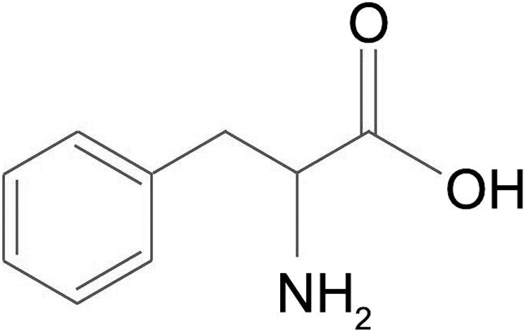
Phenylalanine (Phe) is an essential amino acid found in foods, where it plays a vital role in the biological process of proteins and enzymes. Its chemical structure is shown in Fig. 1. Outside the Earth, amino acids have been detected in dust traces left by the Comet 81P/Wild 2 and collected by the Stardust spacecraft (Sandford et al., 2006; Elsila et al., 2009). Similar astrophysical materials that are present in comets and meteorites rich in complex molecules could have fallen onto early Earth; therefore, organic extraterrestrial molecules could have stimulated pre-biological processes and formed nucleobases and other molecular building blocks of life (Oba et al., 2019).
The
Glavin et al. (2010) found several amino acids in a fragment of the Almahata Sitta meteorite. Extraterrestrial nucleobases were identified in 12 meteorites (Callahan et al., 2011). Ionizing irradiation (UV photons and energetic particles) is possibly the primary process of synthesizing cosmogenic complex molecular species observed in comets and meteorites (Nuevo et al., 2014). The physical–chemical evolution of these molecular materials, when exposed to cosmic radiation, may be partially understood by performing laboratory simulations under similar environmental conditions (i.e., temperature, pressure, and radiation). In particular, the survival capacity of molecules exposed to high doses of cosmic radiation can be estimated from these measurements.
The vast majority of ionization and destruction of molecular species occurs at low temperatures (∼30–150 K) inside the diffuse interstellar medium (ISM). Nevertheless, the rare but interesting warmer spots where preformed prebiotic material in icy conditions is transported to higher temperature regions should not be excluded. Examples of this include dense interstellar clouds, protostellar disks, intermediate circumstellar envelopes, and comets/meteorites. Dense interstellar clouds are generally cold (10–100 K) and mainly constituted by hydrogen, but they may include small molecules such as H2O, CO, and CO2; the densest regions of the cloud may collapse into stars that heat up their environments (McKee and Ostriker, 2007).
Protostellar disks exhibit an extensive temperature range; they are very hot near the center, hot in the disk far from the symmetry plane, and very cold at peripheral regions; inside the disk, grains recovered by ices are forced to move under a temperature gradient (Henning and Semenov, 2013). The intermediate dense circumstellar envelopes around dying stars contain dust with molecules rich in C, H, O, and N that can subsist at temperatures between 100 and 1000 K (see van Dishoeck et al., 2013, and references therein).
At last, comets and meteoroids, which likely exist outside the Solar System with highly eccentric orbits, synthesize ices into prebiotic material and bring it close to the star such that it is irradiated at relatively high temperatures (Mumma and Charnley, 2011, and references therein).
Besides the analysis of extraterrestrial prebiotic molecules, another interest in amino acids exposed to ionizing radiation lies in understanding the physical and chemical mechanisms for applications in radiotherapy. The chemical effects of irradiation in the bulk of phenylalanine (Phe) in aqueous matrix were studied by Chrysochoos (1968), Krajnik et al. (1995), Maskos et al. (1992), Wang et al. (1993), Orzechowska et al. (2007), Gerakines et al. (2012), and Ludwig et al. (2018). The charged desorbed fragments of Phe during ion irradiation were addressed by Guthier et al. (1983), Leite et al. (1992), and Lemoine et al., (2013). The studies presented thus far, however, provide insufficient information on how radiolytic effects in Phe depend on the projectile energy.
At room temperature in the present study, the projectile energy released when the ion–solid interaction affects the radiolytic destruction of solid Phe was analyzed. Following previous findings (Vignoli Muniz et al., 2017; da Costa et al., 2020, 2021), current data confirmed the power-law that connects the destruction cross-section of irradiated molecules with the projectile electronic stopping power, Se , in the sample material.
This simple mathematical dependence is quite useful to estimate the Phe half-life in extraterrestrial environments. In addition, we sought in the present study to analyze the synthesis of new compounds during the Phe radiolysis. For cryogenic temperatures, which are relevant for astrophysics, it has been reported that, when cooling down: (1) infrared (IR) bands become narrow and integrated absorbances for non-irradiated samples increase by a factor of 2 (e.g., valine [Val] infrared absorbance at cryogenic temperatures, da Costa et al. (2019)) and (2) for MeV ion beams, cross-sections increase typically two to five times [Review by da Costa et al. (2021)—Table 1 and Fig. 12; and by Gerakines et al. (2012)—Table 5, for 0.8 MeV H+].
d,l-Phenylalanine Infrared Bands at 300 K: Assignments and Band Strength
From Olsztynska et al. (2001), Mary et al. (2005), and Hernández et al. (2010): overtones and combination, ot. & c; asym, asymmetric; br, broad; def, deformation; i.p., in-plane;
Galactic Cosmic Ray Parameters and Phe Rate Destruction Under Galactic Cosmic Rays Irradiation
Meyer et al. (1998). Relative abundances: Hydrogen flux is set to 1.000 × 10⁶ ions/cm2/s.
Webber and Yushak (1983).
GCR = galactic cosmic rays.
2. Sample Preparation and Experimental Setup
Phe films were prepared according to the procedures from the work of Souza-Corrêa et al. (2019). The films were made by deposition in a vacuum chamber by using an Edwards thermal evaporator. Phe powder was placed in a Mo boat, heated by a current of 27 A, sublimated, and deposited on ZnSe substrates under residual gas pressure of 2 × 10⁻⁶ mbar during deposition. Films of ∼241 and 346 nm (measured by a 6 MHz quartz crystal microbalance) were then produced. Thereafter, the Phe samples were inserted into an analytical chamber; and after pumping down, residual pressure was in the high 10−6 mbar range, but during irradiation, this increased to 2 × 10⁻⁵ mbar.
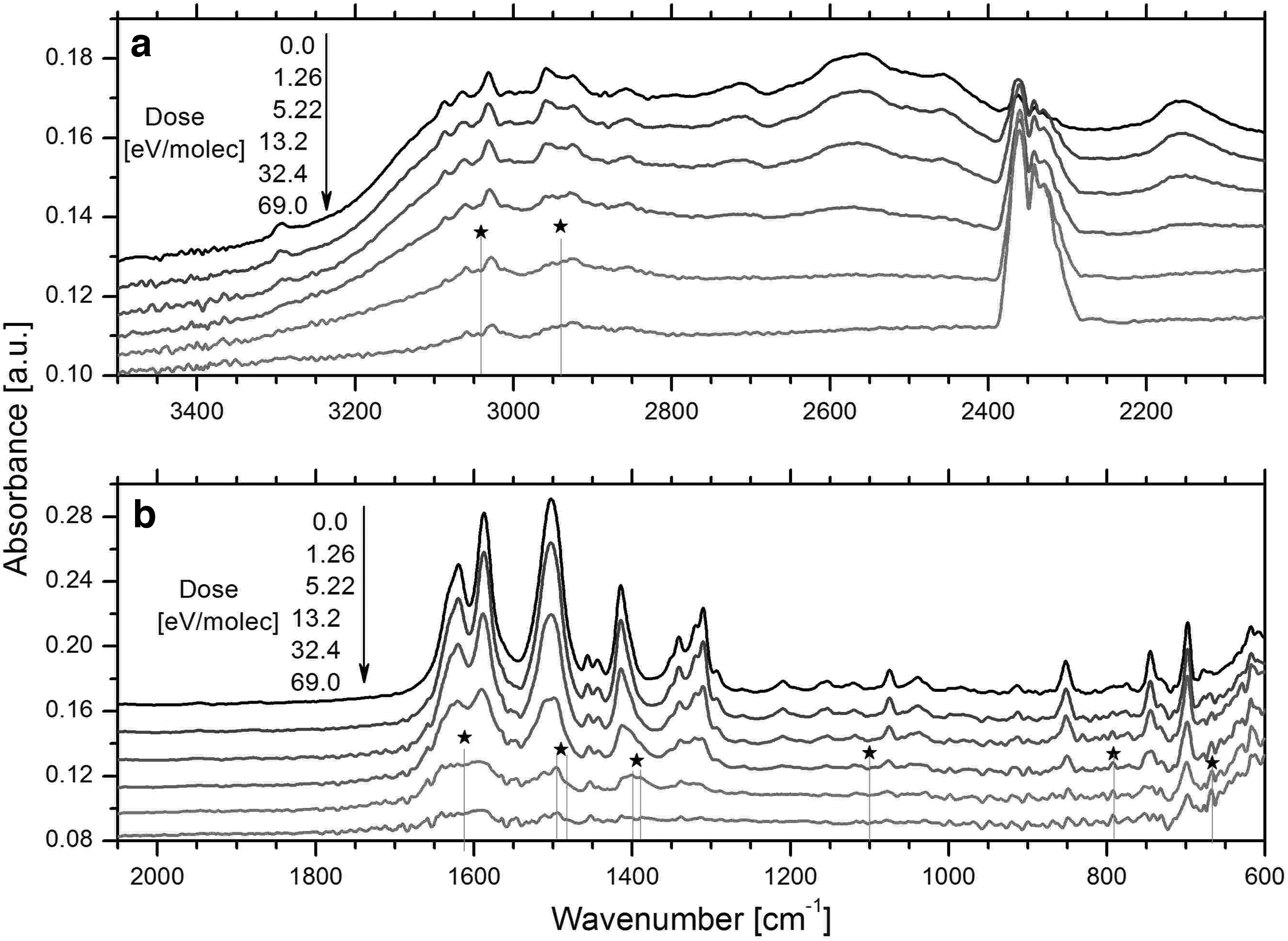
Infrared spectroscopy was employed to monitor the Phe degradation after steps of irradiation. The IR spectra were acquired by a JASCO FTIR-4100 spectrometer, with 2 cm−1 resolution and 70 scans/spectrum in the 3500–600 cm−1 wavenumber range. Mid-infrared spectra, in the transmission mode, are shown in Fig. 2; observed absorptions bands are presented in Table 1, as well as their assignments (Mary et al., 2005).
Spectra of irradiated
To have a perpendicular incidence of the ion beam and the IR radiation into the Phe film, the sample holder was rotated back and forth by 90° for each irradiation step. Phe was irradiated, at room temperature, with the following projectiles and energies: H+ 2 MeV, H+ 0.5 MeV, He+ 2 MeV, N+ 0.5 MeV, and N+ 2 MeV. The ion beams, with fluxes from 4.8 to 120 × 1010 ions/cm2/s, were delivered by the Van de Graaff electrostatic accelerator of the Pontifícia Universidade Católica do Rio de Janeiro (PUC–Rio) (Pilling et al., 2013). Electronic stopping powers, Se , were computed by the SRIM software (Ziegler et al., 2010). The ion beam and sample characteristics are set out in Table 2.
Ion Beam, Its Electronic Stopping Power (S e ) and Ion Flux, Sample Thickness, and Initial Column Density
The apparent destruction cross-section,
3. Results
3.1. Phenylalanine IR spectroscopy
The Phe IR spectrum is well known. Table 1 presents the
Figure 2 shows a sequence of infrared spectra obtained during the Phe N+ 2 MeV irradiation, that is, panel (a) and (b) for the 3500–2100 and 2100–600 cm−1 wavenumber regions, respectively. At the top, the first spectrum of the sample was acquired before irradiation; the others correspond to the absorbed doses listed on the left side of the figure. The irradiation absorbed dose is calculated by D = Se F, with Se being the electronic stopping power of each projectile ion in Phe, and F the fluence. The irradiation doses increased until 200 eV/molec, typically over the course of 2–7 h, with fluxes that are displayed in Table 2. Quantitative analysis was performed by analyzing the evolution of the integrated absorption of selected bands (see Section 3.2). New IR features appear and are treated in Section 3.4.
3.2. Data analysis
The evolutions of five Phe band absorbances, for six ion beams, were analyzed as a fluence (or dose) function. Figure 3a illustrates the normalized integrated absorbance decays of the five bands for the 2 MeV N+ ion beam irradiation. The exponential behavior of each decay is promptly seen, but it is also clear that slopes are not the same for the different bands. This effect has been previously reported (da Costa et al., 2020), and the adopted procedure is to define the average slope in semilog scale (line) as the apparent cross-section.
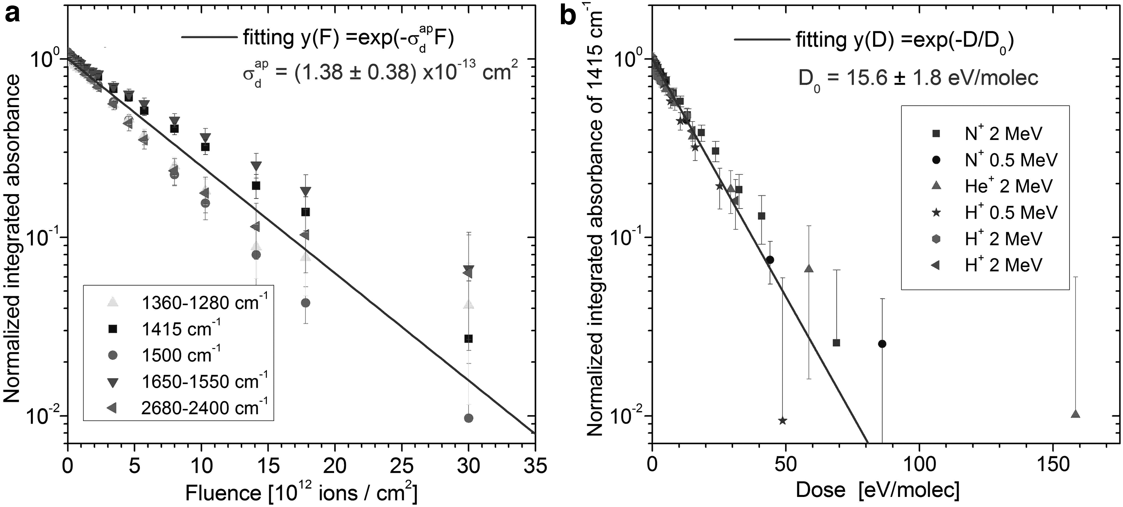
Figure 3b illustrates the integrated absorbance decay of a particular band (1415 cm−1) for the six ion beams as a function of the absorbed dose. The representation as a function of dose and not of fluence aims at merging all the decay lines into an average one.
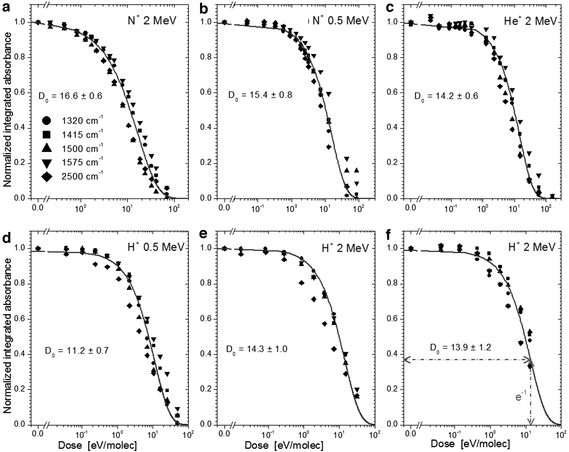
Figure 4 presents an overview of the obtained results: the dependence of the integrated absorbances on the absorbed dose. The absorbance evolution as a function of fluence is about the same for all bands, and the line is the fitting using Eq. 4. The systematic deviations shown in Fig. 3a also occur for other ion beams. The relatively small deviations are attributed to the distinct sensitivity of specific vibration modes to chemical environmental changes and/or to the presence of products that vibrate at about the same frequencies (da Costa et al., 2020, 2021). Note that, in general, the 2500 and 1500 cm−1 band absorbances decrease faster during irradiation than the others.
Dependence of normalized integrated absorbances on dose. Since data points originate from five Phe bands, with the fittings corresponding to average absorbances. Phe was irradiated by:
3.3. Cross-section and critical dose measurement
The Beer–Lambert law states that the column density of a thin sample at a fluence F, N(F), can be determined by:
where Abvi
(F) is the band strength (A-value) at the frequency vi
,
The decrease rate of the precursor column density is due to radiolysis and sputtering:
where σd
is the destruction cross-section of the Phe chemical dissociation. The Phe sputtering yield Y(F) is assumed to be
The solution of Eq. 2 is:
Considering
which directly expresses the integrated absorbance dependence on the absorbed dose D(F). Current data are analyzed by using this equation. Fittings are displayed in Fig. 4, for the selected bands 1325, 1415, and 1500 cm−1, and for the region 2680–2400 cm−1. Averaging over all bands, the obtained parameters D
0 and
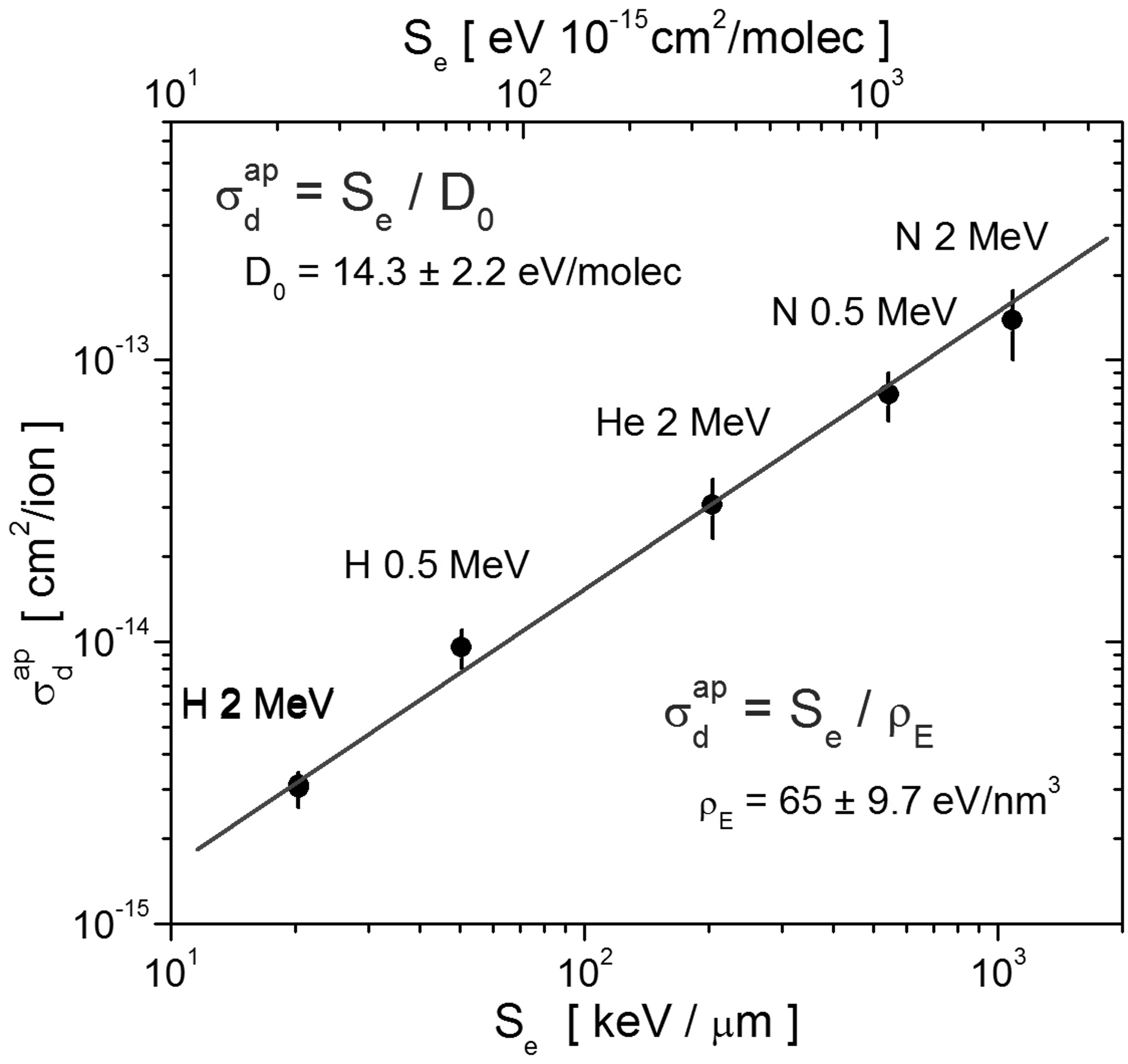
Proportionality between the apparent destruction cross-section (Table 2) and the electronic stopping power:
The parameter D 0 appears as the mean (or critical) dose for causing radiolysis/sputtering in Phe independently of the projectile type or its energy. Since the volume of a Phe molecule is 0.213 nm3, the energy density ρE , required for the Phe removal is ρE = D 0/Vm = 65 ± 9.7 eV/nm3.
3.4. Synthesizing new molecular species
Figure 6 shows two features that occurred after Gaussian-shape decomposition of the 1430–1380 cm−1 wavenumber region: The 1414 cm−1 band diminishes continuously from (a) → (b) →(c), whereas the other band moves from 1410 to 1395 cm−1; this shift suggests molecular rearrangements.
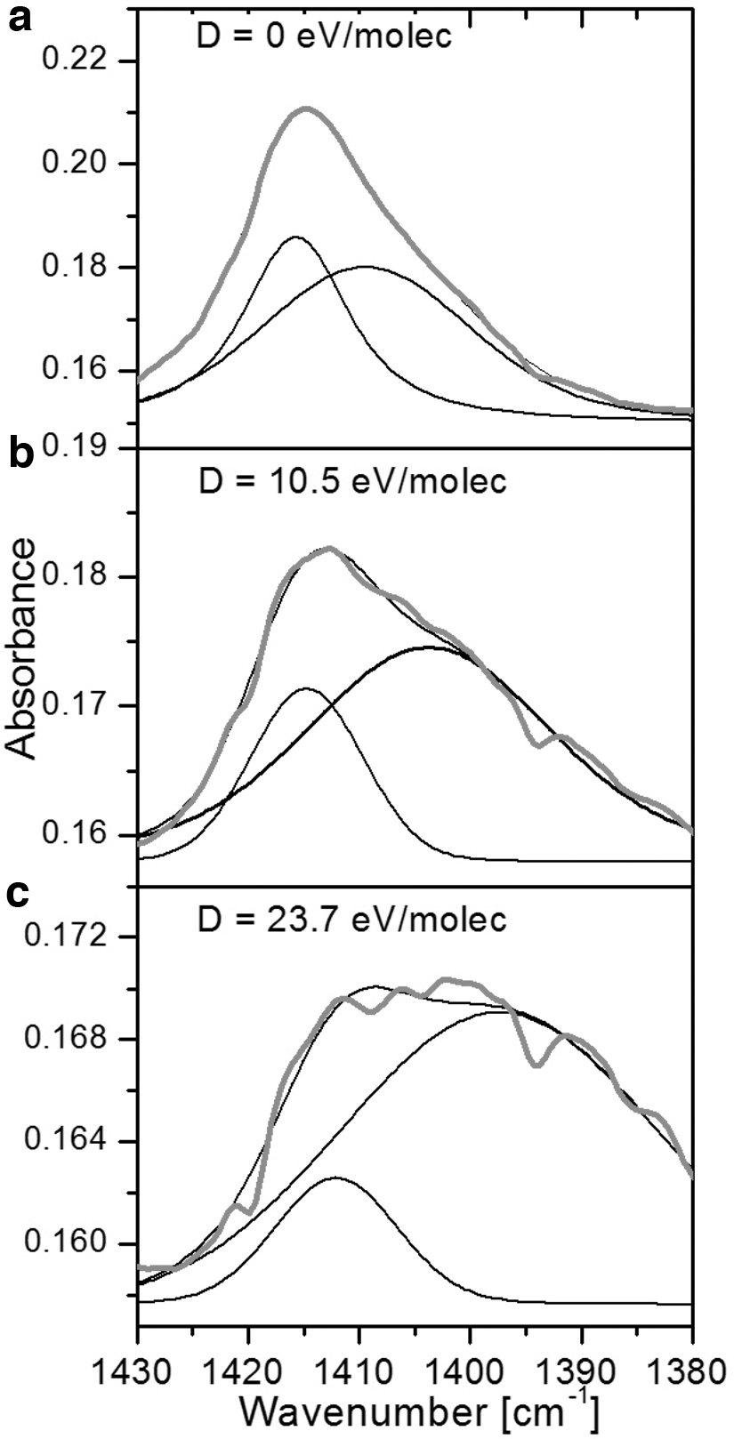
Decomposition of the 1430–1380 cm−1 feature into two Gaussian-shape bands. The band around 1415 cm−1 decreases and redshifts as doses increases:
In all current measurements, the rising of a feature around 2350 cm−1 can be observed; this band corresponds to gaseous CO2 contamination outside the chamber (Fig. 2). Although the CO2 absorbance increased with time, it is not expected that this band is due to the Phe radiolysis. CO and CO2 are possible products of the Phe radiolysis, but they diffuse and desorb when irradiation is performed at 300 K; however, a small amount of them may remain trapped in the sample. Finally, Fig. 7 presents the 668 cm−1 band for increased doses (0, 10.5, and 23.7 eV/molec.), which should correspond to CO2, but may also include a contribution due to the C6H6 vibration mode. In addition, weak infrared bands emerged in spectra of the irradiated sample (see Fig. 1), but noise signals prevented their analysis; these little bands are mainly located in the regions 2960–2900 and 1640–1620 cm−1, which confirms the observations of Gerakines et al. (2012).
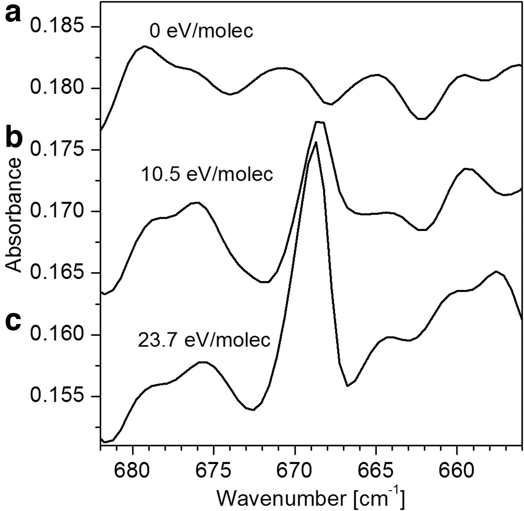
Absorbance of the 669 cm−1 band increases during irradiation at doses of
The new bands observed in the Phe spectra just after irradiation are listed in Table 3, with possible attributions to glycine (COOHNH2CH), alanine (COOHNH2CHCH3), methylamine (CH3NH2), ethylamine (C2H5NH2), and benzene (C6H6). Synthesized products were observed by Gerakines et al. (2012) in the IR spectra of irradiated Phe at low temperatures (15, 100, and 140 K). They assigned the new features to ethylamine and methylamine molecules.
New Infrared Bands Attributed to Products: Glycine (COOHNH2CH), Alanine (COOH NH2CHCH3), Methylamine (CH3NH2), Ethylamine (NH2CH2CH3), and Benzene (C6H6)
The observed bands that correspond to each candidate molecules are marked with the checkmark ✓ symbol.
4. Discussion
4.1. Band evolution
Figure 3 presents the integrated absorbance of selected Phe IR features. Note that some integrated absorbances decrease at a different rate on irradiation. For example, in Fig. 3a, the 1575 cm−1 band absorbance decreases slower than the 1500 and 2500 cm−1 ones. σ = Se n /ρE .
As shown in Fig. 6, the profile deformation of the 1411 cm−1 band (at doses of 0, 10.5, and 23.7 eV/molec.) may be attributed to both synthesized molecules and structural changes of solid Phe. Although the absorbance of Phe bands could be affected by the appearance of the synthesized molecules, it should be noted that structural modifications of solid Phe may also alter absorbance.
Pilling et al. (2013) and da Costa et al. (2020) observed significant structural modifications during irradiation of solid glycine and Val amino acids, respectively. They proposed that the absorbance change at the beginning of irradiation may have been caused by a molecular rearrangement that induces structural processes in the sample, such as compaction, amorphization, or even crystallization. Since the selected band absorptions decrease with the destruction of cross-sections that are slightly different, the fittings relative to all bands in Fig. 4 deliver an average destruction cross-section.
Figures 4c and f indicate a small absorbance increase at the beginning of irradiation (before the absorbed dose of 0.1 eV/molec). Previous works (particularly on ice radiolysis) pointed out that IR absorption of precursors may increase despite their destruction by the incident radiation (de Barros et al., 2011; Andrade et al., 2013). The opposite effect has also been reported; too fast of an absorption decrease can occur at the first irradiation intervals (Dartois et al., 2015; Souza-Corrêa et al., 2019).
The beam-induced molecular rearrangement is attributed to this phenomenon because (1) the absorption of certain bands increases whereas for others it decreases (e.g., Pereira et al., 2020); (2) absorbance decay corresponds to a relatively high cross-section (much higher than the radiolysis ones); and (3) it is achieved with a low dose (when the concentrations of products are negligible compared with the precursor one). The molecular rearrangement may happen via different processes, namely, compaction (porous or cracks are eliminated from the processed material), crystalline phase transitions (amorphous–crystalline or crystalline–crystalline), intermolecular transitions (modification of cluster distributions), and intramolecular transitions (modification of conformer distributions). At least some of these rearrangements are vibrational band dependent, which means they may enhance the absorption for certain bands and attenuate it for others.
The selected five absorbances used to describe the Phe radiolysis exhibit different decreases in Figure 3a. The rate of these integrated absorption bands may be due to structure alterations, as mentioned earlier. Therefore, these discrepancies need to be interpreted with caution since errors in integrated absorbance measurements could also be made when new features of unidentified species grow up next to integration limits that alter the baseline.
Figure 3b compares the normalized integrated absorbances for the 1415 cm−1 absorption band of the six experiments as a function of the absorbed dose. No significant alteration in the integrated absorbances is noted for D < 50 eV/molec; above that, data are in detection limit.
4.2. Phe radiolysis and sputtering
Due to the 0.5–2 MeV energy of the H+, He+, and N+ ion beams, the projectile–Phe interaction occurs mostly by electronic excitation since the stopping power relative to electronic collisions is two orders of magnitude higher than the nuclear one (knock-on collisions). Irradiation by energetic ions removes Phe molecules from the sample by two processes as follows: (1) radiolysis, when intramolecular bonds are broken and the molecular fragments delivered along the projectile track synthesize new species; (2) sputtering/desorption, when charged fragments, intact molecules, new molecular species, and clusters are ejected (Leite et al., 1992).
Radiolysis happens over the whole region traversed by the beam (column density N 0), but sputtering occurs only in the first monolayers, where the two processes are entangled. Assuming, in a first approximation, that radiolysis and sputtering are uncoupled, the precursor's disappearing rate writes dN/dF ≈ −σd N(F) − Y(F), Eq. 2. Both quantities, σd and Y(F), depend on the projectile-solid interactions, which, in turn, depend on the projectile energy.
Based on this, the apparent destruction cross-section is defined as
The Fourier transform infrared (FTIR) technique monitors the number of intact precursor molecules during the irradiation, but it is not able to inform whether the missing molecules disappeared by radiolysis or by sputtering. Further information is necessary for this questioning. The sputtering of frozen gases was extensively studied in the 1980s and 1990s. The linear-cascade theory is a successful model for describing the dependence of the sputtering yield on projectile energy and temperature (Schou, 1987; Sigmund, 1987; Johnson, 1996).
For low projectile energy, knock-on collisions are the dominant mechanism, and Y is proportional to Sn . As the projectile energy increases, electronic sputtering dominates, and Y becomes proportional to Se n , where n may be 1 or 2, according to the system (e.g., Famá et al., 2008; Johnson et al., 2013). In general, for high stopping powers, the same projectile causes, in the material surface, two ionizations that are too close to each other, which cooperates for enhancing particle ejection; that is the origin of the non-linear dependence on Se .
As examples, for MeV H+ impacting on solid N2, Y ∼ Se if S e < 10 eV per 1012 N-atoms cm2, and Y ∼ Se 2 for higher stopping power (Schou, 1987). For He+ impacting on H2O ice, Y ∼ Sn if EHe < 4 keV and Y ∼ Se 2 for higher projectile energy. Seperuelo Duarte et al. (2010), analyzing the interaction of 50–500 MeV nitrogen ions on CO ice, reported Y ∼ Se 2 for Y over six orders of magnitude.
With regard to amino acid or DNA base targets, the sputtering yield dependence on Se has not been established to date. The existing data, which are scarce, show that the apparent cross-section varies as Se n , where n = 1 within 10%, n = 0.936 for Val, da Costa et al. (2020), or n = 1.08 for adenine (Vignoli Muniz et al., 2017). For high Se , the Se 2 sputtering contribution should predominate over the radiolysis linear contribution, but experiments with 230 MeV S15+ (Vignoli Muniz et al., 2017) and 90 MeV I14+ (Salehpour et al., 1984) ion beams do not support this option. At this juncture and with the current results (Figs. 3 and 4 show the absorbance dependence on dose) included, the conclusion is that the sputtering contribution is either negligible with respect to radiolysis or its dependence on Se is also linear.
4.3. Apparent cross-section dependence on temperature
Given that the measured cross-section includes sputtering and radiolysis, the temperature may affect these two processes differently. Thermal expansion varies over inter- and intramolecular distances, which can change physical chemistry properties, in particular (1) sublimation and beam-induced sublimation; (2) mobility of defects and products; and (3) diffusion and chemical reaction velocities. These characteristics are expected to be modified by the sample temperature as well as by temperature increase (or local thermal spikes) due to the energy deposited by the beam (stopping power and beam flux). In contrast, electronic and nuclear (knock-on) sputtering yields should not change once most of the secondary particles are energetic enough to be disturbed by the local temperature (Schou, 1987).
Infrared (FTIR or Raman) spectroscopy offers information with regard to temperature effects on the molecular structure of solids. In particular, when cooling down the sample, the positions of vibrational lines shift, integrated absorbances increase, bandwidths become narrower, and peak shapes may be more structured (Zeidler et al., 2013, 2015; da Costa et al., 2019). Sample temperature acts via three mechanisms: (1) dynamical effects, (2) energy distribution of states, and (3) geometric effects. Dynamical effects have to do with molecular velocity effects.
As the temperature decreases, molecules move with lower velocity and photon absorption is stricter (Doppler Effect) such that bandwidth decreases; moreover, molecular rotation is constrained, and molecular polarizability is increased (higher absorbance). With regard to energy distribution of states, the population of conformers changes as temperature changes and, therefore, their absorbances change accordingly. As for geometric effects, these operate on the anharmonicity shape of the lattice potential energy; the thermal expansion of the solid changes its mass density and, more importantly, its optical constants (i.e., its dielectric function).
Consequences are the shift of band lines and the variation of bandwidths (connected with the damping of oscillators); in semi-quantum models, phonons dissipate the oscillator energies and interact with each other, which determines their lifetimes, the vibrational damping, and bandwidths (Xie et al., 2021).
The ultimate question is how temperature affects the apparent cross-section: This is related to solid relaxation just after the projectile passage; precursor molecules are ejected from the surface or dissociated in the bulk. For sputtering induced by the keV ion (Schou, 1987; Famá et al., 2008) and 0.1–3 MeV He ion (Besenbacher et al., 1982) projectiles, experimental data show that sputtering yields of condensed gases are independent of the sample temperature until thermal sublimation begins. Since amino acids at high vacuum pressures do not sublimate at room temperature, such findings suggest that their sputtering yields should not vary significantly in the 10–300 K range.
For radiolysis, diffusion and mobility of products or defects are affected by local temperatures; further, recombination and chemical reactions depend on excitation densities, which, in turn, depend on both stopping power and spatial distribution of projectile trajectories inside the solid. So far, sputtering yield variation of a factor ∼2 has been reported for the 15–140 K range for 0.8 MeV H+ beam (Gerakines et al., 2012) and a decrease by one order of magnitude for the 14–300 K range for 46 MeV Ni11+ beam (Portugal et al., 2014).
The
Gerakines et al. (2012) irradiated Phe with 0.8 MeV H+ and found D 0 = 33 eV/molec at 15 K, 48 eV/molec at 100 K, and 30 eV/molec at 140 K. Figure 8 compares their results with that of the current work at 300 K. The higher D 0 values at low temperature may be a consequence of a lower molecular fragment mobility (lower chemical reaction velocities), which requires more energy per molecule to separate the fragments permanently.
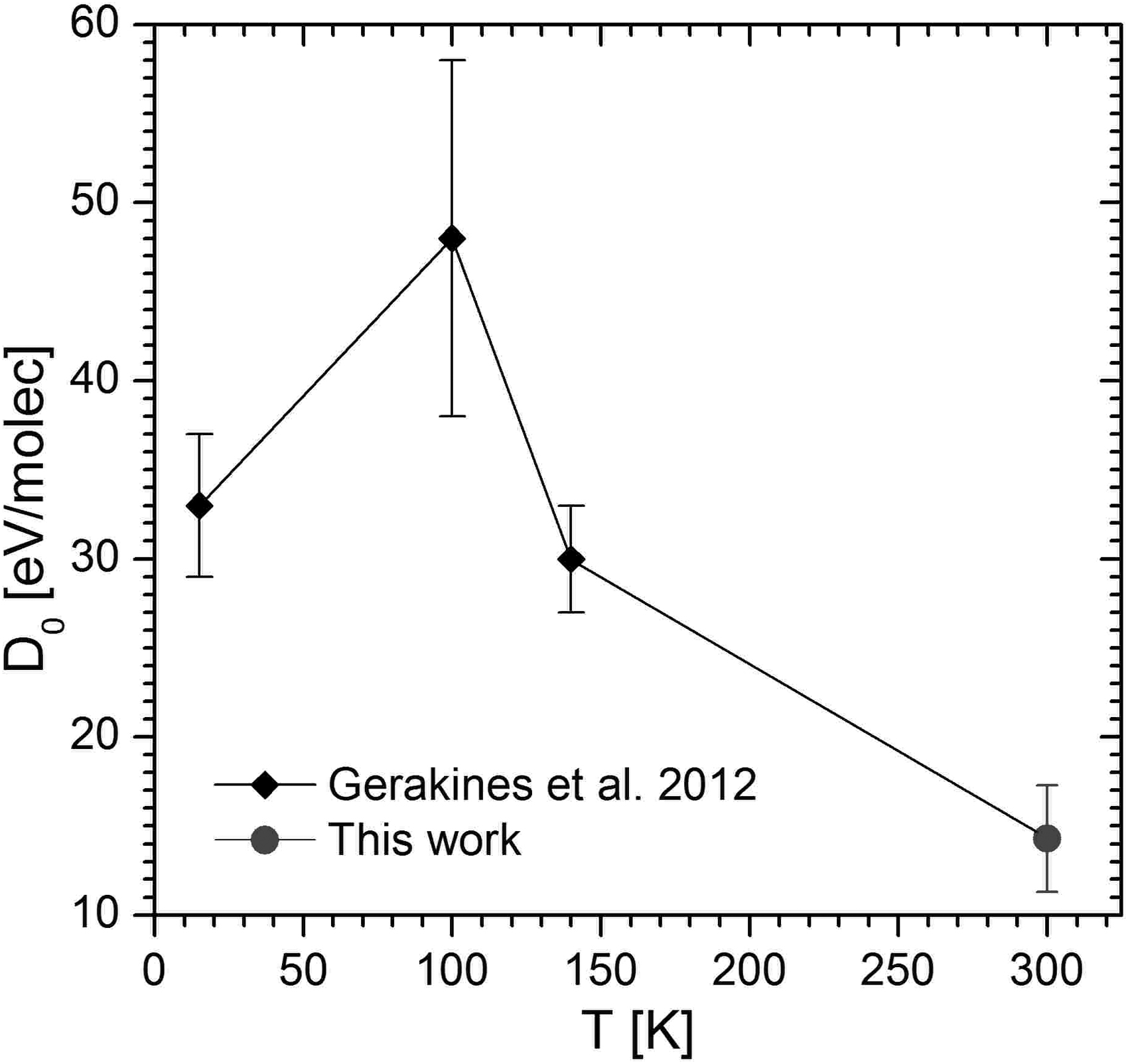
Critical dose as a function of temperature irradiation. Data below 300 K were taken from Gerakines et al. (2012). The measurement at 300 K is reported in the current work.
Moreover, volatile synthesized molecules remain in the bulk and affect chemical reaction pathways or precursor reformation. In contrast, the behavior observed in Fig. 8 is consistent with that reported in the work of Souza-Corrêa et al. (2019): For glycine irradiated by energetic ions, absorbed doses decrease as its temperature increases.
Table 4 compares critical doses and absorbed energy densities obtained for some amino acids. Vignoli Muniz et al. (2017) found the dependence
Molecular Weight (M), Density (ρ), Molecular Volume (V m ), Critical Dose (D 0), and Absorbed Energy Density (ρ E ) for the Glycine, Valine, Adenine, and Phenylalanine, with σ ap d = (S e /D 0) N
References: da Costa et al. (2021) for Gly, da Costa et al. (2020) for Val, Vignoli Muniz et al. (2017) for Ade.
These values correspond to target temperatures ≤20 K; their data were reviewed considering n = 1.
In general, it is expected that larger amino acid molecules are destroyed more efficiently than the smaller ones and according to their structures; however, large molecules are more resilient against radiolytic destruction in the sense that most of their chemical properties are preserved after absorbing a moderate dose. A method with which to estimate the molecular stability is to determine their absorbed energy density ρE (Table 4). For Phe, in particular, the molecular volume of which is 0.21 nm3, the energy density required for the Phe removal is ρE = 65 eV/nm3, the lowest quantity reported in Table 4. Such a result indicates that, among these listed species, the Phe molecule is the most fragile against ionizing irradiation.
4.4. Synthesized species
The spectra displayed in Figs. 6 and 7 exhibit features obtained by Gaussian–shape decomposition that might belong to newly synthesized molecules that remained in the bulk sample. The IR features created by the radiolysis are presented in Table 3 and should correspond to synthesized molecules.
Measurements of secondary ion desorption induced by MeV projectile impact certainly help to identify the molecular species synthesized through the ion track (Andrade et al., 2013). In the case of Phe, positive ion desorption was reported by Guthier et al. (1983), and positive and negative desorbed ions were studied by Leite et al. (1992). Together, these studies identified the most abundant charged fragments ejected after an energetic projectile impact on Phe surface.
For positive ion species, their masses are: 28, 30, 44, 73, 74, 77, 90, 120 and 166 u, attributed to: CO+, CH3NH+, CO2 +, CON2-CHCH2 +, COOHNH2C+, C6H5 +, COOHNH2CHCH3 +, NH2CHCH2C6H4 +, and COOHNH2CHCH2 C6H4 +, respectively. In the negative ion mass spectrum, the masses of the most abundant desorbed species are 28, 50, and 74 u, corresponding very likely to CN−, (COOHNH2CHCH2C)2−, and COOHNH2C−, respectively. Peaks attributed to ethylamine and glycine were also present in the negative mass spectra studied by Leite et al. (1992).
Gerakines et al. (2012) suggested that methylamine and ethylamine could be synthesized from irradiated Phe. However, they could not measure their column densities, possibly due to the IR technique's limitations. A more detailed investigation of synthesized molecules should reveal all the molecules formed in the radiolysis of Phe. For example, a high-performance liquid chromatography apparatus coupled with a high-resolution mass spectrometer was used to identify complex molecular species (Oba et al., 2019).
5. Astrophysical Considerations
A relevant astrophysical implication based on destruction cross-section knowledge is half-life estimates. As an illustration, the Phe endurance on galactic cosmic rays (GCR) in the ISM is calculated.
The first step is to select the six most abundant GCR ionic species and their energy distributions. They are as follows: H+, He+, Cn+, On+, Nen+, and Fen+, labeled as j = 1 to 6. Isotropic radiation is assumed, and their flux densities are those of the Webber and Yushak (1983) model, as adopted by Shen et al. (2004).
E is energy per nucleon of the cosmic ray ion, E 0 is a constant that defines the energy per nucleon where the distribution undergoes the maximum, and Cj is a coefficient proportional to the absolute abundance of the j-component. Usually, E 0 ranges between 200 and 600 MeV/u for all j. It is important to note that E 0 does not depend on the projectile mass only if E is the energy per nucleon. Integrating Eq. (5) for all energies, one gets dϕ/dΩ j = ηCj (ions/cm2/s/sr), where η 400 = 1.53 × 10⁻⁵ for E 0 = 400 MeV and η 300 = 2.48 × 10⁻⁵ for E 0 = 300 MeV.
The ϕj dependence on E 0 does not change the relative flux of the CR species but alters the absolute value. For low GCR energy flux densities (E < E 0), it alters by a factor of 2.4 and, for the total flux, by ∼50%. We adopted E 0 = 400 MeV/u in the current calculation.
The second step is to determine the Cj coefficients from experimental data. The second column of Table 5 lists the GCR source relative abundances reported in the work of Meyer et al. (1998). For absolute flux density, Webber and Yushak (1983) propose, for j = 1, CH = 9.42 × 10⁴ particles cm−2 s−1 sr−1 (MeV/u)1.7. Assuming GCR isotropic radiation impinging on the object just from one side, Eq. 5 can be integrated over 2π sr, so that the factor 2πCH = 5.92 × 10⁵ ions/cm2/s (MeV/u)1.7 is obtained. Making use of the reported relative abundances, the 2πCj coefficients are determined for the main GCR constituents and displayed in the third column.
Integrating again Eq. 5 over all projectile energies per nucleon, the absolute ϕj values are calculated and shown in the fourth column of Table 4. For comparison, in the fifth column, average fluxes of GCR detected at 1 AU are displayed; they are quoted from the work of Bennett et al. (2013), who estimated the GCR fluxes at ∼80 AU to be one order of magnitude higher. Such moderate agreement allows considering that Eq. 5 and the proposed Cj coefficients are a good analytical description of the GCR fluxes; they have been used in the current calculations.
The third step is the destruction rate calculation of pure Phe thin films, which is defined as:
where σ ap d,j (E) = a Phe Se (E). The σ ap d,j (E) ϕ(E) dependence on the energy per nucleon for each of the selected GCR species is displayed in Fig. 9. It should be noted that GCR protons are responsible for the highest destruction rate over all the energy range; the second most destructive GCR species are He ions, at low energy, and Fe ions, at high energy.
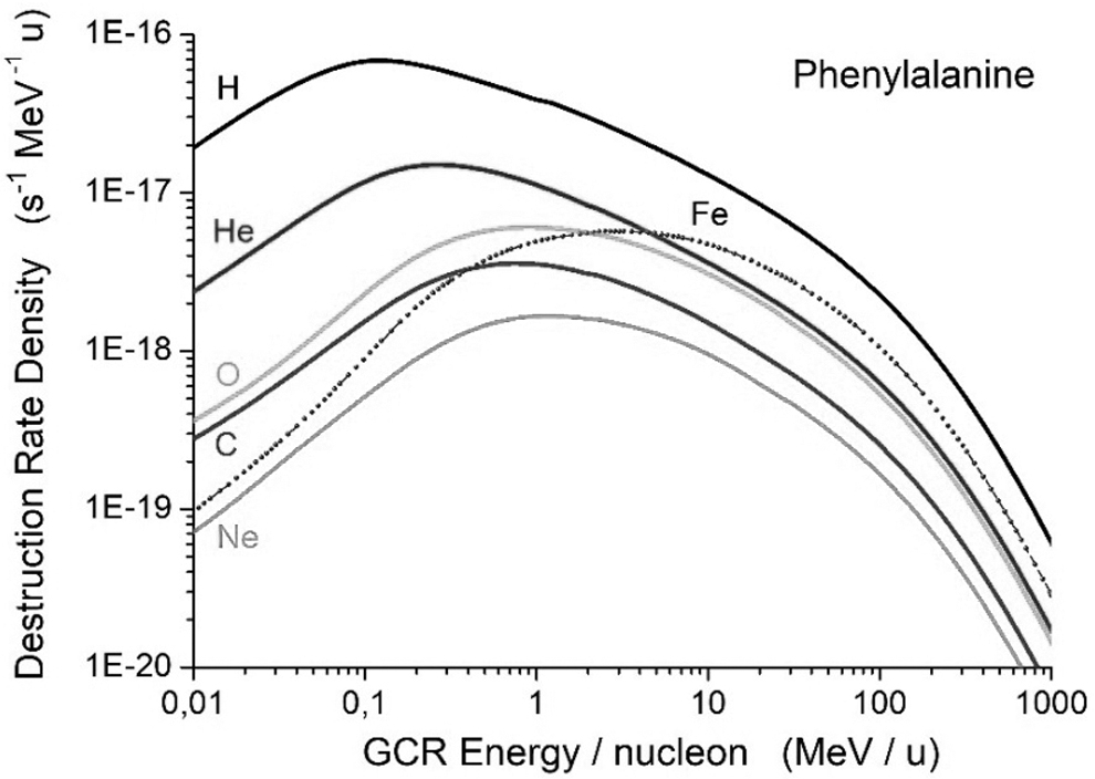
Dependence of the Phe destruction rate density of each GCR species on their energy/nucleon. GCR, galactic cosmic rays.
The predicted partial destruction rates, Rj , and the corresponding partial half-lives, τj = ln(2)/Rj , are presented in the last two columns of Table 5. The (total) half-life, given by τ 1/2 = ln(2)/R, is calculated to be 10 million years. Likewise, Gerakines et al. (2012) estimated a τ 1/2 = 14 million years for Phe irradiated at 10 K; this value is according to Fig. 8 since the critical dose is also a measurement of the Phe radioresistance at different temperatures.
In comparison with previous results, the predicted total half-life for Phe is practically the same as that calculated for adenine by Vignoli Muniz et al. (2017). On the other hand, the partial half-lives for Phe are expected to be 23% lower than those obtained for Val by da Costa et al. (2020), once the constant 1/ρ Phe is found to be ∼1.3 greater than 1/ρ Val. Because the constant “1/ρ” is proportional to Rj , it indicates that any GCR constituent should destroy Phe 23% faster than Val. However, we found a ratio τj (Val)/τj (Phe) ∼ 1.3/Mj (where Mj is the ion's mass), evidence that a mistake happened in their Val Rj calculations. The discrepancies hinge on 1/Mj , and so it is not too difficult to discern that the origin of the misunderstanding comes from the definition of the E 0 constant, as discussed earlier.
The earlier calculation demonstrates the feasibility of half-life estimates for molecular solids. More realistic materials such as mixtures of amino acid with water ice or silicates can be performed by implementing a similar procedure.
6. Conclusions
The main conclusions are: Column densities of the essential α-amino acid Phe irradiated by the ion beams (H+ 2 MeV, H+ 0.5 MeV, He+ 2 MeV, N+ 0.5 MeV, and N+ 2 MeV) decay exponentially with fluence or dose (Fig. 4). The apparent destruction cross-section of Phe radiolysis is proportional to the electronic stopping power of the ionizing radiation (Fig. 5). The inverse of the proportionality constant, at room temperature, is found to be the critical apparent dose Do
ap = (14.3 ± 2.2) eV/molec. This value corresponds to the molecular energy density ρE
≈ 65 eV/nm3, which expresses the minimum average energy density to eliminate Phe molecules from the sample by radiolysis and/or by sputtering.
Do
depends on sample temperature. The D
0 value obtained at 300 K is a factor ∼2.7 lower than those reported by Gerakines et al. (2012) for Phe irradiated at lower temperatures (Fig. 8). The energy density ρE
= 65 eV/nm3 measured for Phe means that this molecule is more fragile than adenine (80 eV/nm3), Val (100 eV/nm3), or glycine (120 eV/nm3), when exposed to ion beam irradiation. The IR spectra of irradiated Phe samples (Figs. 6 and 7) exhibit new bands attributed to synthesized molecules (Table 3). Some of the formed molecular species were also observed in the mass spectrometry experiments performed by Guthier et al. (1983) and Leite et al. (1992). Destruction rate calculations predicted that GCR protons rule the Phe destruction in the ISM. Destruction rates due to He and Fe ions are about 30% and 40% of that of protons, respectively. The Phe half-life in the ISM is predicted to be around 10 million years (Table 5) at 300 K and 20–30 million years at 15–150 K (assuming that destruction cross-sections are 2–5 higher).
Footnotes
Author Disclosure Statement
No competing financial interests exist.
Funding Information
Part of this work was supported by the Direccion de Investigacion de la Universidad de Cuenca, DIUC. The authors acknowledge the Brazilian agencies FAPERJ (E-26/202.843/2018 and E-26/200.413/2020) and CNPq (PDS n° 118349/2017-1) for partial financial support. This study was financed in part by the Coordenação de Aperfeiçoamento de Pessoal de Nível Superior—Brasil (CAPES)—Finance Code 001.
Abbreviations Used
Appendix
Associate Editor: Lewis Dartnell