
Abstract
Identification of spectroscopic fingerprints that correspond to relevant molecules/minerals in a Mars-like environment is a crucial search in astrobiology. Therefore, we studied the stability of Gly·MgSO4·5H2O under Mars-like surface conditions and compared it to the behavior of epsomite and glycine. Gly·MgSO4·5H2O has been identified as a molecule of astrobiological interest since an amino acid and water molecules, which are essential for life, are part of its structure. Furthermore, this compound may form by the interaction of sulfate minerals with glycine-bearing aqueous solutions, and both could be present on Mars.
The main analyses were performed by using in situ Raman spectroscopy, a ground-breaking technique for NASA and ESA Mars planetary missions. We have integrated a Raman spectrometer in a Planetary Atmosphere and Surfaces Chamber (PASC) and have identified the processing of molecules exposed to a simulated martian atmosphere, UV irradiation, and temperature.
Our results show that pressure is critical to provoke amorphization of Gly·MgSO4·5H2O, and the release of glycine from the compound; the stabilization effect at low temperature and stability of Gly·MgSO4·5H2O is greater than to glycine and epsomite.
The strategy employed here allows us to evaluate the effect of diverse simulated martian environmental conditions on molecular preservation by using Raman spectroscopy.
1. Introduction
Amino acids are the building blocks of proteins and therefore have been extensively studied to explore the emergence of life on early Earth. In addition, these molecules are important for searching for extraterrestrial life, since it is generally assumed that if any such life exists, it is based on the same fundamental biochemistry as that found on Earth (Cockell, 2015; Bhattacharya and Lichtman, 2016). Likewise, amino acid studies can provide clues for investigating the hypotheses that describe how Earth life could come from Mars (McKay, 2010).
This idea has been supported by the detection of extraterrestrial glycine, the simplest amino acid. Glycine has been found in comets (Elsila et al., 2009) and in meteorites along with other organic compounds (Glavin et al., 1999; Botta and Bada, 2002). The latter study shows that glycine is one of the two more predominant amino acids, along with glutamic acid, in the martian meteorite Nakhla. Despite this evidence of martian glycine, neither this amino acid nor others have been detected at the martian surface (Eisenstein, 2005). The most promising results in this regard are those provided by the Sample Analysis at Mars (SAM) instrument on board the Mars Science Laboratory Curiosity rover. The SAM instrument analyzed samples collected from the top 5 cm of the martian regolith and drilled mudstones by heating them to 875°C and monitoring the volatiles released from the samples with a mass spectrometer (Mahaffy et al., 2012). The analysis of the results indicates the existence of organic compounds (Glavin et al., 2013; Freissinet et al., 2015; Millan et al., 2016; Franz et al., 2017; Eigenbrode et al., 2018) by the detection of fragments derived from the high-temperature decomposition of the original material, which are compatible with those found in martian meteorites (Steele et al., 2018).
In addition to pyrolysis, the original organic material has been subjected to previous geological processes, which are influenced by martian mineralogy. Indeed, it is widely recognized that studies not taking into account the effects of mineral phases cannot be considered very realistic since the interactions of organic molecules with minerals may completely change their reaction pathways (Fornaro et al., 2018); that is, the catalytic and/or protective properties of several minerals have been previously reported (Martínez-Frías et al., 2006; Mateo-Marti et al. 2019).
Among the minerals studied, sulfate minerals have attracted great attention due to their occurrence on Mars and their geochemical implications. Landed and orbital missions have detected these minerals in large quantities and widely spread on the surface of Mars. For instance, jarosite, KFe3+ 3(SO4)2(OH)6, was identified by the Opportunity rover (Klingelhöfer et al., 2004), and the OMEGA spectrometer of the Mars Express orbiter mission identified kieserite (MgSO4·H2O), gypsum (CaSO4 ·2H2O), and other polyhydrated sulfates with different cations (Gendrin et al., 2005). Kieserite and other Mg polyhydrated sulfates are the most common and abundant sulfates observed thus far on Mars (Wang et al., 2016).
Regarding the chemical implications, like other minerals, sulfates have shielding properties for organic molecules (Amaral et al., 2006), and it is worth noting that the degree of hydration can shed light on the hydrologic evolution of the planet and provide information about its current water reservoirs (Wang et al., 2006).
In the present work, we are interested in the study of glycine and hydrated magnesium sulfates due to the mentioned importance, but it is important to note that the study is focused not on the isolated molecules but on the ternary system of glycine-MgSO4-water.
Specifically, we are focused on the Gly·MgSO4·5H2O molecule, which has been recently identified as a molecule of planetological interest, as this compound may form by the interaction of martian kieserite with glycine-bearing aqueous solutions (Howard et al., 2016).
This compound and the one with three water molecules in its structure, Gly·MgSO4·3H2O, (Howard et al., 2016) are the only known structurally characterized Mg(II) materials containing both glycine and sulfate compounds (Srinivasan, 2020). Its crystal structure was reported in 2007 (Elayaraja et al., 2007).
In this structure, the glycine molecule exists in the zwitterionic form, which is a common chemical form of amino acid compounds with inorganic salts. The nonhydrogen atoms of the glycine achiral molecule are coplanar. The unit of this magnesium sulfate complex with glycine consists of two cations, [Mg(H2O)6]2+ and [Mg(C2H5NO2)2(H2O)4]2+, and one SO4 2- anion. The Mg(II) atoms in both cations are in an octahedral coordination environment. The Mg(II) atom is coordinated in the first cation by six water molecules and in the second cation by two O atoms from two glycine ligands and four water molecules (Fig. 1A). These cations pack as alternate layers parallel to the ab plane, with the sulfate anions lying between them. The complex cations and sulfate anions are linked to form a three-dimensional network by O—H···O and N—H···O hydrogen bonds (Fig. 1B).
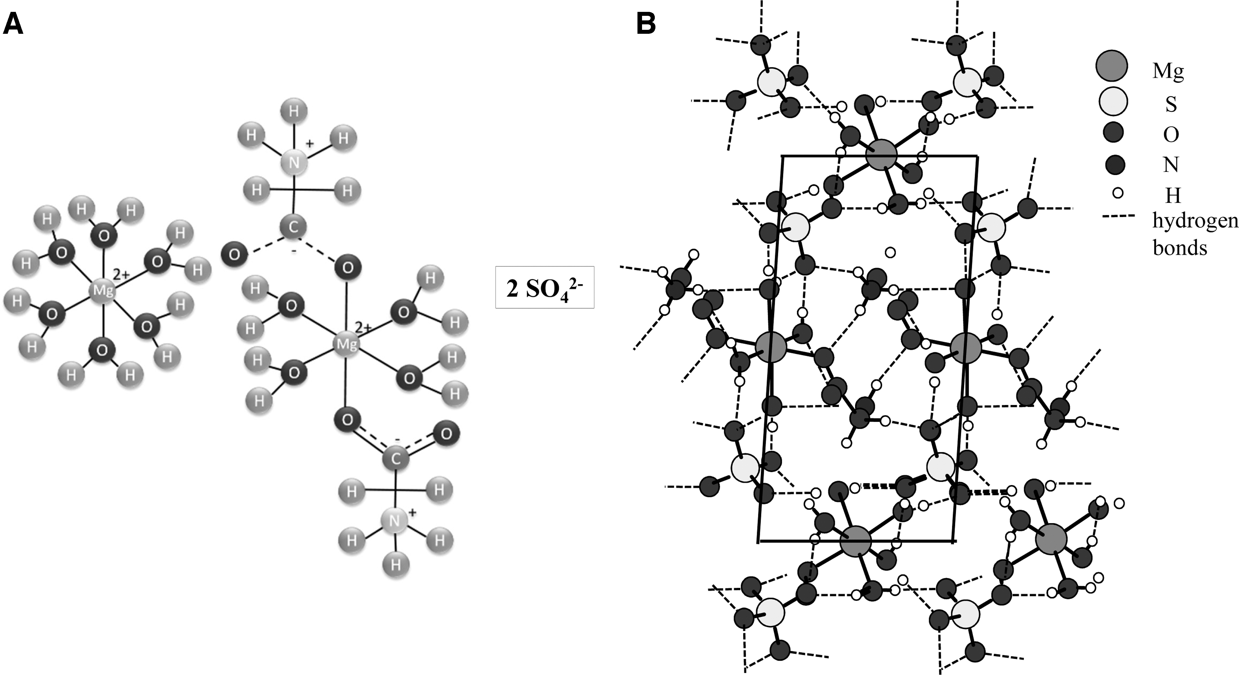
Molecular sketch (
The described structure was proven by Tepavitcharova et al. (2012), who used single-crystal X-ray diffraction (XRD) and vibrational spectroscopies (FTIR and FT Raman). Furthermore, these authors studied its crystallization and thermal behavior at high temperatures ranging from 293 to 773 K.
Howard et al. (2016) characterized the Gly·MgSO4·5H2O and Gly·MgSO4·3H2O compounds by XRD, neutron powder diffraction, and Raman spectroscopy. Moreover, they studied the stability of the pentahydrate molecule between 298 and 478 K by in situ XRD, obtaining that pentahydrate leads to trihydrate at high temperatures.
In this work, we study the stability of Gly·MgSO4·5H2O exposed to simulated martian surface environmental conditions—CO2 atmosphere, UV irradiation, and low temperature—by in situ Raman spectroscopy analysis. To the best of our knowledge, there are no data about the behavior of this molecule under simulated martian conditions and no published studies on the vibrational spectra of this material under these conditions.
Raman spectroscopy has been chosen as the main technique because it has been widely recognized as a valuable technique for planetary exploration due to its unique characteristics, such as (i) high capability to detect organic and inorganic compounds, (ii) sample manipulation is not required, (iii) it allows the analysis of very small sample amounts, and (iv) it is a nondestructive technique (Jorge-Villar and Edwards, 2006). Therefore, Raman spectroscopy has been selected as a technique that will be part of the upcoming NASA and ESA Mars planetary missions. For example, the Raman Laser Spectrometer (RLS) is a part of the ExoMars 2020 mission, key instruments devoted to identifying mineral phases at the grain scale in crushed sample material to determine their composition and establishing the presence of carbon (inorganic/organic) analysis of samples collected underneath the martian surface (Vago et al., 2017; Rull et al., 2018). Likewise, in the current Mars 2020 rover, two instruments, SuperCam and Scanning for Habitable Environments with Raman and Luminescence for Organics and Chemicals (SHERLOC), are also focused on Raman-based organic and mineral detection (Martin et al., 2020).
Therefore, our goal here is, on the one hand, to study the chemical evolution of this particular molecule under simulated martian planetary conditions by in situ Raman spectroscopy. For this aim, we performed a systematic study of each condition separately (CO2 pressure, temperature, and UV radiation) to uncouple and compare the different effects. On the other hand, we provide quality Raman spectra of compounds (as a database) for current and upcoming Mars planetary missions to help with future chemical assignment and detection on the martian surface.
Therefore, the work is structured as follows: In Section 2, the experimental procedures used in this study are described. Then, in Section 3, the results obtained are presented, where they have been organized into three parts to analyze the effect of each studied condition separately: (1) the effect of the atmosphere (99% CO2 and 0.6% H2O at a pressure of 7 mbar) at room temperature; (2) the effect of UV irradiation under previous atmospheric conditions; and (3) the low-temperature effect. The discussion presented in Section 4 analyzes and evaluates the results shown in the previous sections for all conditions and presents a comparative study of the processes identified for each molecule under the different studied conditions. The conclusions of this work are presented in Section 5.
2. Materials and Methods
2.1. Sample preparation
Glycine amino acid (Gly, NH2-CH2-COOH, purity ≥99%) provided by Sigma-Aldrich was dissolved in Milli-Q water (total organic content lower than 5–10 ppm and resistivity higher than 18 mΩ·cm−1) at saturation concentration; thereafter, epsomite and MgSO4·7H2O from Sigma-Aldrich were added at equimolar concentrations. The slow evaporation at room temperature for 2 weeks gives rise to Gly·MgSO4·5H2O crystals.
After crystallization, the samples were prepared in disc form. Therefore, approximately 200 mg of each solid was ground with an agar mortar; then samples were pressed at high pressure with a hydraulic press from 8 to 10 tons for ∼15 min to form a compact pellet from the compound.
The synthesized Gly·MgSO4·5H2O before pelletization and the raw material (glycine and MgSO4·7H2O) were analyzed by several techniques: Raman spectroscopy, infrared spectroscopy and X-ray diffraction (XRD). The results of these analyses can be found in Supplementary Figs. S1–S3 and Tables S1–S6.
2.2. Characterization techniques
2.2.1. X-ray diffraction
X-ray diffraction characterization was performed by means of a Bruker D8 Advance diffractometer using Cu Kα1 radiation (λ = 1.54056 Å) operating at 45 kV and 40 mA. A Bragg–Brentano configuration geometry was used. The 2θ range covered was from 10 to 40 at 0.041 scanning steps.
2.2.2. Infrared spectroscopy
Infrared spectra were obtained with a thermo-Nicolet spectrometer. Spectra (4 cm−1 resolution and 64 scans) were collected in the mid-infrared region (400–4000 cm−1) using an XT-KBr beamsplitter and a DTGS-ATR detector.
2.2.3. Raman spectroscopy
Raman spectra were acquired by using a B&WTek i-RamanTM, ExemplarPro model, with a green HeNe laser with a wavelength of 532 nm and an operation power of 100 mW. The spectral range available to this spectrometer is from 65 to 4000 cm−1, with a pixel resolution of 2.99 cm−1.
This equipment can be coupled to two different probes. The first one is set up inside the Planetary Atmosphere and Surfaces Chamber (PASC) through a CF-40 flange to measure under simulated martian conditions, and the other one can be used to measure outside the chamber at room conditions. Raman spectra were acquired with a typical exposition time of ∼200 s and 3 accumulations. All acquired spectra were recalibrated by using a Neon emission light and baseline corrected by the method described in Sanz-Arranz et al. (2017).
2.3. Planetary Atmosphere and Surfaces Chamber (PASC)
The target molecules and compounds were characterized before and after being exposed to simulated martian conditions (99% CO2 and 0.6% H2O at a pressure of 7 mbar, UV radiation [200–400 nm], and temperature of 293 and 250 K) inside the PASC at the Centro de Astrobiología. The planetary simulation chamber PASC is an ultrahigh vacuum chamber 500 mm long and 400 mm in diameter with a total volume of approximately 62,800 cm3 and standard CF flanges.
Samples were placed in a covered gold sample holder at the end of the He cryostat. Before setting martian atmospheric conditions, the chamber was pumped down to a high vacuum level of approximately 6 × 10⁻⁵ mbar to remove the atmospheric gases inside the chamber and reach a clean vacuum environment. Then, CO2 gas was introduced from the gas manifold inside the chamber to increase the partial pressure of the chamber to a 7 mbar value to simulate martian atmospheric conditions.
For the UV radiation experiments, a UV deuterium lamp (Hamamatsu C130) was placed perpendicular to the sample, and the UV radiation entered the system through a quartz window. The UV flux measured at the sample position, (F) obtained by integration of the irradiance over the 200–400 nm wavelength range, is 2270 mW−2 and corresponds to a photon flux of Φ = 2.3 × 10¹⁴ photons cm−2 s−1, approximately 10 times lower than the UV flux on the martian surface.
To achieve the low working temperature of the sample (271 K), it was regulated by a commercial closed-cycle helium cryostat (Advanced Research Systems, Inc.), cooling system, connected to the sample holder, and it can be adjusted from 15 to 323 K. Additionally, Raman spectroscopy (532 nm) was coupled to the chamber, allowing in situ characterization of the samples under study (see Supplementary Fig. S5).
2.4. Linkam stage
The Linkam TSH600 stage used in this work is a heating and cooling stage that allows in situ characterization of samples from ambient temperature (295 K) to 1773 K by using Raman spectroscopy. The mechanical design and electronics of the Linkam stage provided precise control and temperature stability better than 0.2 K. Spectra were collected with the Raman probe for RT measurements. This probe can focus the laser excitation source on the sample surface (housed in the sample holder) through a silica window (used in place of the standard quartz optical window) on the top of the stage. Then, the scattered radiation is collected in backscattering geometry by the probe.
3. Results
The influence of environmental conditions on the behavior of molecules is critical for their stability, reactivity, and possible degradation. Furthermore, the identification and characterization of the accurate compound is influenced by these conditions; the outcome of complete or partial simulated settings separately is also taken into account to discriminate its significance. Therefore, glycine, MgSO4·7H2O, and Gly·MgSO4·5H2O molecules were characterized by in situ Raman spectroscopy under simulated martian conditions (99% CO2 and 0.6% H2O at a pressure of 7 mbar, UV radiation in the 200–400 nm range, and temperature of 271 K). We performed a systematic study of each condition separately to uncouple the different effects due to exposure to several experimental parameters. With this aim, we first studied the effect of the Mars atmosphere (99% CO2 and 0.6% H2O at a pressure of 7 mbar) at room temperature. Then, we studied the effect of UV irradiation under previous atmospheric conditions, and the sample was irradiated with a UV lamp for up to 24 h. The last experiment was focused on the low-temperature sample effect. The sample material was exposed to a simulated martian atmosphere (99% CO2 and 0.6% H2O at a pressure of 7 mbar) at 271 K under UV irradiation for 24 h.
3.1. Mars atmosphere influence
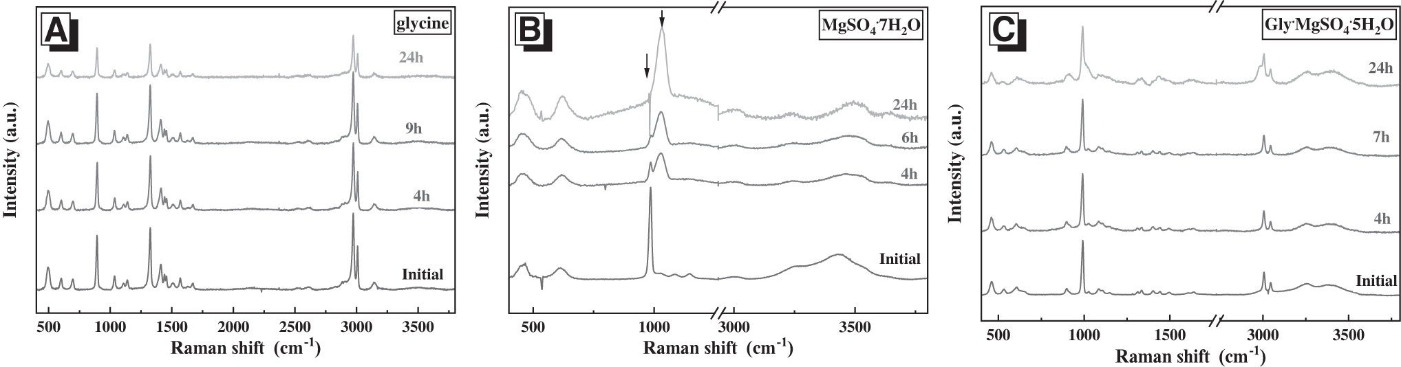
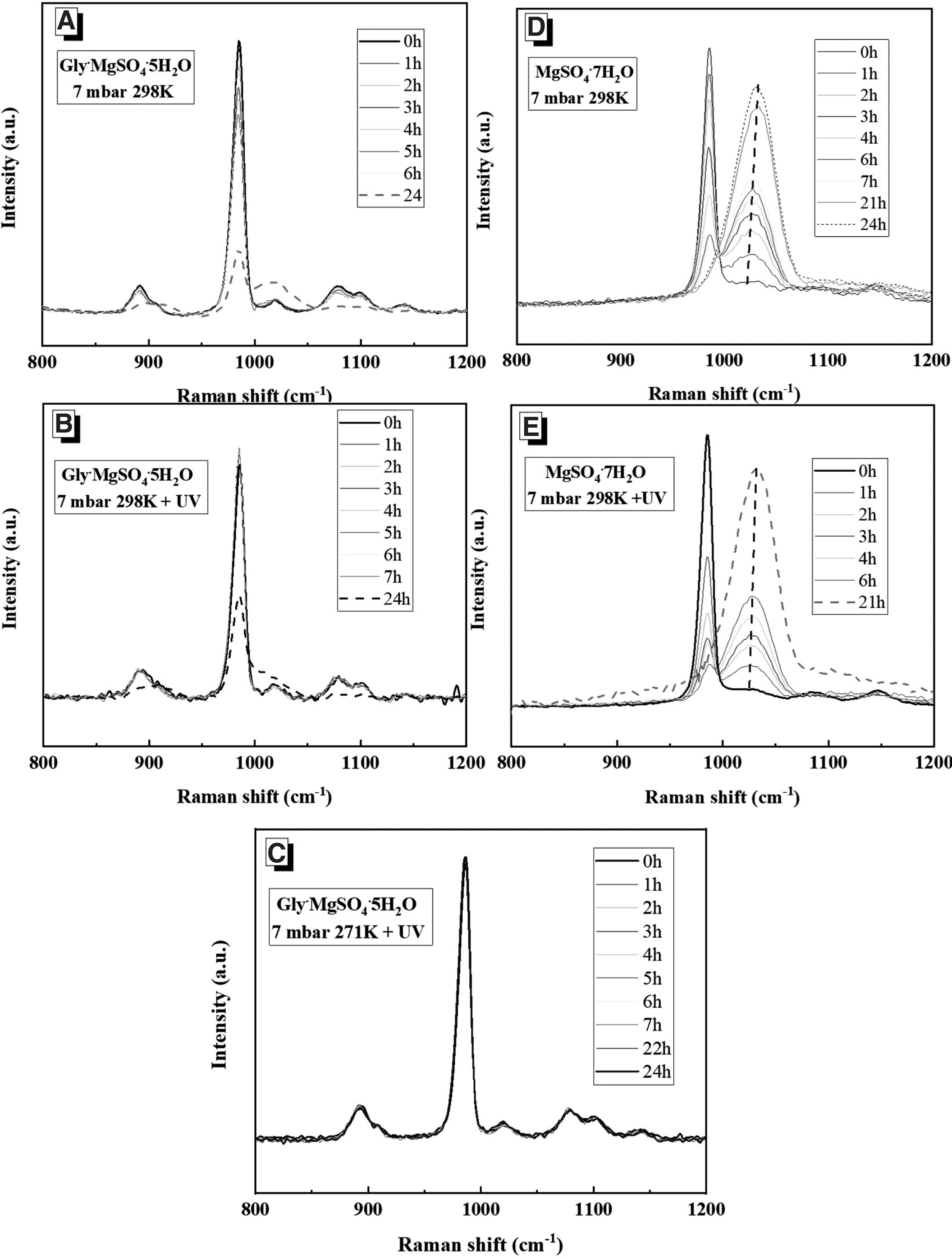
In situ Raman spectroscopy measurements of (A) glycine, (B) MgSO4·7H2O, and (C) Gly·MgSO4·5H2O at 7 mbar pressure of 99% CO2 and 0.6% H2O at room temperature are presented in Fig. 2.

Time evolution of in situ Raman spectra of (
Glycine spectra (Fig. 2A) do not show any changes after 24 h of exposure to simulated martian atmospheric conditions, indicating high stability for this organic molecule under simulated martian atmospheric conditions. By contrast, the hydrated salts MgSO4·7H2O (Fig. 2B) and Gly·MgSO4·5H2O (Fig. 2C) spectra vary as a function of exposure time under these conditions.
The main observed changes in the epsomite (MgSO4·7H2O) spectra are (1) a significant decrease in the intensity of ν1 SO4 (at ∼980 cm−1), which essentially disappears at a certain point between 7 and 21 h, and (2) a more striking appearance of a new broadband at ∼1020 cm−1, which continuously increases in intensity and shifts to higher frequencies (see Fig. 2B). These Raman features have been previously recognized; see Wang et al. (2006, 2009, 2011), Wang and Zhou (2014), and Chou et al. (2013), who identified it as a result of amorphization of the mineral phase due to a dehydration process. In the mentioned studies, the authors perform numerous dehydration experiments at different temperatures and under Mars-relevant pressure. Raman spectra were acquired from intermediate and final products of the amorphization of epsomite at 21°C, 0°C, and 8°C. The results show that the continuous dehydration of epsomite causes the amorphous MgSO4·H2O phase, characterized by the appearance and growth of a very broad Raman peak that follows the development of dehydration and shifts from 1025 to 1034 cm−1.
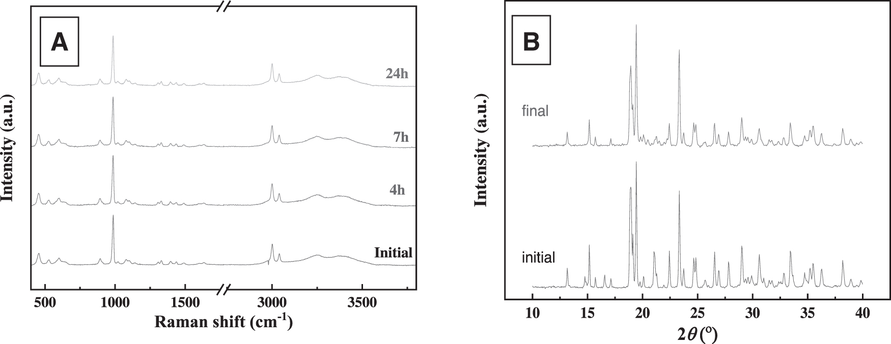
For the Gly·MgSO4·5H2O compound, a similar behavior was found in our experiments, showing a drop in the ν1 SO4 intensity and appearance of the broadband at ∼1020 cm−1. Therefore, we identify this behavior as a result of an amorphization process of the sample. To verify this assumption, the sample was analyzed by ex situ XRD after exposure to simulated martian atmospheric conditions for 24 h (Fig. 3). The characteristic peaks in the diffractogram become less pronounced, which is in accordance with an amorphization process. In addition to this epsomite-like behavior, a relevant change of the bands at higher frequencies corresponding to the stretch bands of glycine CHs at long exposure times of ∼24 h should be noted. CH stretching bands decrease, and a new band appears at ∼2974 cm−1. This behavior highlights a possible structural change of glycine (C2H5NO2) in the [Mg(C2H5NO2)2(H2O)4]2+ cation under the studied conditions.
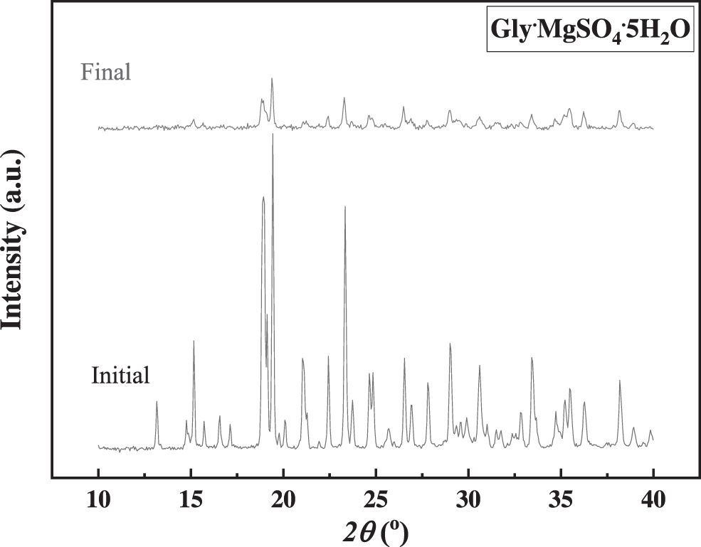
XRD pattern of Gly·MgSO4·5H2O (Initial) and after 24 h exposure at simulated martian atmospheric conditions (99% CO2 and 0.6% H2O at a pressure of 7 mbar [Final]).
3.2. Mars atmosphere and UV irradiation influence
To evaluate the effect of the addition of UV radiation for 24 h on the samples, (A) glycine, (B) MgSO4·7H2O, and (C) Gly·MgSO4·5H2O at 7 mbar pressure of 99% CO2 and 0.6% H2O and room temperature (296 K), in situ Raman spectroscopy measurements were performed (see Fig. 4).
Time evolution of in situ Raman spectra of (
The UV/atmosphere irradiated glycine spectra do not show any additional Raman bands or band extinction; however, the intensity of the bands decreases as a function of the UV exposure time, which can be explained by the degradation/desorption of glycine, as would be expected after UV irradiation (Poggiali et al., 2020). Moreover, the relative intensity between the bands does not suffer significant variations; thus, similar decreases in the intensity of all the bands could be related to molecular desorption due to the UV irradiation effect (Foti et al., 1991). No deeper analysis has been performed, as it is not the aim of the present study, and many works have addressed the behavior of glycine under UV radiation conditions (Ehrenfreund et al., 2001; Kate et al., 2005; Orzechowska et al., 2007; Johnson et al., 2012; Poch et al., 2014).
MgSO4·7H2O and Gly·MgSO4·5H2O compounds show similar behavior to that in the absence of UV radiation, as can be observed in the obtained Raman spectra (see Fig. 4B, 4C) and in the X-ray diffractograms (Fig. 5).
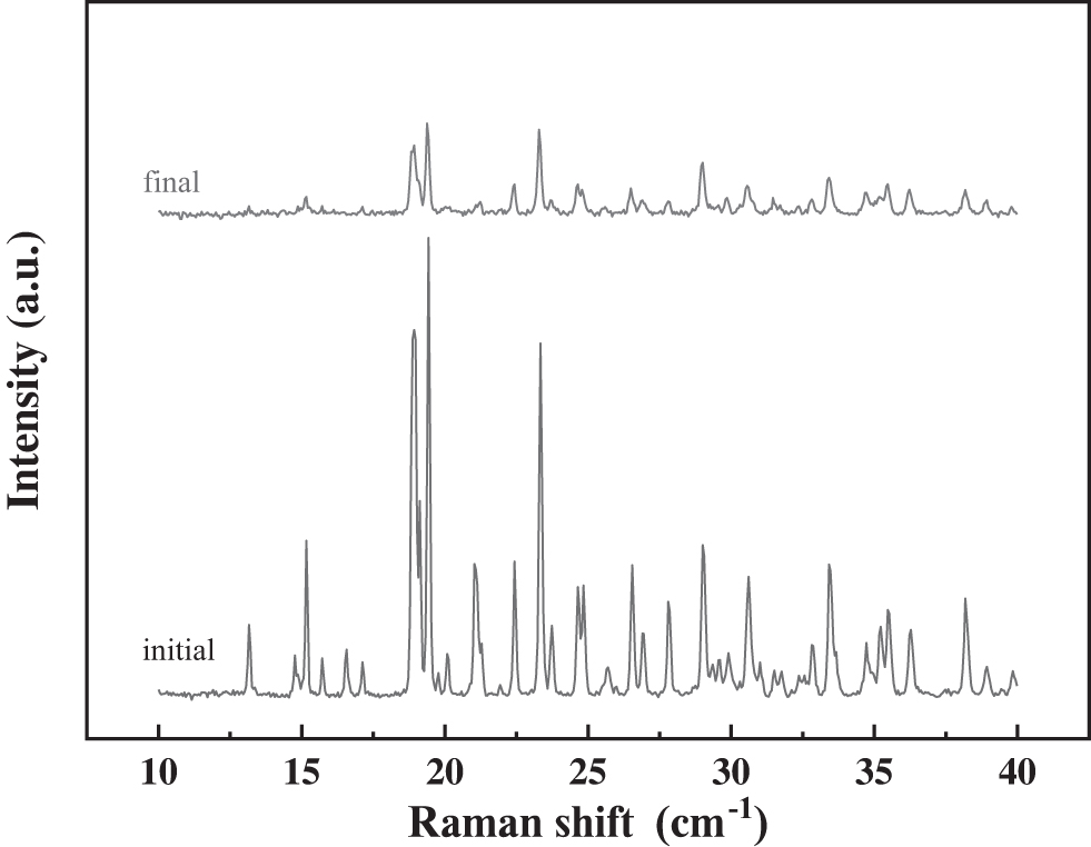
XRD pattern of Gly·MgSO4·5H2O (initial) and after exposure to 24 h at simulated martian atmospheric conditions (99% CO2 and 0.6% H2O at a pressure of 7 mbar) at room temperature and UV irradiation (final).
3.3. Mars atmosphere + UV irradiation and temperature influence
Once we had analyzed the target molecule (Gly·MgSO4·5H2O) under simulated conditions of a martian atmosphere, with and without UV radiation, we added the low-temperature effect to the samples exposed under the same conditions. Figure 6 shows the in situ Raman spectroscopy measurements and X-ray diffractogram of Gly·MgSO4·5H2O at 7 mbar pressure of 99% CO2 and 0.6% H2O, exposed to UV radiation at 271 K.
(
The fact that no spectral feature differences are revealed between any of the Raman spectra as shown in Fig. 6A suggest that low-temperature conditions (271 K) stabilize the Gly·MgSO4·5H2O molecule. Furthermore, Fig. 6B shows similar XRD patterns of the molecule for the initial and final states, proving the absence of the amorphization process in this case, which does not take place at low temperatures, and confirming the stabilization of the Gly·MgSO4·5H2O molecule at low temperatures.
4. Discussion
Having analyzed the in situ Raman spectra obtained from Gly·MgSO4·5H2O that was exposed to different simulated martian conditions over 24 h and identified the main processes that take place (above), we now evaluate these processes by comparing the effect of each condition on the Gly·MgSO4·5H2O, glycine and epsomite molecules.
With this aim, we present a quantitative Raman analysis from the experiments described above. Thus, a detailed band-profile analysis of the spectra was carried out. First, a second derivative analysis allowed us to obtain the Raman frequency of each band; second, the Raman frequencies obtained were used as a fixed value in a Voigt fit. An example of the profile analysis of MgSO4·7H2O at selected times is given in the Supplementary Information (Fig. S4).
In the following figures, we present the results of these analyses for the main bands. On the one hand, we analyzed the behavior of the amorphization process due to the effect of the low-pressure (7 mbar) vacuum conditions; for that, we analyzed the intensity of the broadband at ∼1020 cm−1 of the Gly·MgSO4·5H2O molecule as a function of exposure time to vacuum conditions compared with that of the epsomite compound. On the other hand, to evaluate the possibility of a structural conformational change of glycine (C2H2NO2) in the [Mg(C2H5NO2)2(H2O)4]2+ cation, the νs(CH) and νa(CH) stretching band intensities of this complex were deeply analyzed, therefore providing a plausible explanation for the appearance of the new band at ∼2974 cm−1.
4.1. Amorphization process
Raman spectra of Gly·MgSO4·5H2O at the studied conditions in the medium wavenumber range (800–1200 cm−1) are depicted in Fig. 7A, 7B, 7C compared with those corresponding to epsomite (7D, 7E).
Raman spectra in the medium wavenumber range (800–1200 cm−1) at different exposure conditions as a function of time of Gly·MgSO4·5H2O (
As can be noted in this figure, the loss of crystallinity, evidenced by the appearance and growth of a new broadband and the decreasing intensity of the ν1 SO4 peak (at ∼980 cm−1), occurs in all conditions studied for both molecules, except at low temperature. Note also that the broad band shifts from 1020 to 1032 cm−1 for epsomite (dashed line in Fig. 7D, 7E). Thus, we plotted the intensity of the broadband, hereafter referred to as νamorphous SO4 (Fig. 8), by considering the calculated initial intensity of the ν1 SO4 band in each experiment as 100% (maximum intensity); that is,
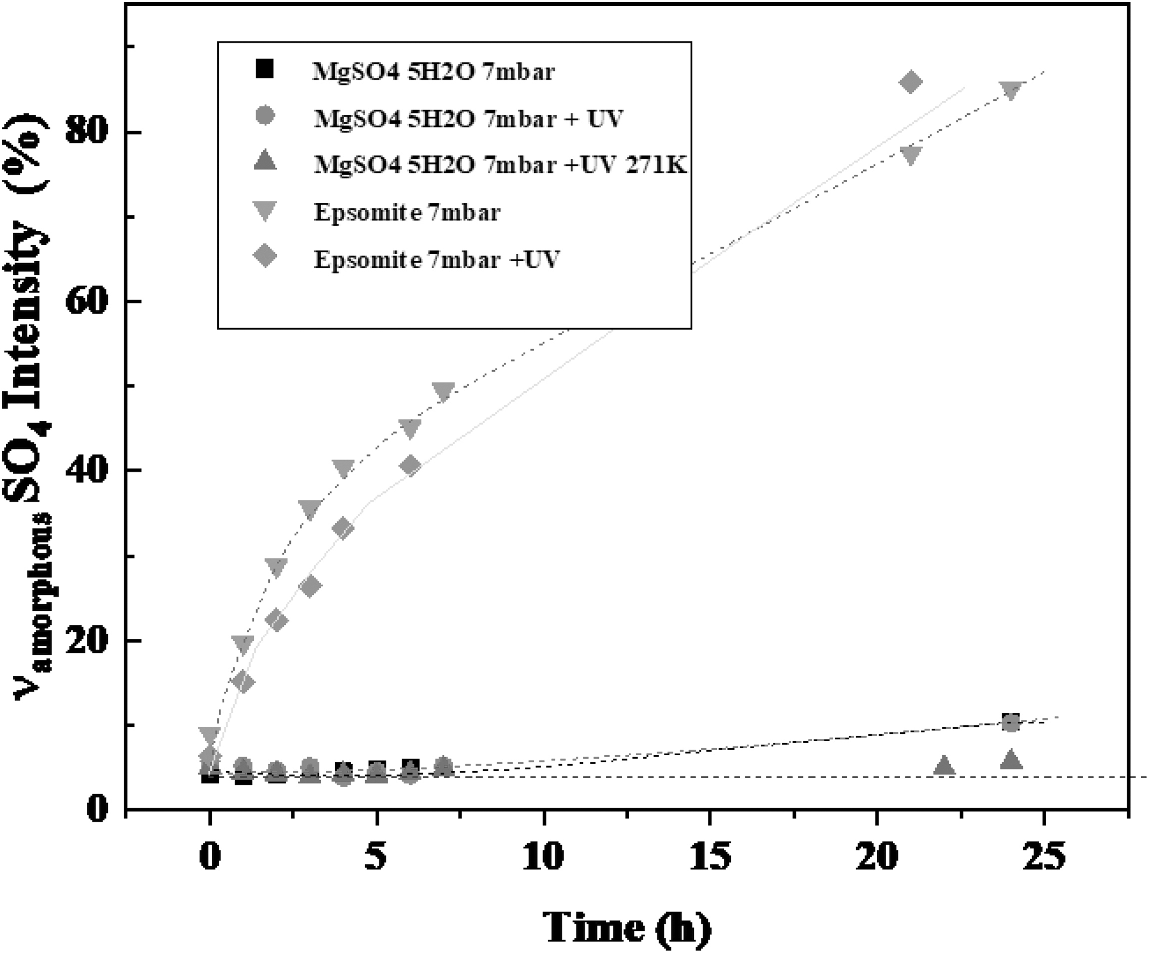
Intensity evolution of ν1 SO4 to νamorphous SO4 % with time at different simulated martian conditions (see legend).
Following Eq. 1, the results are plotted in Fig. 8, which indicates that the intensity of the νamorphous SO4 band increases more rapidly for epsomite (a 70–80% increase in intensity, in 24 h) than for Gly·MgSO4·5H2O (a 10% increase in 24 h), whereas at low temperature for Gly·MgSO4·5H2O this band does not alter whatsoever its intensity. In addition, UV radiation does not seem to affect the stability of the compound at the studied times or the conditions. Therefore, it is remarkable that the presence of Gly in the structure of the compound drives the molecular behavior, protecting the compound from dehydration and partially blocking the amorphization process. On the other hand, the low temperature even increases this protective effect from dehydration, completely avoiding the amorphization process.
4.2. Evaluating the νs(CH) and νa(CH) stretching bands: Conformational changes
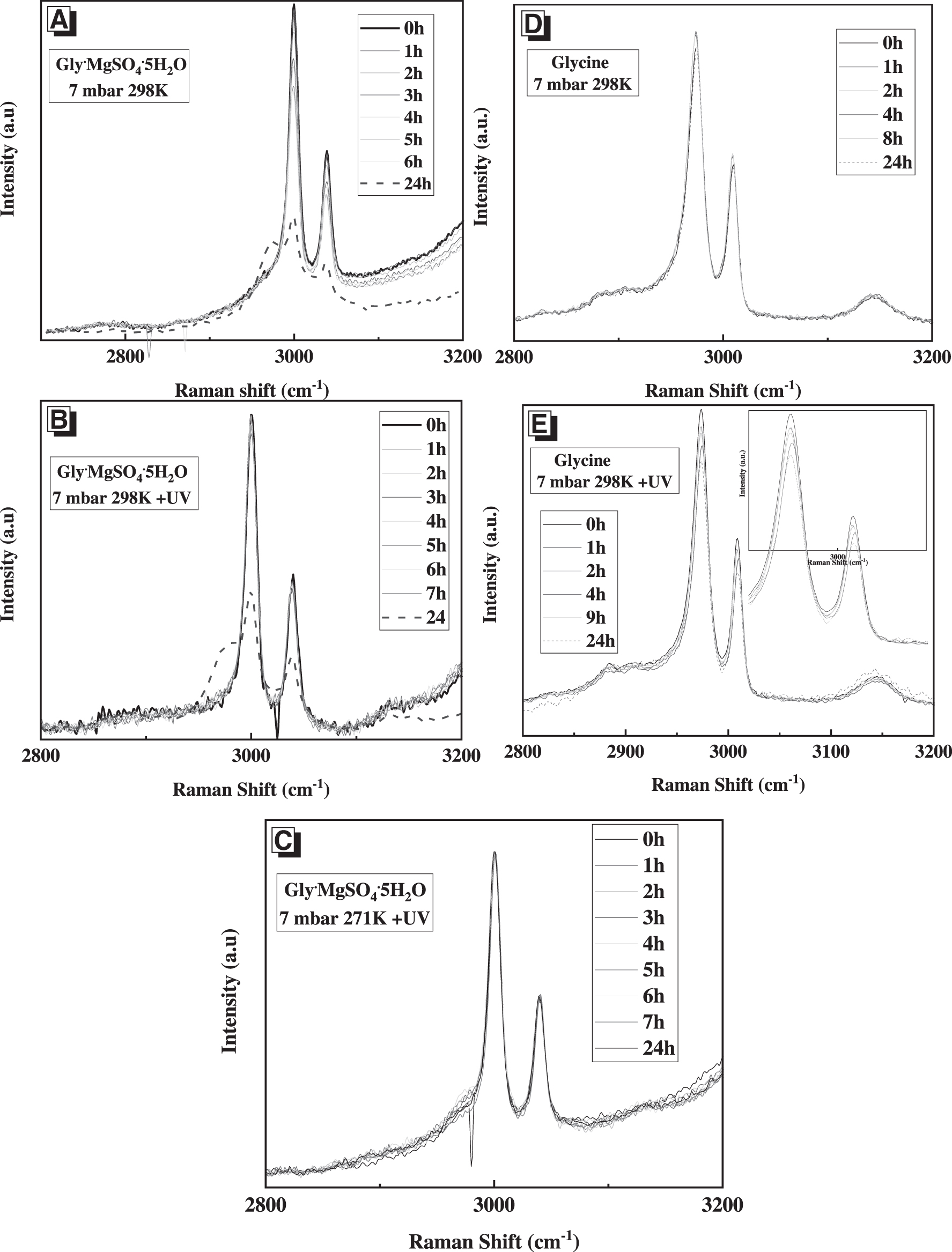
The CH stretching Raman band region (2800–3200 cm−1) of Gly·MgSO4·5H2O is shown in Fig. 9A, 9B, 9C compared with those corresponding to glycine (9D, 9E).
Raman spectra in the range from 2800 to 3200 cm−1 of Gly·MgSO4·5H2O (
Figure 9 shows the spectra corresponding to glycine under a Mars atmosphere (Fig. 9D). No change was detected after 24 h, whereas it is remarkable that in the spectra corresponding to Gly·MgSO4·5H2O under the same conditions (Fig. 9A) the appearance of a new band at ∼2974 cm−1 at long times (i.e., 24 h) is detected. Moreover, the behavior of glycine and the Gly·MgSO4·5H2O complex in the presence of UV irradiation was also different (see Fig. 9B, 9E). Therefore, glycine in the presence of UV radiation suffers degradation, which becomes evident for the decrease in the νs(CH) and νa(CH) stretching band intensities (see inserted graph in Figs. 9E and 4), but for the Gly·MgSO4·5H2O complex, no effect of UV radiation was detected, since the spectra obtained were equal to those obtained in the absence of UV radiation (comparison between Fig. 9A and 9B); that is, the changes observed in these graphs are due to the 7 mbar pressure (see Fig. 2C).
Figure 10A displays the comparison of the obtained Raman spectra of Gly·MgSO4·5H2O after exposure to a simulated martian atmosphere for up to 24 h, with the typical spectra of gamma and alpha glycine. Figure 10B shows the expanded spectra in the CH region and the peak fit of the CH stretching bands. The appearance of this new band at ∼2974 cm−1 can be attributed to the isolated glycine (both α- and γ-); note that no appreciable differences can be found for these two allotropes in the CH bands. Therefore, the appearance of this new spectral feature can be related to the 7 mbar vacuum exposure conditions. Then, the destabilization of Gly·MgSO4·5H2O takes place, and it seems to release glycine molecules to the surface for a long time, providing a fingerprint at ∼2974 cm−1 for the success of the glycine release process.
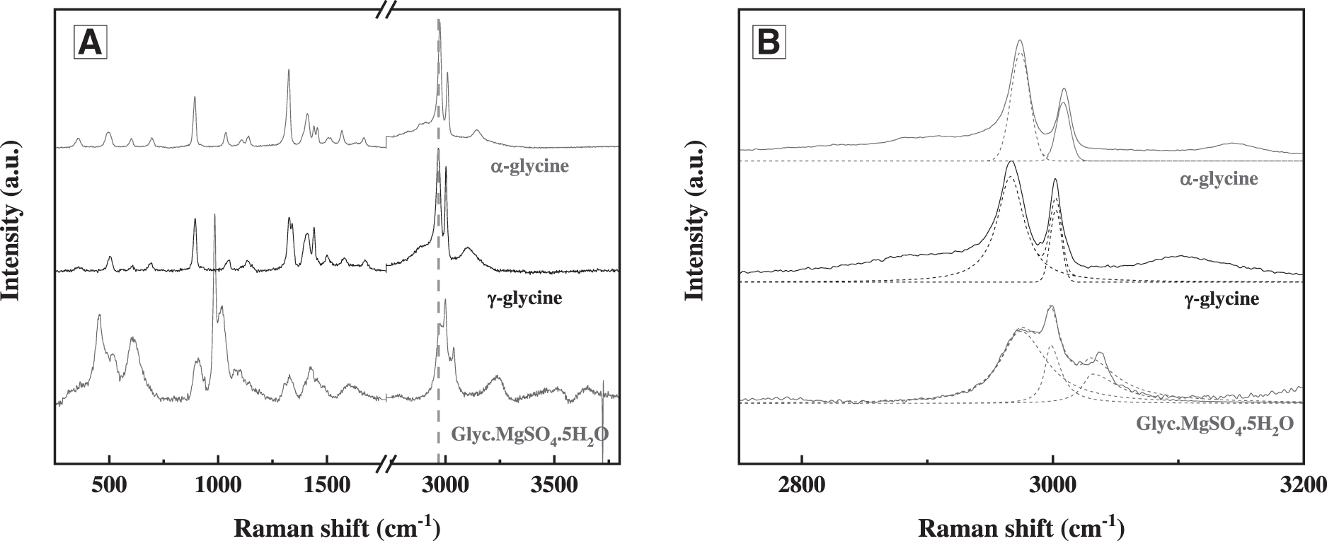
Comparison of the Raman spectra of Gly·MgSO4·5H2O under simulated martian atmosphere up to 24 h with α- and γ-glycine at normal temperature and pressure conditions (
To confirm the destabilization of Gly·MgSO4·5H2O and the release of glycine, we performed a different dehydration process due to high temperature instead of the simulated martian atmospheric conditions. Thus, a small amount of Gly·MgSO4·5H2O was placed in the Linkam stage and heated to 85°C. Then, in situ Raman spectra were acquired as a function of time over 2 days. Figure 11 shows the evolution of the Raman spectra due to the described conditions (85°C). The appearance of the band (named in this work νamorphous SO4), which appears as a shoulder at ∼1020 cm−1 after 17 h of exposure, is remarkable (see arrow in Fig. 11). Moreover, the vs(CH) band at 2974 cm−1 related to the glycine release process also appears in the spectra at similar exposure times. Longer exposure times increased the intensity of the relevant bands, which was previously attributed to the characterization of the amorphization process and glycine release process, favoring and increasing the achievement of the process. Therefore, at high temperature, the same Raman spectral features are observed as those reported previously under martian atmospheric conditions. This means that dehydration of the compound due to temperature or vacuum conditions causes the destabilization of Gly·MgSO4·5H2O, constraining the compound to easily release glycine to the surface.
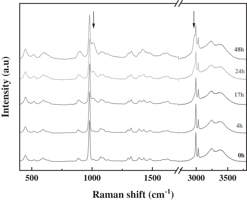
Time evolution of the Raman spectrum of Gly·MgSO4·5H2O during heating experiments in a Linkam thermal stage at 85°C as a function of time up to 48 h.
In summary, Gly, epsomite (MgSO4·7H2O), and Gly·MgSO4·5H2O compounds have been exposed to different environmental conditions, and the effect caused for each condition is summarized in Table 1.
Summary of the Effect Caused for Each Simulated Martian Condition on the Studied Molecules
99% CO2 and 0.6% H2O at a pressure of 7 mbar.
It has been proven that low temperatures stabilize the molecules, and UV degrades glycine molecules but not epsomite or Gly·MgSO4·5H2O compounds under the studied conditions, that is, at the exposure times and pressures. The most remarkable finding is the effect of the exposition to 7 mbar vacuum conditions which causes (like epsomite) amorphization of the complex but for Gly·MgSO4·5H2O also provokes the release of glycine molecules; this process has been carefully monitored and described by a Raman spectral fingerprint of the molecule. Contrary to glycine, the exposure of Gly·MgSO4·5H2O to a pressure of 7 mbar did not provoke any change.
5. Conclusions
We performed Raman spectroscopic characterization of the Gly·MgSO4·5H2O molecule under Mars-like surface conditions. The partial discrimination of the different physical parameters helps to understand molecular behavior. Raman spectra enabled us to monitor in situ the effect of vacuum, UV irradiation, and temperature conditions on Gly·MgSO4·5H2O and compare the results with those obtained for glycine and epsomite molecules. A very important conclusion is that 7 mbar pressure is critical to provoke dehydration and amorphization of the epsomite and Gly·MgSO4·5H2O molecules. In addition, the release of glycine due to amorphization is identified for the latter molecule (Gly·MgSO4·5H2O). The work presented here highlights the greater stability of Gly·MgSO4·5H2O compared to glycine and epsomite against both processes: against the amorphization process due to the simulated martian atmosphere and against the degradation process due to UV irradiation. Specifically, the results show that Gly·MgSO4·5H2O does not suffer degradation due to the UV radiation observed by the isolated glycine molecule, and the amorphization process due to the low pressure of 7 mbar is less significant for Gly·MgSO4·5H2O than for isolated epsomite. At low temperature, the previous processes do not take place, which gives molecular stability to the studied system (Gly·MgSO4·5H2O).
Mimicking the complex martian geochemical environment is still an enormous challenge; nevertheless, the selection of different environmental conditions could help discriminate under which conditions molecule/mineral compound preservation would or would not be negligible.
Our studies provided new insights into Gly·MgSO4·5H2O compound detection on the martian surface due to its stability against the dehydration process and its being more stable than glycine amino acid against UV irradiation conditions. Gly·MgSO4·5H2O detection would be a crucial statement from the astrobiology context, as amino acids and water form part of its molecular structure. Raman spectroscopy is a successful technique to discriminate Gly·MgSO4·5H2O, epsomite minerals, and glycine molecules under martian conditions, as confirmed in this work.
We also report the success of the PASC and in situ Raman spectroscopy as powerful tools for characterizing and studying chemical processes occurring in molecules exposed to simulated martian conditions. This equipment also offers the significant possibility of discriminating between the effects of individual physical parameters and selected combinations thereof. Therefore, these studies confirm the significance of the PASC simulation chamber facility as an established instrument to emulate martian planetary conditions and assess different multidisciplinary astrobiological studies, which could contribute to a better evaluation of the habitability of the martian surface and expand the powerful capabilities of this planetary-environment simulation chamber.
Furthermore, the stability and characterization of certain minerals and compounds on planetary surface conditions will be a requirement for the success of future planetary and space missions with astrobiological interest. Moreover, the findings of each space mission must be verified at terrestrial laboratory facilities to validate and confirm the in situ measurements (rover) or orbital observations of the martian surface. In this context, Raman in situ spectroscopy inside simulation chambers has been shown to be a necessary requirement to reinforce future planetary space missions and to validate in situ measurements from orbital or rover observations. Moreover, our results could help the ExoMars rover mission increase the scientific value and efficiency of sample analysis by the ExoMars rover, providing Raman database spectra of mineral samples under simulated martian conditions.
Footnotes
Acknowledgments
The authors used the research facilities of the Centro de Astrobiología (CAB) and were supported by the Instituto Nacional de Técnica Aeroespacial “Esteban Terradas” (INTA), by projects PID2019-104205GB-C21 and PID2019-107442RB-C32 from the Spanish Ministerio de Ciencia, Innovación y Universidades and by the Spanish State Research Agency (AEI) project MDM-2017-0737 Centro de Astrobiología (CSIC-INTA), Unidad de Excelencia María de Maeztu. Additionally, the authors are grateful to María Teresa Fernández from CAB for performing the XRD measurements.
Supplementary Material
Supplementary Data
Supplementary Figure S1
Supplementary Figure S2
Supplementary Figure S3
Supplementary Figure S4
Supplementary Figure S5
Supplementary Table S1
Supplementary Table S2
Supplementary Table S3
Supplementary Table S4
Supplementary Table S5
Supplementary Table S6
Abbreviations Used
References
Supplementary Material
Please find the following supplemental material available below.
For Open Access articles published under a Creative Commons License, all supplemental material carries the same license as the article it is associated with.
For non-Open Access articles published, all supplemental material carries a non-exclusive license, and permission requests for re-use of supplemental material or any part of supplemental material shall be sent directly to the copyright owner as specified in the copyright notice associated with the article.