




Abstract
Maintaining an optimal leaf and stem orientation to yield a maximum photosynthetic output is accomplished by terrestrial plants using sophisticated mechanisms to balance their orientation relative to the Earth's gravity vector and the direction of sunlight. Knowledge of the signal transduction chains of both gravity and light perception and how they influence each other is essential for understanding plant development on Earth and plant cultivation in space environments. However, in situ analyses of cellular signal transduction processes in weightlessness, such as live cell imaging of signaling molecules using confocal fluorescence microscopy, require an adapted experimental setup that meets the special requirements of a microgravity environment. In addition, investigations under prolonged microgravity conditions require extensive resources, are rarely accessible, and do not allow for immediate sample preparation for the actual microscopic analysis. Therefore, supply concepts are needed that ensure both the viability of the contained plants over a longer period of time and an unhindered microscopic analysis in microgravity. Here, we present a customized supply unit specifically designed to study gravity-induced Ca2+ mobilization in roots of Arabidopsis thaliana. The unit can be employed for ground-based experiments, in parabolic flights, on sounding rockets, and probably also aboard the International Space Station.
Introduction
It is generally accepted that the response to a given gravitropic stimulus in plants is initiated by a rolling movement of statoliths, which are specialized starch containing plastids within gravity sensing cells called statocytes (for review see Takahashi et al., 2021). Further steps of the subsequent transduction chain, which is partly in common with the light-induced phototropic response, entail the establishment of an asymmetrical lateral auxin gradient causing an unequal cell elongation on the top and bottom of roots and shoots, which ultimately leads to a parallel alignment of the plant axis to the gravity vector (for review see Nakamura et al., 2019).
One important signaling element has proved to be multiple Ca2+-release events at different timepoints within this process (Plieth and Trewavas, 2002). To unravel and distinguish light from gravity-induced signaling events, one goal of our studies was the analysis of light-induced signaling under different microgravity (μg) conditions by monitoring Ca2+ fluxes in situ. This approach, however, requires prolonged exposure of samples to μg, which is currently only available aboard the International Space Station (ISS). Thus, a suitable growth and supply system adjusted to the specification of the FLUMIAS (FLUorescence MIcroscopy At Space)-ISS live-cell imaging structured illumination laser-based microscope system will be necessary for this kind of approach (Thiel et al., 2019).
Several systems tackling this problem have already been developed (Guichard et al., 2020; Moussus and Meier, 2021; Yanagisawa et al., 2021). One important disadvantage of these systems is that a continuous supply of fluid is required to support the growing seedlings, which makes the overall supply susceptible to disruption. In this study, we describe the design, production, and performance tests of a supply unit aimed to omit this problem by providing an internal nutrition source. This design, therefore, might be a suitable alternative for long-term fluorescence analysis under μg conditions and appropriate for the current available conditions of the storage and experimental container as part of the FLUMIAS device.
Materials and Methods
Supply unit and three-dimensional print
Supply units (Fig. 1A–C) were created by using an open-source parametric three-dimensional (3D) modeler software (FreeCAD, version 0.20.1). Detailed sizes of each element are presented in Supplementary Data S9–S12. The 3D model was rendered by using the Standard Triangle Language-(STL) format. Converting the 3D model into printing instructions (slicing) was done in Ultimaker™ CURA, version 4.4.1 (Ultimaker). For critical slicing settings, see Supplementary Data S1. A Creality™ Ender-5-Pro fused deposition modeling (FDM)-3D-Printer was used for printing.
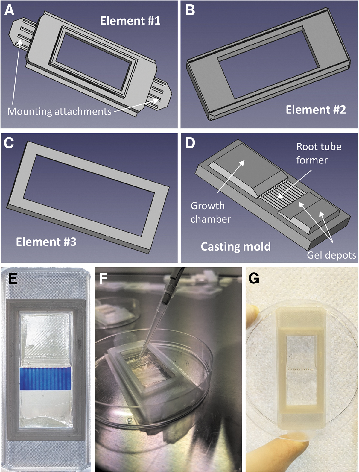
Schematic pictures of all 3D printable elements required to assemble a fully functional supply unit.
Please refer to supporting information for the applied customized Cura-printing Profile. Transparent polylactic acid (PLA) filament was used for printing. Gcodes are provided in the Supplementary Data S9–S12. Cover slips are made of optically clear vinyl plastic, 0.157 mm in thickness (SPI Supply, West Chester, PA).
All experiments were performed with Arabidopsis thaliana (L.) Heynh., ecotype Columbia wild type plants expressing constitutively R-GECO1, an established red fluorescing Ca2+ sensor (Waadt et al., 2017). Seedling preparation and plant maintenance were previously described in detail (Dümmer et al., 2016; Rath et al., 2020). Before further processing, seeds were sterilized by rinsing them in Sterilium™, followed by 70% (v/v) ethanol and finally by a mixture of 95% (v/v) ethanol and 5% (v/v) sodium hypochlorite. The growth chamber of the supply unit (see Section 3) was cast with 2.0% (w/v) Phytagel containing half-strength Murashige and Skoog salts medium (MS-medium; Sigma, St. Louis, MO) with 25 mM MES, adjusted to pH 5.8, 1 mM MgCl2, and 0.1% (w/v) sucrose.
Seeds were applied to the growth chamber, which was equipped with additional almost liquid (0.2% Phytagel) MS medium. Subsequently, the supply unit was sealed with two transparent coverslips (see Supplementary Data S2 and S3 for details). After seedling application, the supply units were placed in a vertical position at temperatures as indicated in Section 3. Seedlings were either dark grown or illuminated with constant white light [10 μmol/(m2·s)].
Fluorescence analysis and quantification
Images of R-GECO1 (Keinath et al., 2015) wild type and mutant plants containing a red fluorescent cytosolic calcium reporter (R-GECO1) were recorded either with a conventional vertical Leica SP5 confocal microscope at the CALM facility at Philipps-University Marburg, Germany, or with a Nikon Eclipse 80i horizontal confocal microscope equipped with a 360° pivotable table by using a Cyber Shot camera (Sony) provided by the DLR ground-based facility (GBF) in Cologne, Germany (Fig. 2A). Settings for the confocal microscopic analysis of calcium dynamics in the root tips are shown in the Supplementary Data S4.
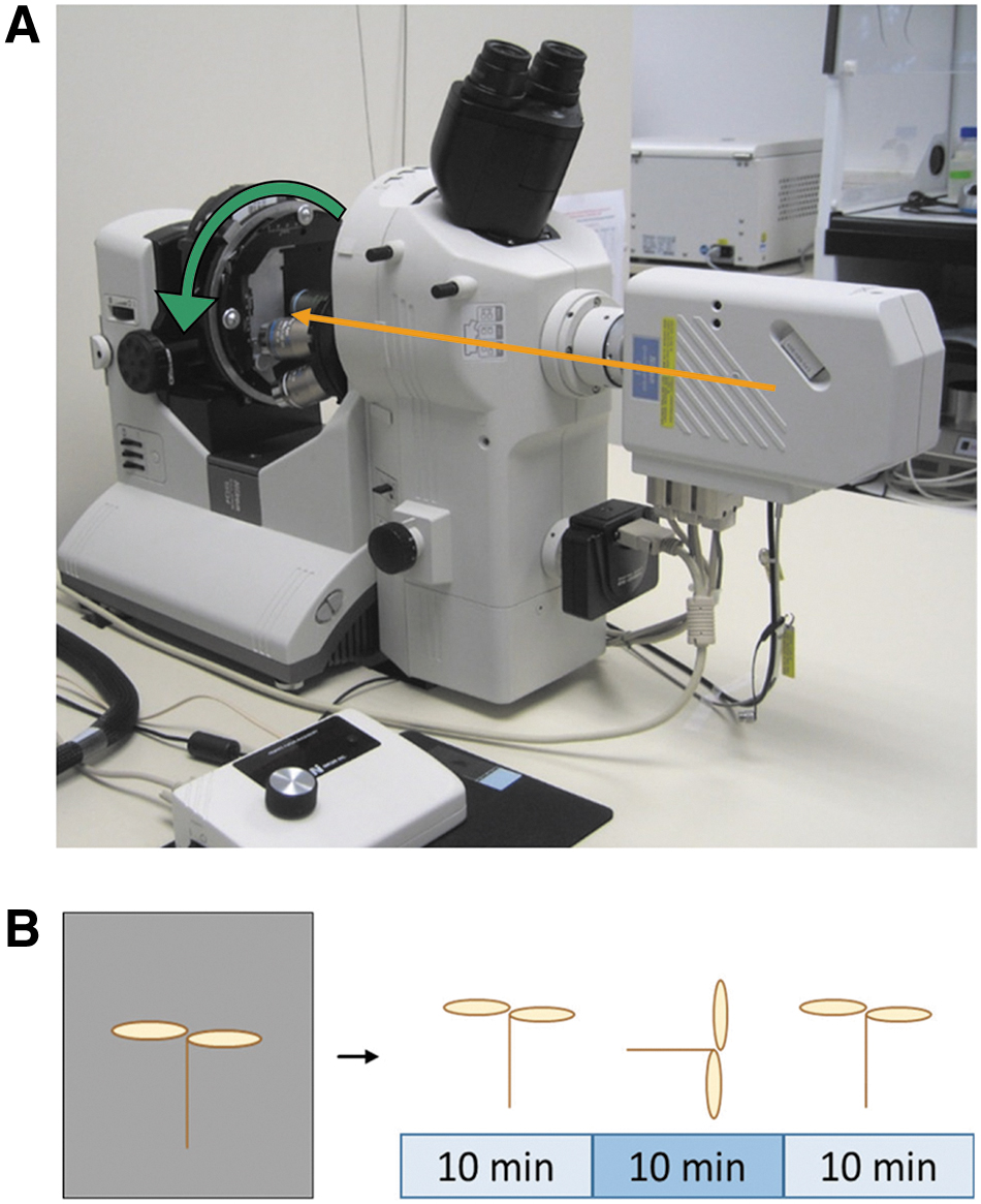
Experimental setup for an in situ fluorescence analysis of Ca2+ fluxes in Arabidopsis seedlings after gravitropic stimulation (for details see Section 2). Fluorescence was monitored constantly by confocal analysis of vertically grown seedlings for ∼10 min.
Fluorescence evaluation was performed using Fiji (Schindelin et al., 2012) as part of ImageJ software (National Institute of Health, USA open source). Measurements resulted in a mean value for each region of interest (ROI) and for each frame for the respective fluorescence brightness. Mean values were converted to percentage based on the maximum value of each data set.
Experiments were mainly performed using the Nikon horizontal confocal microscope at the DLR GBF in Cologne, Germany. In this case, the supply unit was attached to the rotatable object table of the microscope such that the seedlings were oriented vertically with respect to the gravitational vector but could be tilted 90° from this position for gravitropic stimulation (Fig. 2A). After focusing on the root tip region (resulting in a delay of 45–90 s), Ca2+-induced fluorescence was observed in the vertically oriented root for 3 min. Supply units were positioned vertically to Earth's gravity vector before they were also analyzed on a Leica SP5 confocal microscope placed horizontally on the object stage of the microscope.
This orientation shift caused a gravitropic stimulation for the applied seedlings. Specimen positioning was set to ensure focusing on the root tip region but also to encompass root hairs in the differentiation zone. These microscopic adjustments caused a delay of 45–90 s before fluorescence analysis. Further experimental procedures included three steps: (1) fluorescence analysis of the vertically oriented root tips for ∼10 min, (2) rotation of the object table by 90° (gravitropic stimulation) for another 10 min, and (3) rerotation of the stage so that the roots were returned to their initial vertical position (Fig. 2B). Ca2+-induced fluorescence was observed throughout the period.
Results
Design and production of the supply unit
As mentioned above, the plant supply container consists of three parts (Fig. 1A–C), which can be easily assembled on the basis of a click–connect system. Both sides of the supply unit contain a window fitting the two coverslips, thus allowing passage of excitation and emission light for confocal analysis as well as a supporting blue/red light for photosynthesis. A casting mold (Fig. 1D) required for suitable agar formation within the growth chamber was developed to provide space for both developing leaves and tubes for coordinated root growth. The mold also incorporates an implementation of a specified place for seed deposition (Fig. 1E–G).
Further details as to how to assemble the supply unit are provided in the Supplementary Data S2 and S3. To test the stability of the system during launch, we applied supply chips to either 14g and 56g acceleration on a centrifuge with the centrifugal force perpendicular to the seedling. Summarizing the outcome after centrifugation, no visible damage could be detected to the supply unit, within the agar support structure, or to the orientation of the applied seeds (Supplementary Data S5).
Root growth kinetics
Root length development and capability of the developing root to enter the growth tube were analyzed within a time frame of 18 days at 21.5°C (Fig. 3A). Approximately 95% of the applied seeds germinated, and >80% germinated seeds were subsequently able to grow correctly into the predefined root canal. Root lengths were investigated from seedlings that were kept for various periods of time at 6°C before incubating them at 21.5°C. Thus, root lengths were determined from plants that were kept at 6°C for 7, 14, and 34 days before raising the temperature to 21.5°C for 7 days (Fig. 3B).
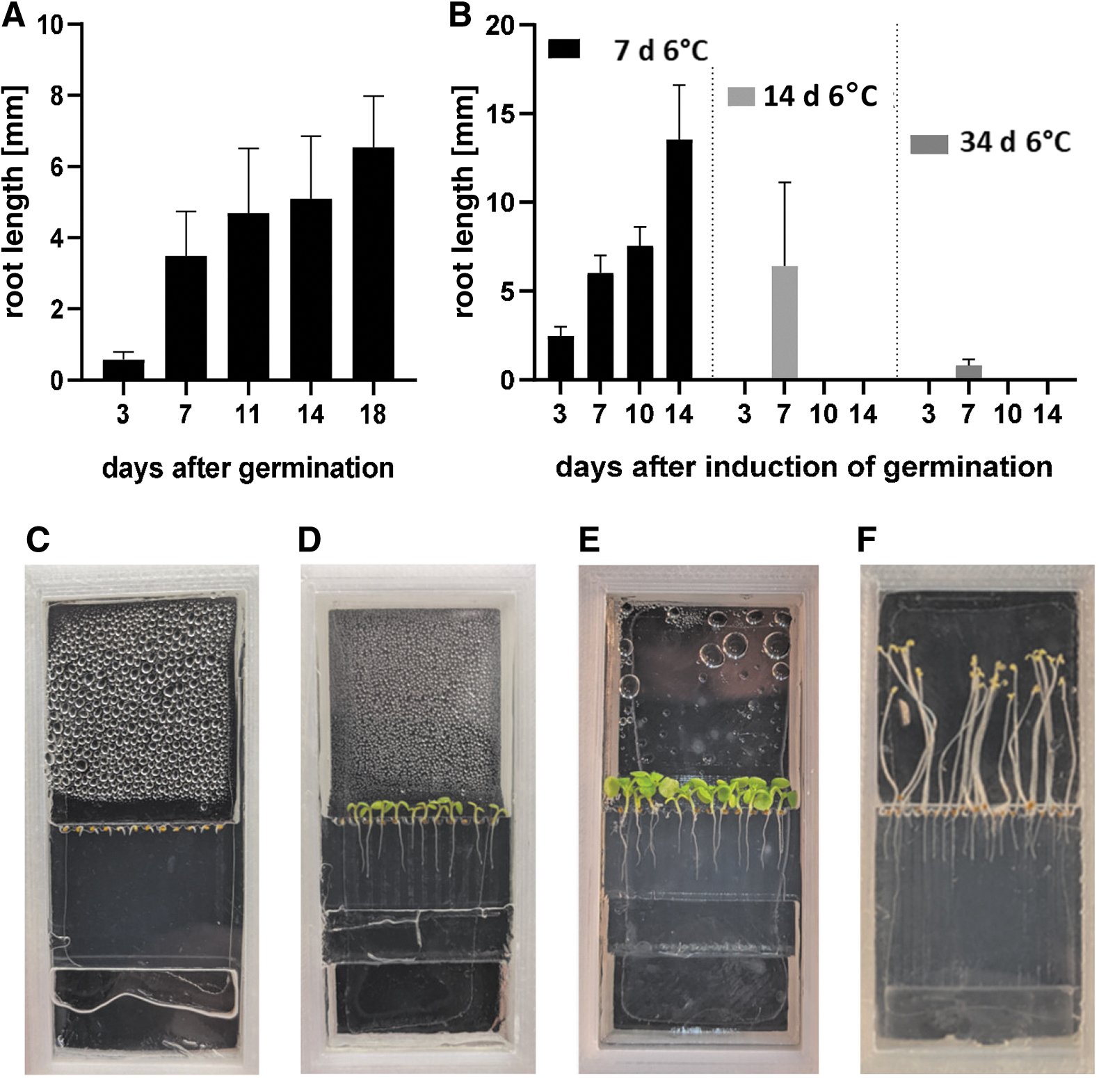
Root length development.
The results revealed that seedlings kept cold (6°C) for 7 days showed a similar or better growth than those grown permanently under ambient (21.5°C) temperature conditions. Even after 14 days under reduced temperature conditions followed by 7 days at ambient conditions, seedlings showed similar growth rates. After 34 days at 6°C, however, root growth was strongly reduced (2 mm), although plants were subsequently kept at 21.5°C for 7 days. Summarizing our data, we conclude that delaying germination by up to 2 weeks is possible without affecting the root growth rate. Examples for root development are presented in Fig. 3C–F.
Seedlings grown in supply units were analyzed for their ability to maintain Ca2+-induced fluorescence over time. Initially, the overall fluorescence decay in vertically grown root tips was determined by measuring the fluorescence over a period of ∼10 min (Supplementary Data S6). Next, two independent physiological parameters were investigated: First, the accumulation of Ca2+ ions within the root tip after a given gravitropic stimulus (Fig. 4 and Supplementary Data S7) was observed, and, second, the fluorescence oscillation rate in root hairs (Fig. 5 and Supplementary Data S8) was analyzed.
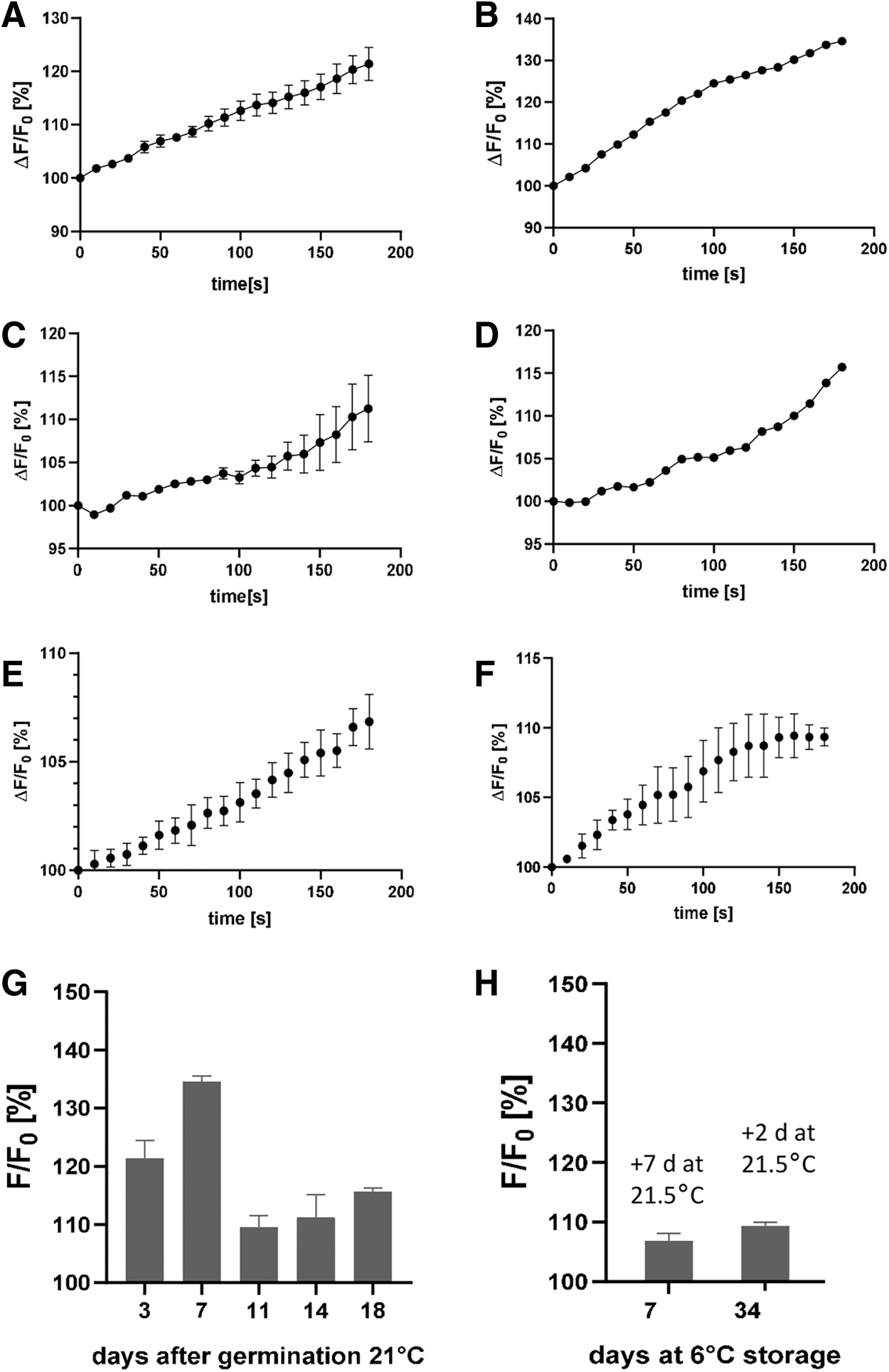
Relative fluorescence accumulation in root tips after gravitropic stimulation. Seedlings were analyzed after
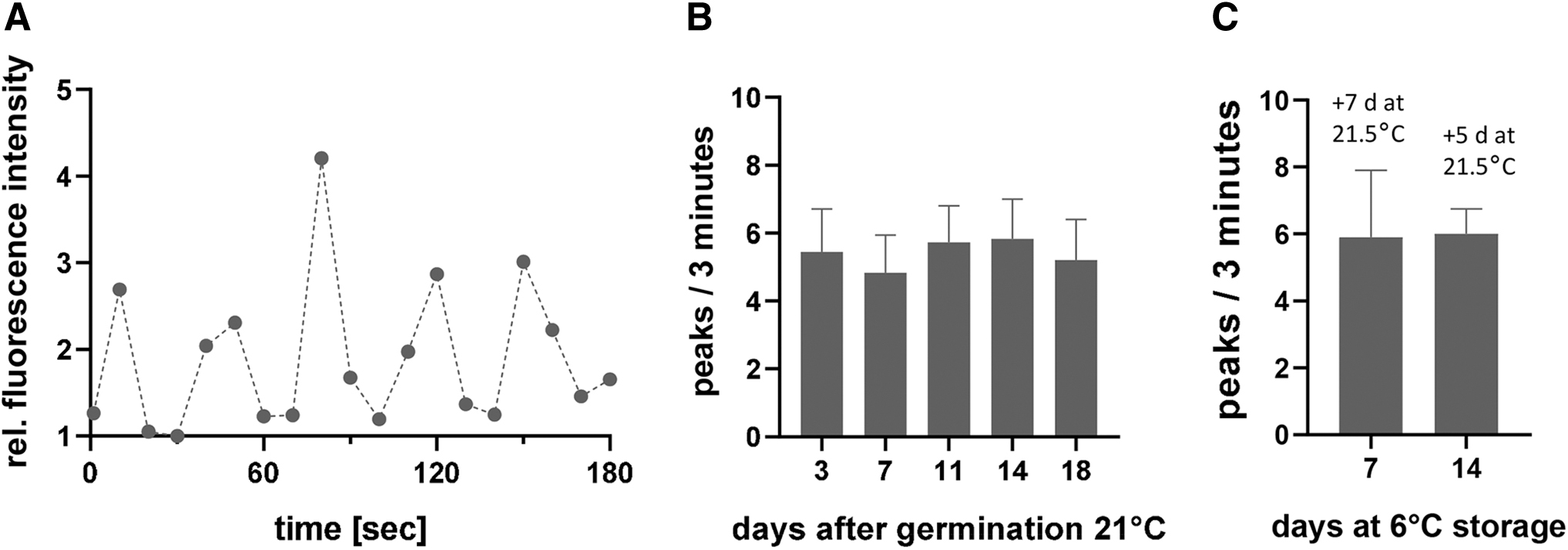
Analysis of the Ca2+-induced root hair fluorescence.
Despite considerable calcium fluorescence in several regions of the root (e.g., in root hairs, epidermal cells, and the central cylinder), only a subset of root tips showed a gravity-induced fluorescence increase over time. However, we chose to focus on this gravity-dependent effect because the accumulation of fluorescence over time only occurred after replenishment of the R-GECO1 pool (Supplementary Data S6), therefore, proving it to be a robust indicator of cell and tissue signaling and hence seedling vitality.
To verify whether our system is suitable to perform such experiments, we tilted the supply unit loaded with 7 days old Arabidopsis seedling by 90°. After a gravitropic stimulation, that is, by tilting seedlings by 90°, Ca2+-indicator fluorescence increased quickly in root tips (Fig. 4). We were interested in determining how long seedlings were able to respond with a detectable Ca2+-dependent fluorescence signal to a gravitropic stimulus when grown within the supply unit. Therefore, the increase of the fluorescence signal in root tips after tilting by 90° was quantified in 6–21 roots. The resulting mean values of all investigated root fluorescence values are shown in Fig. 4A–F.
Using fresh roots and supply units, similar experiments at 21.5°C were performed 7, 11, 14, and 18 days after germination, respectively. Starting with an average increase of 20%, the relative fluorescence increase after 7 days was ∼35%, followed by 10% after 11 days, 15% after 14 days, and 18% after 18 days, respectively (Fig. 4G). The fluorescence signals of the roots remain vital for at least 18 days. Even when seedlings were kept at 6°C for up to 34 days before incubating them either for 7 or 2 days at 21.5°C, only a slight loss in the intensity was found (Fig. 4H).
Ca2-dependent fluorescence in root hairs
Ca2+-dependent fluorescence signaling within root hairs was determined over a period of 18 days in similar conditions as described above. Under these conditions, root hairs revealed an oscillating calcium emission peaking approximately every 35 s (Fig. 5A and Supplementary Data S8). This effect has already been described by Monshausen et al. (2008). The fluorescence oscillation rate remained stable within the investigated period of at least 18 days (Fig. 5B), regardless of whether the root hairs were kept continuously at 21.5°C or at 6°C followed by cultivation at 21.5°C (Fig. 5C). We conclude from these data that the supply unit can sustain a stable environment suitable for in situ analysis of Ca2+ ions for at least 18 days.
Gravity-induced Ca2+-ion dynamics in root tissue
Our scientific goal was to analyze and understand how gravity and blue light alter Ca2+ signaling in plants, especially in root tips. It has already been reported that both environmental stimuli have an impact on local Ca2+-ion concentration in roots and shoots (Vanneste and Friml, 2013). To test whether Arabidopsis seedlings grown in our supply unit respond in a similar manner, Ca2+ fluorescence in root tips of 7-day-old seedlings was examined after a given gravitropic stimulus. Ca2+ fluorescence was analyzed in vertically oriented roots (0°; control), after gravitropic stimulation (90° rotation) and after reorientation to a vertical position.
All phases were observed for ∼10 min each, with recordings every 8 s. For a quantitative comparison of Ca2+-induced fluorescence fluxes, two ROIs were defined on the left and right sides of a vertically growing root within the elongation zone. One side, shown in orange, becomes the physiologically lower side after tilting (Fig. 6). The relative fluorescence signal differences were calculated over time and compared with each other. To summarize the outcome, it could be shown that the analyzed roots respond to a given inclination angle as reported in previous studies.

For monitoring the Ca2+ flux, two ROIs within the elongation zone of the root tip of 7-day-old light grown Arabidopsis seedlings were defined. ROI 1 (left; blue) will become the physiological upper side after tilting, ROI 2 (right; orange) will become the lower side. Over ∼10 min, the fluorescence decay (Supplementary Data S6) in each ROI was analyzed. Please note that only in ROI 2 of horizontally orientated roots, the Ca2+ decay was reversed, indicating an accumulation of Ca2+ ions, which is in line with previous observations. ROI, region of interest.
In this contribution, we present a self-made and easy-to-assemble supply system for growth and maintenance of plant seedlings under hyper and μg conditions. The individual components can be produced in a conventional 3D printer. A finally assembled supply unit provides a closed system with a sterile nutrient-supplied plant agar in which the plants can grow autonomously. The system allows sufficient gas exchange to keep the plants in a physiologically active state for the duration of the study. The crucial point for the usability of such a system is the physiological fitness of the plants after a prolonged incubation time to respond properly to a given environmental stimulus.
To test this, three independent physiological parameters were analyzed after various incubation times: root growth kinetics (Fig. 3), endogenous induced Ca2+ fluorescence dynamics (Fig. 4), and Ca2+-induced fluorescence response after gravitropic stimulation (Fig. 6). From the results obtained, it can be concluded that at least up to 18 days after germination, incubation of Arabidopsis seedlings within the supply unit had no obvious effect on the growth rate and root development. Thus, plants can be kept in a stable stand-by mode up to this timepoint before starting an experiment. Eighteen days cover the time between late access, transportation, and installation of the supply unit in the FLUMIAS-ISS device, including a buffer for an unintended delay, as can sometimes occur before scheduled rocket launch.
During that time, the F/F0 ratio dropped moderately from 120% to 115%. These findings were further confirmed when seedlings were kept at 6°C for up to 34 days followed by a brief growth period of 2 days at 21.5°C. In this case, the F/F0 ratio dropped from 120% to 110% (Fig. 4). Measurement of a gravity-independent oscillatory Ca2+ signal in root hairs (Fig. 5) after 18 days, which indicates the vitality of the roots, supports these findings, regardless of whether the roots were stored at 6°C before the standard treatment or not.
Finally, an on-ground analysis of spatial and temporal Ca2+-induced fluorescence dynamics after gravitropic stimulation in root tissue was performed with seedlings kept in the supply unit for 7 days to gain further experience with plant handling (Fig. 6). The results obtained were consistent with previous observations that an imbalance of Ca2+ ions could be observed in the elongation zone in a horizontally oriented root. Specifically, it could be shown that more calcium ions could be observed in the lower side of a vertically oriented root than in the physiological upper side.
It is hypothesized that this event triggers asymmetric root growth in the direction of the gravity vector by redirecting PIN (PIN-FORMED) proteins that subsequently lead to a redistribution of auxin (Adamowski and Friml, 2015). Again, this observation provided evidence that roots grown within the supply unit react normally within the analyzed time frame, which would be a prerequisite for any investigation of seedlings aboard μg platforms like the ISS. In addition, the supply unit has proven to be quite durable. It could cope with gravitational forces of a rocket launch unaffected and kept the agar and the seedlings undamaged (Supplementary Data S5).
Summarizing the outcome, we conclude that the presented supply system is a robust containment system that (1) can ensure normal development of plant seedlings over a given time frame even under extreme gravity conditions and (2) allows a consistent in situ measurement of Ca2+-induced fluorescence in root tips. We look forward to testing our supply system under real flight conditions and hope that it will provide us with new exiting data, which will increase our understanding of how plants react on the molecular level if exposed to altered gravity conditions.
Material Availability
There are no restrictions on materials or data. All data are available in the main text or in the Supplementary Data S1–S12.
Footnotes
Acknowledgments
We are greatly indebted to Paul Galland for his highly valuable and helpful comments and suggestions. Thanks to Yannick Lichterfeld (DLR Cologne) for introducing us to the DLR GBF equipment, Agnes Damm for skillful technical assistance, and the group of Karin Schumacher (COS, Heidelberg, Germany) for providing Arabidopsis seeds expressing R-GECO1. We are grateful to Lee Herrick and Ralf Anken for their editorial work on the article.
Authors' Contributions
M.R. designed and developed the supply unit, performed the confocal microscopic experiments, provided the fluorescence raw data, and contributed to the article. M.D. developed a script for the compilation of fluorescence data, contributed significantly to the data analysis and figure plotting, and wrote parts of the article. J.H. performed the initial adjustment of the supply unit and supervised the experiments at the DLR facility. C.L. was involved in project administration, funding acquisition (ESA CORA-GBF 2020-003), and experiment implementation in Cologne. C.F. was responsible for funding acquisition, conceptualization, and supervision of the project. C.F. also validated the data and wrote the article.
Author Disclosure Statement
No competing financial interests exist.
Funding Information
This study was financed by the DLR/BMWi funded project 50BW2134 and European Space agency project CORA GBF 2020-003 granted to C.F.
Supplementary Material
Supplementary Data S1
Supplementary Data S2
Supplementary Data S3
Supplementary Data S4
Supplementary Data S5
Supplementary Data S6
Supplementary Data S7
Supplementary Data S8
Supplementary Data S9
Supplementary Data S10
Supplementary Data S11
Supplementary Data S12
Associate Editor: Petra Rettberg
Abbreviations Used
References
Supplementary Material
Please find the following supplemental material available below.
For Open Access articles published under a Creative Commons License, all supplemental material carries the same license as the article it is associated with.
For non-Open Access articles published, all supplemental material carries a non-exclusive license, and permission requests for re-use of supplemental material or any part of supplemental material shall be sent directly to the copyright owner as specified in the copyright notice associated with the article.
