




Abstract
Solar radiation that arrives on the surface of Mars interacts with organic molecules present in the soil. The radiation can degrade or transform the organic matter and make the search for biosignatures on the planet's surface difficult. Therefore, samples to be analyzed by instruments on board Mars probes for molecular content should be selectively chosen to have the highest organic preservation content. To support the identification of organic molecules on Mars, the behavior under UV irradiation of two organic compounds, undecanoic acid and L-phenylalanine, in the presence of vermiculite and two chloride salts, NaCl and MgCl, was studied. The degradation of the molecule's bands was monitored through IR spectroscopy. Our results show that, while vermiculite acts as a photoprotective mineral with L-phenylalanine, it catalyzes the photodegradation of undecanoic acid molecules. On the other hand, both chloride salts studied decreased the degradation of both organic species acting as photoprotectors. While these results do not allow us to conclude on the preservation capabilities of vermiculite, they show that places where chloride salts are present could be good candidates for in situ analytic experiments on Mars due to their organic preservation capacity under UV radiation.
Introduction
Over the past decades, one of the primary goals of Mars exploration has been to establish whether Mars ever fulfilled possible criteria as a habitable planet. Among these criteria, detection of organic matter is key, both for the emergence and the sustainability of life. The first investigations for organic molecules on Mars were performed by the Viking twin landers of NASA's Viking mission. However, despite the identification of low molecular weight molecules, including chlorohydrocarbons (Biemann et al., 1976), the presence of these organic compounds on Mars was first attributed to terrestrial contaminants (Biemann et al., 1976, 1977). However, the detection of oxychlorines in different regions of Mars (Hecht et al., 2009; Glavin et al., 2013; Meslin et al., 2022) and laboratory experiments reproducing the pyrolytic sequence of the Viking GC-MS have shown that chlorinated compounds are very likely produced from the interaction between martian hydrocarbons and perchlorates present in the soil during thermal degradation analysis (Navarro-González et al., 2010; Guzman et al., 2018). Since then, different organic molecules have been identified with the Sample Analysis at Mars (SAM) instrument suite on board the Mars Science Laboratory (MSL) Curiosity rover (Freissinet et al., 2015; Eigenbrode et al., 2018; Szopa et al., 2020; Millan et al., 2022) and possibly with the Scanning Habitable Environments with Raman and Luminescence for Organics and Chemicals (SHERLOC) instrument on board the Mars 2020 Perseverance rover (Scheller et al., 2022; Sharma et al., 2023). However, organics on the surface of Mars are continuously exposed to harsh environmental conditions. Among them, the radiation environment on Mars is known for its critical implications on the organic matter present in the soil. The heterogeneous ozone spatiotemporal distribution (Perrier et al., 2006) and the overall thin atmosphere of Mars (Jakosky et al., 2017) reduce the atmospheric absorption of UV light, which can penetrate down to a few microns or millimeters depth into the surface (Schuerger et al., 2012; Fornaro et al., 2018c; Carrier et al., 2019). UV photons can dissociate molecular bonds, produce ionic species, and excite molecules. These properties mean that UV radiation can stress organic compounds (Sagan, 1973) and lead to their decomposition and transformation. Past and current probes on Mars have analyzed samples at the surface, where organic matter is the most exposed to UV light, and in the first few millimeters or centimeters below the surface (Abbey et al., 2019; Moeller et al., 2021), where UV does not penetrate but can still affect freshly exposed subsurface material on very short timescales. Studies on the impact of UV radiation on organic molecules in the martian context have, therefore, been of critical importance to interpreting the in situ data collected by probes as well as preparing missions' next analytical targets.
In this effort, understanding the environment in which organic matter evolves on the martian surface is necessary. Minerals play a crucial role in those processes experienced by organic molecules on Mars, which influence their chemical evolution. The preservation state of organic molecules is often regulated by their interaction with the mineral phase in which they are embedded. Investigations on the catalytic and protective properties of different martian minerals under Mars-like conditions have been carried out and reviewed by Fornaro et al. (2018c), who concluded that, in several paleoenvironments on Earth, long-term preservation of terrestrial biosignatures is attributed to sedimentary materials, notably phosphates, silica, clays, carbonates, and metalliferous materials. However, the straightforward classification of martian minerals as catalytic or protective is not possible, since the behavior of minerals under martian conditions depends on the organic molecules involved and their specific interactions with the mineral surface sites. Thus, it is important to investigate the response to UV irradiation of specific molecule-mineral complexes.
The Compact Reconnaissance Imaging Spectrometer for Mars (CRISM) spectra taken at various locations of the landing area chosen for the future ExoMars Rosalind Franklin mission are consistent with material bearing Fe/Mg-rich clay minerals (Carter et al., 2016). The closest match is vermiculite, a phyllosilicate phase with a structure intermediate between mica, smectite, and saponite (Mandon et al., 2021). Vermiculite was also detected at Jezero Crater where the Perseverance rover landed in February 2021 (Dehouck et al., 2023). Phyllosilicates are characterized by a high organic sequestration and preservation potential (Ehlmann et al., 2008), which makes them desirable targets for investigations on Mars. Vermiculite is a 2:1 phyllosilicate, which means that it is composed of tetrahedral sheets [X4O10]n-, where X is Si4+, Al3+, and Fe3+, and an octahedral sheet composed of two planes of closely packed O2- and OH- anions with the central cations being Mg2+, Fe2+ or Fe3+, Al3+. The negative layer charge of vermiculite results from the substitution of Si4+ by trivalent cations in the tetrahedral positions. The general formula can be written as X4(Y2–3)O10(OH)2M·nH2O, where X are the cations in the tetrahedral positions, Y the ones in the octahedral, and M refers to the exchangeable cations positioned in the interlayer space that can be Mg2+, Ca2+, Ba2+, Na+, and K+ (Valášková and Martynkova, 2012). The interlayer space thickness depends on the cations in the interlayer and on the interlamellar water molecules. The hydration properties of vermiculite depend on the interlayer cations, especially in Mg2+ for being the major cation and to a lesser extent in the minor ones Ca2+, Na+, and K+ (Valášková and Martynkova, 2012). This structure confers to vermiculite several properties that promote the adsorption of biomolecules such as its high surface area to interact with, through electrostatic interactions, hydrophobic interactions, or Van der Waal's forces. Its structure also provides a good cation exchange capacity as the interlayer cations can be exchanged by some organic cations. Finally, this structure allows other kinds of intercalation of molecules in the mineral interlayers. Water molecules in the interlayer space of vermiculites can be displaced by polar molecules, and some neutral organic ligands can form complexes with the interlayer cations (Lagaly et al., 2013).
Among the inorganic species present in the martian soil, salts are important components of the fine-grained regolith on Mars, the presence of which may also affect the preservation of the organic molecules, among which chloride salts have been identified on Mars through IR spectrometry (Osterloo et al., 2008) and at Gale Crater, where the Curiosity rover operates, with the ChemCam instrument (Thomas et al., 2019). Moreover, a change in the oxychlorine signature along the path of Mount Sharp advocates for the presence of chloride-bearing material among the chemical phases (Clark et al., 2020). Geologic environments that contain saline minerals are potential areas of biological activity. Although the long-term viability of microorganisms within salt is still questioned, salt crystals and brine inclusions appear to provide excellent environments for the long-term preservation of biomolecules (Farmer and Des Marais, 1999). For these reasons, this study aims to understand the possible effect of the presence of two types of chloride salts, magnesium chloride (MgCl2) and sodium chloride (NaCl), in the detection and preservation of the organics adsorbed on the mineral. Sodium and magnesium were identified as important elements in the soils at all landing sites, with mass fractions ranging from 0.5% to 5% (Karunatillake et al., 2007). The adsorption of molecules onto minerals is a complex process in which a variety of physical and chemical interactions, such as cation exchange, electrostatic interactions, hydrophobic/hydrophilic affinity, hydrogen bonding and Van der Waals forces, can take part (Yu et al., 2013). The adsorption of amino acids by minerals is also likely affected by ionic strength and interactions between different salts (Zaia, 2012). For example, in the case of electrostatic adsorptions, the increase of the ionic strength would bring a competitive ion exchange that would result in a decrease of the adsorption. However, ion strength has no influence on covalent bonding. On the other hand, salt ions can destroy the hydration layers around the molecules and thus increase the exposure of the hydrophobic groups and increase the hydrophobic interaction and adsorption (Yu et al., 2013).
In this work, the interaction, stability, and detection of two organic compounds, undecanoic acid and L-phenylalanine in vermiculite, are studied by means of Fourier transform infrared (FTIR) spectroscopy. Undecanoic acid was chosen as a biomarker as it belongs to the fatty acid family, constituents of cell membranes (Nagy and Tiuca, 2017), and crucial sources of metabolic energy for life as we know it (Schoors et al., 2015). Moreover, carboxylic acids are the most likely oxidation products of organic molecules on martian surface environmental conditions (Benner et al., 2000). Amino acids such as L-phenylalanine play a crucial role in the structure, metabolism, and physiology of cells as constituents of peptides and proteins. Interestingly, amino acids were detected in several meteorites and comets (Cronin and Pizzarello, 1983), which makes their presence through exogenous sources on the surface of Mars likely and, therefore, of high astrobiological interest (Fornaro et al., 2013). In this work, the catalytic/protective behavior of vermiculite and chloride salts over undecanoic acid and L-phenylalanine under UV irradiation is studied by monitoring the IR spectra changes, and a degradation half-lifetime coefficient is calculated for each IR band. The results are extrapolated to calculate the half-lifetime degradation of the molecule under the martian UV flux. Density Functional Theory simulations were used to help with the vibrational modes assignments of IR bands, and other techniques such as X-ray powder diffraction (XRPD) and time-of-flight secondary ion mass spectrometry were used to assist with the interpretation of the results.
Materials and Method
Sample preparation
Germ- and bacteria-free vermiculite was purchased from Sigma-Aldrich in a solid form of 2–3 mm grains. To favor molecular adsorption, the vermiculite was ground in a Planetary Ball Mill PM100 from Retsch for 30 min using a velocity of rotation of 200 rpm and intervals of 2 min.
The vermiculite was then baked in an oven at 500°C for over 3 h to pyrolyze the natural organic compounds present in it. Moreover, 500°C is below the temperature at which the hydroxyl groups present in the octahedral layer of the vermiculite irreversibly react to form oxide and water; thus the structure of the vermiculite remained intact.
The undecanoic acid (purity >97%, Sigma-Aldrich) and the L-phenylalanine (purity >99.5%, Sigma-Aldrich) were then adsorbed onto vermiculite by mixing the vermiculite powder with a solution of undecanoic acid or L-phenylalanine solubilized in ethanol (Analar Normapur 96% v/v) and milli-Q water, respectively. Each sample was prepared to contain 10 wt % of the organic molecule to be able to differentiate the band in the IR analyses and to study the degradation of these bands under UV irradiation. Moreover, to study the effect of the salts, sodium or magnesium chloride (purity >97%, Sigma-Aldrich) dissolved into milli-Q water were added to some of the samples (10 wt %). The suspensions were kept for 3 h on a stirring plate to favor the establishment of physicochemical interactions between the organic molecules and the mineral phase (Fornaro et al., 2018b). The samples were then dried in an oven at 50°C.
Sample characterizations
To understand and explain the UV degradations or preservations of organic molecules adsorbed onto the mineral matrices, it is essential to identify the IR bands of the organic compounds that can be detected in the studied sample. Then it is crucial to assign the vibrational modes involved in these bands and to discern whether there is a shift in their position with respect to the pure organic to understand the interactions between the organic molecules and the mineral.
Infrared measurements
The characterization of the samples was performed with a VERTEX 70v FT-IR spectrometer (Bruker) equipped with a Praying Mantis Diffuse Reflection Accessory (Harrick) to carry out diffuse reflectance infrared Fourier transform spectroscopy (DRIFTS) analysis in the mid-infrared (MIR) range (8000–400 cm−1) using 200 scans for each spectrum with an instrumental resolution of 4 cm−1. The samples were analyzed in a chemically inert environment with a constant flux of dinitrogen (N2) to avoid disturbances in the spectra from atmospheric CO2 and O2.
Density Functional Theory (DFT) simulations
Assignment of the near infrared (NIR) spectra was supported by anharmonic computations, which allows for prediction of frequencies and intensities of overtones and combination bands employing the Generalized second-order Vibrational Perturbation Theory (GVPT2) approach (Barone, 2005; Barone et al., 2014; Bloino, 2015; Bloino et al., 2015). All computations have been performed by means of DFT employing the B3LYP functional (Becke, 1993) with the double-zeta basis sets SNSD (Barone et al., 2014), and including Grimme's dispersion correction D3 (in conjunction with Becke-Johnson damping) (Grimme et al., 2010, 2011; Ehrlich et al., 2011). This approach has been well tested for structural and spectroscopy properties of biological molecules of astrochemical interest (Fornaro et al., 2014, 2015, 2018b; Biczysko et al., 2018; Zhao et al., 2021).
In GVPT2 computations, the recommendations outlined in the work of Bloino et al. (2016) were followed, and default criteria for anharmonic resonances were exploited. Moreover, the Large Amplitude-Motion (LAM) vibrations have been excluded from VPT2 by means of LAM-free VPT2 scheme (Biczysko et al., 2018). Vibrations with harmonic frequencies below 100 cm−1 have been considered as LAMs.
All calculations were performed with Gaussian 16 suite of computer codes (Frisch et al., 2016) and using GaussView to visualize the normal modes and analyze in detail the outcome of vibrational computations and IR spectra. The DFT simulations were performed for the NIR assignments of undecanoic acid in this work and were previously performed in the same way for L-phenylalanine in a former work by Fornaro et al. (2020).
X-ray powder diffraction
The powders for the XRPD analysis were ground in an agate mortar and left in a stove at 363 K for several days to eliminate the eventual moisture absorbed by the vermiculite. The powders were compacted with paraffin oil inside the stove and modelled as a ball of ca. 0.45 mm diameter (less than the diameter of the X-ray beam). Each ball was glued on a glass capillary and mounted on the goniometer head of the instrument. The XRPD patterns were collected at room temperature with the Atlas S2 Rigaku-Oxford Diffraction Gemini R-Ultra diffractometer equipped with a mirror monochromatized Cu-Kα (1.5418 Å) radiation. Each powder pattern was collected by rotating the sample 100 degrees, with an exposure time of 100 s.
Time-of-flight secondary ion mass spectrometry
The non-radiated samples, sitting in the radiation holder, were mounted directly on the time-of-flight secondary ion mass spectrometer (ToF-SIMS) sample holder by clamps, using cleaned tweezers (heptane, acetone, and ethanol, in that order) in a laminar flow hood. After mounting the samples, the sample holder was then directly introduced into the ToF-SIMS.
Analysis of samples was performed in a ToF-SIMS IV instrument (ION-TOF GmbH, Germany) located at RISE Research Institute of Sweden in Borås in Sweden. Samples were analyzed by rastering a 25 keV Bi3+ beam over a 200 × 200 μm2 area for 250 s. The analyses were performed in both positive and negative mode at high mass resolution (bunched mode: m/Δm ≥ 5000 at m/z 30, Δl ∼5 μm) with a pulsed current of 0.1 pA. As the samples were insulating, the sample surface was flooded with electrons for charge compensation.
Ultraviolet irradiation experiments
UV measurements
The UV irradiation experimental setup is constituted by a Newport Oriel 300 W Xenon arc discharge lamp with a spectral range measured between 200 and 930 nm. The light is focused directly on the sample through an optical fiber of 800 μm spot size inserted into the sample compartment to monitor the infrared spectra in situ during the UV irradiation. The optical fiber was measured to start emitting radiation at 230 nm. With this setup, the irradiated spot on the sample presents an area of 7.07 mm2, and the UV flux on the sample is of 2.75∙1017 photons/s∙cm2 in the 200–400 nm spectral range as measured through a single monochromator Spectro 320 scanning spectrometer (Instrument System). This experimental setup allows for monitoring the degradation kinetics in situ by infrared spectroscopy analysis in an inert nitrogen atmosphere as described above without significantly heating the sample due to low irradiance.
UV data treatment
The L-phenylalanine samples were irradiated for a total of 1 h and 10 m (4200 s), and the undecanoic acid samples were irradiated for a total of 5.5 h (19800 s). These total irradiation times were selected after previous longer irradiation experiments. They are the maximum time after which longer irradiation times did not change the degradation evolution of the studied bands of each molecule. For each sample, the bands that correspond to the molecule (L-phenylalanine and undecanoic acid) were integrated with the integration tool available in the OPUS software (Bruker) and normalized to obtain the relative area of the band after each irradiation time.
The degradation curves obtained for each band were fitted by using a first-order kinetics boundary-decay function (Eq.1):
where A(t) is the area of a band at irradiation time t, AM is the area of the band pre-irradiation, b is the degradation rate, a corresponds to the fraction of molecules affected by the UV flux, whereas c refers to the fraction of molecules that are not affected by the UV flux. This is because the beam of the IR laser penetrates deeper into the sample than UV radiation of the lamp.
From the degradation rate obtained for each band (b in Eq. 1), the half-lifetimes (t
1/2) of degradation for the different bands of the pure organic molecules and for the organic molecules adsorbed in the mineral were calculated according to Eq. 2:
The degradation cross section (σ) was calculated from the degradation coefficient (b) and the total flux per area of irradiation of the UV lamp used for irradiation (
The degradation cross section (σ) obtained for each band of the organic compounds in the different analyzed samples is a characteristic parameter of the molecule that can be used to estimate the degradation half-lifetimes under martian conditions. Assuming a martian flux per area of 1.4∙1015 photons/s·cm2 (Patel et al., 2002) in the 190–325 nm spectral range as the martian
Undecanoic acid on vermiculite
Influence of interaction with vermiculite on the IR vibrational features of undecanoic acid
As shown in Fig. 1, when undecanoic acid is adsorbed on vermiculite, some of the characteristic bands of the pure molecule cannot be detected. Among the bands of the pure undecanoic acid that can still be observed in the adsorbed sample, four bands due to nonfundamental vibrational modes in the region >4000 cm−1 and seven bands due to fundamental vibrational modes in the 3000–1380 cm−1 range were observed. The NIR region (>3000 cm−1) is of special interest for Mars rover exploration missions as the SuperCam IR spectrometer on board the NASA Mars 2020 Perseverance rover is analyzing rocks at Jezero Crater on Mars in the 7692–3846 cm−1 spectral range (Fouchet et al., 2022), and the MicrOmega Ma-MISS (Mars Multispectral Imager for Subsurface Studies) instruments on board the future ESA ExoMars Rosalind Franklin rover will investigate the vibrational properties in the 10526–2740 cm−1 (Bibring et al., 2017) and 22000–4000 cm−1 (Coradini et al., 2001) spectral ranges, respectively, of martian samples acquired at various depths in the subsurface.
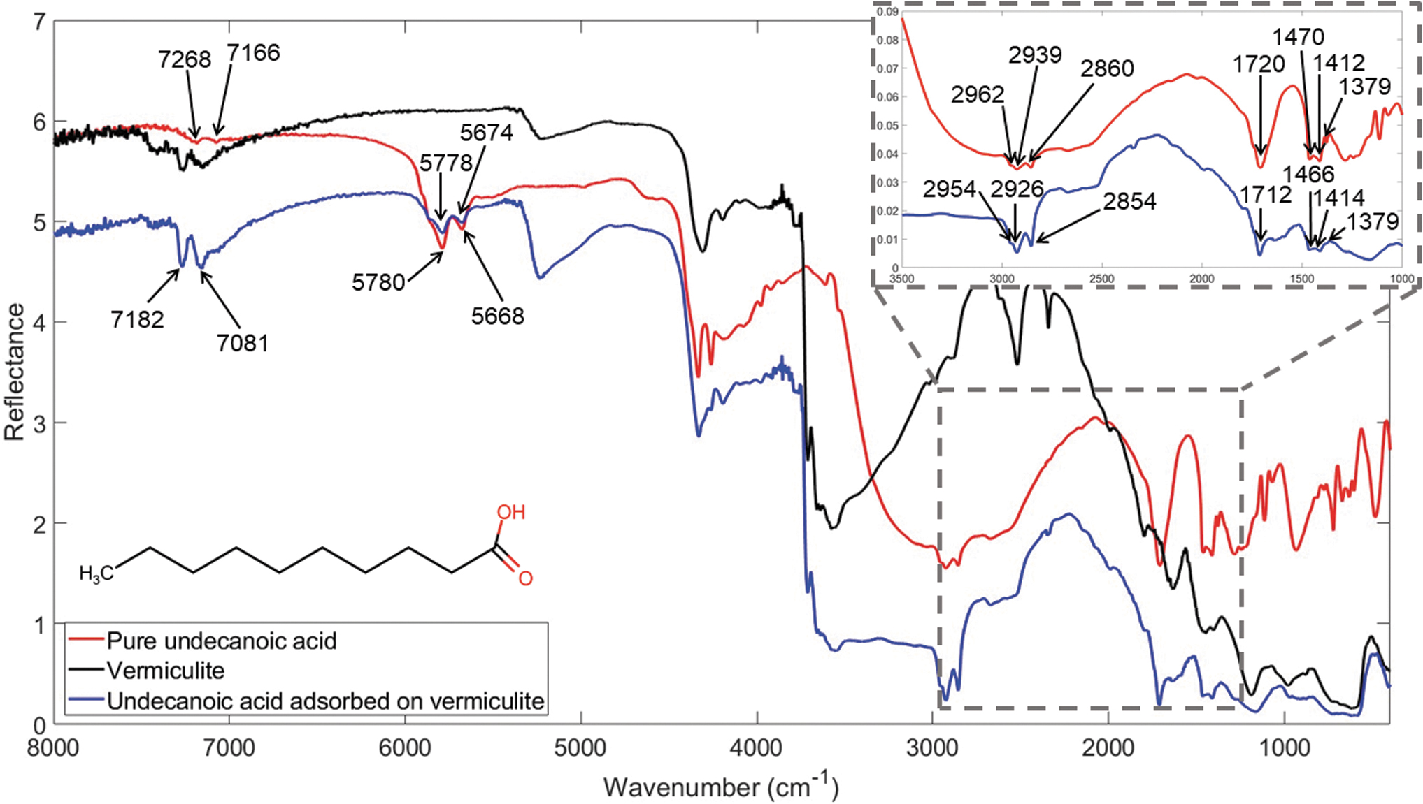
Infrared spectra of vermiculite (black), pure undecanoic acid (red) and 10 wt % undecanoic acid adsorbed on vermiculite (blue). Bands characteristic of the undecanoic acid molecules detected in both the pure and the vermiculite sample are shown with their wavenumbers.
Based on the NIR study on saturated and unsaturated carboxylic acids by Grabska et al. (2017), as well as the NIR computations from this work, the 5780 and 5668 cm−1 bands observed (Fig. 1) are assigned to the first overtones and binary combinations of symmetric stretching modes of the respective CH3 and CH2 groups, as well as to the contribution from the combination mode of asymmetric CH2 stretching in this region (Table 1). NIR simulations yield the doublet with band maxima at 5760 and 5800 cm−1, which originated from several normal modes related to C-H stretches.
Band Wavenumbers (ṽ) and Vibrational Mode Assignments for Pure Undecanoic Acid, Undecanoic Acid Adsorbed on Vermiculite, and Adsorbed on Vermiculite in the Presence of Sodium Chloride (NaCl) or Magnesium Chloride (MgCl2), along with Wavenumber Shifts (Δṽ)awith Respect to the Pure Undecanoic Acid and (Δṽ)bwith Respect to Undecanoic Acid Adsorbed on Vermiculite
ν, stretching; δ, bending; s, symmetric; R, ring. Shifts marked with an asterisk (*) are equal or below the resolution of the instrument (4 cm−1) and therefore not significant.
In the 3000–2850 cm−1 region, the following bands, 2962, 2939, and 2860 cm−1, are assigned to the asymmetric stretching of the CH3 and CH2 groups and to the symmetric stretching of the CH2, respectively (Filopoulou et al., 2021). The band at 1720 cm−1 can be assigned to the C = O stretching vibrational mode that corresponds to the carboxylic functional group of the undecanoic acid (Chapman, 1965). The position of the band at 1720 cm−1 is also indicative of the acidic form of the undecanoic acid (-RCOOH) (Yariv and Shoval, 1982) as expected for the pure solid. Finally, the bands that appear in the 1500–1350 cm−1 region are related to the bending vibrational modes of the C-H groups. More specifically, the 1470 and 1412 cm−1 bands are evidence of CH2 bending, while the 1379 cm−1 band corresponds to the CH3 symmetric bending (Filopoulou et al., 2021). All these assignments are also confirmed by the GVPT2 computations and are listed in the Table 1.
For the pure undecanoic acid, a good agreement was observed between the observed band positions and the ones reported in the literature, while shifts on the vibrational frequencies of some nonfundamental bands were observed when comparing the IR spectra of the pure undecanoic acid and the one adsorbed on vermiculite. Specifically, the nonfundamental bands at 5668 cm−1 present a shift to higher wavenumbers of 6 cm−1. However, due to the complex nature of the band, this shift cannot be simply related to the modification of bonds strength involved in these vibrational modes upon acid adsorption in the mineral. On the other hand, the bands at 2962, 2939, 2860 cm−1 that correspond to the C-H stretching vibrational modes and the band at 1720 cm−1 that corresponds to the C = O stretching present shifts to lower vibrational frequencies (see Table 1). These observations indicate that the molecular bonds become weaker, probably due to their participation in the interaction with the mineral.
Lagaly et al. (2013) observed that the arrangement and orientation of intercalated long-chain compounds between mineral layers are dependent on the Van der Waal's forces established between the alkyl chains. These forces can be strong enough to shift the ending polar groups out of position to form optimal hydrogen bonds. Consequently, the orientation of the acids with short alkyl chains (< C9) is mainly determined by the interactions of the polar groups with the silicate layer, while the longer alkyl chains (> C9) and, therefore, stronger Van der Waal's forces present paraffin-type arrangements (Lagaly et al., 2013). Similarly, the long chain in the undecanoic acid (C11) when adsorbed in vermiculite may be subjected to these Van der Waal's forces, which could explain the shifts observed for the C-H bands at 2962, 2939, and 2860 cm−1. Moreover, acids can be intercalated in vermiculite by displacing the water molecules of the hydrated vermiculite. The OH- and C = O groups of the acid can relocate the water molecules around hard cations such as Na+, Mg2+, and Ca2+ to directly interact with them (Lagaly et al., 2013). According to the work by Yariv and Shoval (1982), there are different possible associations in the interlayer for the fatty acids. The COOH group can interact with the oxygen of a silicate sheet of the mineral or with the structured water (Fig. 2A, 2B). The COOH group can interact with an exchangeable cation through a water bridge (Fig. 2C1, 2C2) or directly with the metal (Fig. 2D1, 2D2). Finally, the COO- group can also interact directly or through a water bridge with the exchangeable cation (Fig. 2E1, 2E2, and 2F1, 2F2, respectively). The predominant associations are highly conditioned by the exchangeable cations and the nature of the fatty acid (Yariv and Shoval, 1982). The absence of bands that correspond to the vibrations of the RCOOH or OH groups in the IR spectra of undecanoic acid adsorbed on vermiculite does not allow for affirmation as to the mechanisms taking place between the mineral phase and the functional groups of the molecule. Only the interactions through the C = O functional group observed at 1712 cm−1 (Fig. 2C2, 2D1, 2D2, 2E1, 2F1) can be inferred due to the shift present for this band in the acid adsorbed on the mineral. Among these possible interactions of the carbonyl group, the most likely is the C2, since a direct interaction with the metal would have resulted in a much larger shift and appearance of a new band.

Different associations that can take place in the interlayer of the mineral for the fatty acids.
The deviations under the resolution of the instrument of 4 cm−1 for the rest of the bands indicate no changes in those vibrational modes when the molecule is adsorbed in the mineral.
To understand the interaction of the undecanoic acid compounds with vermiculite, XPRD measurements were performed. As described above, the intercalation of the undecanoic acid between mineral layers could cause two effects on the crystal structure of the vermiculite: the distortion of the crystal lattice, which results in the enlargement of the cell, or the displacement of the water molecules without modification of the vermiculite skeleton. In the first case, a sensible shift of the peaks in XRPD patterns should be observed together with a change in their relative intensities. In the second one, only differences in the relative intensities are expected. Since the undecanoic acid is present in the structure at low quantities, we do not expect large modifications in the patterns, but they should be significant enough to be observed. The XRPD patterns of the vermiculite (black) and the 10 wt % undecanoic acid adsorbed on vermiculite (red) are reported in Fig. S1.
No modification in the peaks' position can be observed in the XRPD patterns of vermiculite with and without undecanoic acid. In contrast, small differences in the relative intensity of many peaks are perceptible, especially those at 2θ values of 29.80, 30.82, and 31.94 degrees. Thus, we can conclude that the molecules of undecanoic acid were introduced into the structure of the vermiculite without any modification of the mineral skeleton; that is, the molecules lay between the layers and interact with the metal ions present in the interlayers of the vermiculite according to one of the mechanisms shown in Fig. 2. This is consistent with the small shift variations observed in the IR spectra between the pure undecanoic acid and the undecanoic acid on vermiculite.
In the presence of chloride salts, as shown in Fig. 3, the dominating feature of the IR spectrum is the water band located at 3400 cm−1 (Brubach et al., 2005), which corresponds to the stretching mode of the covalent O-H bonds. We also observed a broad band at 5100 cm−1 due to a combination of the stretching and bending modes of the H-O-H group. Finally, the band 1650 cm−1 that corresponds to the H-O-H bending mode is present. The water bands are much more prominent in the presence of MgCl2 due to its higher hygroscopic nature than NaCl.

Infrared spectra of 10 wt % undecanoic acid adsorbed on vermiculite (blue), adsorbed on vermiculite in the presence of sodium chloride (NaCl) (green), and in the presence of magnesium chloride (MgCl2) (magenta). Water bands generated by the hygroscopic nature of MgCl2 are indicated by circles.
The presence of chloride salts during the adsorption process of the undecanoic acid in vermiculite also caused different shifts in the vibrational frequencies of some of the bands of undecanoic acid, as reported in Table 1. When chloride salts are present in the sample, small shifts of the nonfundamental bands are observed with respect to their position in the spectrum of undecanoic acid adsorbed on vermiculite. A 6 cm−1 high-frequency shift of the band at 5778 cm−1 in the presence of NaCl can be appreciated. However, the spectral region most affected by the presence of salts is between 1700 and 1500 cm−1 where a significant low-frequency shift (∼78 cm−1) and splitting of the band originally reported at 1712 cm−1 in the sample without salt is observed (see Fig. 3). Specifically, it splits into two main bands at 1634 and 1587 cm−1 in the sample adsorbed in the presence of MgCl2, and 1645 and 1587 cm−1 in the sample adsorbed in the presence of NaCl. These bands are assigned to the stretching of the undecanoic acid carbonyl (C = O) group, and their splitting in multiple features can be explained considering that, in such a multicomponent system, the carbonyl groups of undecanoic acid molecules can be involved in different kinds of interactions. The significant low-frequency shift of this band indicates that the carbonyl group is involved in very strong interactions such as hydrogen bonds (Fornaro et al., 2016, 2018a), while the broadening may more likely be caused by the presence of water and the contribution of the bending H-O-H mode of the water in that region (Brubach et al., 2005).
Moreover, chloride salts can also change the structure of the vermiculite by their incorporation or even complex the organics and drive them into vermiculite, thus changing the organic-mineral interactions. The XRPD pattern of undecanoic acid adsorbed on vermiculite in the presence of NaCl (Fig. S2) shows evidence of free solid NaCl mixed with the vermiculite sample. Moreover, the pattern is slightly modified in the relative intensities of the peaks, especially in the 2θ range between 28 and 38 degrees (peaks evidenced with the asterisks in Fig. S2). This suggests that the presence of NaCl can have a role in the absorption of the undecanoic acid on vermiculite. On the other hand, the free MgCl2, as well as its hydrates, are not visible in the XRPD pattern, and the whole pattern is greater modified with respect to the pattern without the salt. In addition to the relative intensities of the peaks between 28 and 38 degrees, there is a significant modification of the pattern at low angle and a new small peak at 2θ = 21.6°. Thus, we can conclude that MgCl2 interacts stronger with the vermiculite compared to NaCl, and consequently, it can be hypothesized that MgCl2 has a stronger influence on the adsorption of the undecanoic acid.
The same results can be concluded from the IR spectroscopy by comparing the pure vermiculite IR spectrum with the vermiculite treated with the chloride salts (see Fig. S5). In the case of the vermiculite in the presence of NaCl, no significant changes can be appreciated in the bands related to vermiculite. The only changes are due to the presence of the undecanoic acid and one band at 3400 cm−1 attributable to pure NaCl co-precipitated with vermiculite. These outcomes indicate that part of the Na+ is incorporated with the mineral phase, but a large amount of crystalline NaCl does not interact with the vermiculite. On the other hand, in the presence of MgCl2 changes in the spectra can be appreciated in the bands related to the structure of the vermiculite in the 1300–1000 cm−1 ranges (Fig. S6), which indicates that the Mg2+ ions of the magnesium chloride are incorporated in the vermiculite layers.
The IR bands studied for the UV irradiation experiments were those of the undecanoic acid that were distinguished when adsorbed onto vermiculite and studied in the previous section. In Table 2, the calculated degradation coefficients (b) and the degradation half-lifetimes under laboratory conditions for the bands mentioned are shown together with the obtained degradation cross-section values employed to extrapolate and obtained the results under the martian UV flux.
Degradation Cross Section (σ), Degradation Coefficients (b), and Degradation Half-Lifetimes (t
1/2)Values Calculated for Some IR Bands of Undecanoic Acid and L-phenylalanine Samples after UV Irradiation Experiments under Laboratory Conditions and the Extrapolated Results under Martian Conditions
Degradation Cross Section (σ), Degradation Coefficients (b), and Degradation Half-Lifetimes (t 1/2)Values Calculated for Some IR Bands of Undecanoic Acid and L-phenylalanine Samples after UV Irradiation Experiments under Laboratory Conditions and the Extrapolated Results under Martian Conditions
Among the bands of undecanoic acid that could be appreciated both in the spectrum of the pure molecule and the spectrum of the molecule adsorbed on vermiculite, only two were reported to decrease because of UV radiation in both cases; the one at 1720 cm−1 assigned to the stretching of C = O and the one at 2962 cm−1 assigned to the asymmetric stretching of CH3. The degradation kinetics of these two bands for pure undecanoic acid and adsorbed on vermiculite are shown in Fig. 4. For both bands of undecanoic acid, the degradation curves as a function of time reach a plateau faster when the acid is adsorbed on the mineral matrix. The calculated half-lifetime of degradation under martian flux shown in Fig. 4 and Table 2 is lower in the adsorbed samples, meaning that the acid degrades faster when adsorbed in the vermiculite phase than the pure molecule. This shows that vermiculite has a photocatalizing effect on undecanoic acid under UV irradiation conditions. The band at 1470 cm−1 assigned to the bending of CH2 shows the same tendency in the obtained degradation coefficients (see Table 2) for the pure and the adsorbed sample. However, the overall amount of acid molecules degraded is proportionally higher in the pure (∼60%) than adsorbed on the mineral matrix (less than 10%). This indicates that, while vermiculite acts as a photocatalyzer for the undecanoic acid molecules directly interacting with the UV light, it also provides a mechanical shielding effect.

Degradation kinetics curves for bands at 1720 cm−1 (ν C = O) and 2962 cm−1 (νas CH3) of pure undecanoic acid (red) and undecanoic acid adsorbed on vermiculite (blue) and the corresponding half-lifetimes of degradation under martian UV flux expressed in hours.
Other bands (5780, 5668, and 1412 cm−1) show a decrease in the sample with vermiculite, whereas there is no appreciable change over the resolution of the instrument in the bands of the pure undecanoic acid. This reinforces the hypothesis that vermiculite acts as a photocatalyzer for undecanoic acid molecules. Finally, the bands 2939 and 2860 cm−1 assigned to the asymmetric and symmetric stretching of CH2, respectively, did not show any decrease in the pure or in the adsorbed sample. The band at 1379 cm−1 assigned to the bending of CH3 showed no degradation in the pure acid, but the band was too small in the acid adsorbed to be integrated; therefore, no comparison could be performed.
In the presence of chloride salts, no bands were found to decrease in the presence of MgCl2, and thus no degradation of the acid was observed after the UV light exposure. Instead, some of them were found to increase (see Fig. 5). The reason for this behavior could be explained by the evaporation of water present in the sample caused by exposure to UV. Indeed, molecular bands that fall within the broad water bands increase as the intensity of the water bands decreases due to water desorption. Since no decrease of molecular bands was observed in the presence of MgCl2, an overall photoprotection behavior can be inferred for undecanoic acid in such mineral matrix. When NaCl was present, in most of the bands, no degradation was observed, and the same increasing pattern as in MgCl2 appeared. However, two combination bands (5674 and 5778 cm−1) showed a decrease after the UV irradiation. The band at 5674 cm−1 showed a degradation with a lower half-lifetime than in the sample where the salt was absent (t 1/2 = 2.40 ± 0.7 h with NaCl and t 1/2 = 3.96 ± 0.6 h without salt), which would point at a photocatalyzing effect of NaCl over this band. It is important to note that the intervals almost overlap and the photocatalyzing effect is very small. The band at 5778 cm−1, in contrast, has the same half-lifetime as the band adsorbed on vermiculite without salt. Taking these data into account, it can be said that both salts present mainly a photoprotecting behavior for the undecanoic acid adsorbed onto vermiculite.

Degradation kinetics curves for bands at 5674 and 5778 cm−1 of undecanoic acid adsorbed on vermiculite (blue), with NaCl (magenta), with MgCl2 (green), and the corresponding half-lifetimes of degradation under martian UV flux expressed in hours.
Influence of interaction with vermiculite on the IR vibrational features of L-phenylalanine
When L-phenylalanine is adsorbed on vermiculite, most of the characteristic bands of the pure molecule are not detectable due to the high absorption of the vermiculite. In Fig. 6 the bands of the pure L-phenylalanine that can still be appreciated when the amino acid is adsorbed on vermiculite are indicated by their wavenumbers.
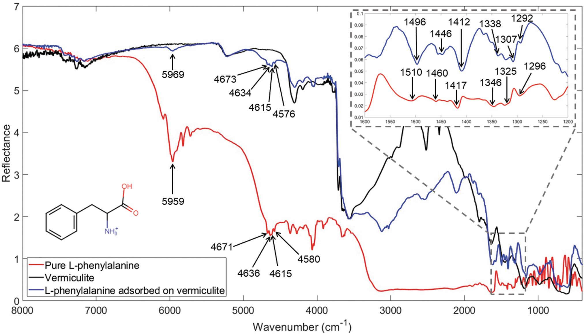
Infrared spectra of vermiculite (black), pure L-phenylalanine (red), and 10 wt % L-phenylalanine adsorbed on vermiculite (blue) with the wavenumbers marked for the bands of the amino acid detected in both the pure and the vermiculite sample.
L-phenylalanine is expected to be in its zwitterionic form (Fig. S3B) in the solid state as well as during the adsorption process of the amino acid onto the mineral once it was dissolved in water (Olsztynska et al., 2001). During the preparation of the sample, the pH was measured at the beginning and at the end of the adsorption process into the vermiculite. The pH slightly varied from 8.21 ± 0.01 to 8.32 ± 0.01; therefore, the adsorption process took place at the pH range where the zwitterionic form is predominant. The zwitterionic form of L-phenylalanine presents the amino group in the protonated state, NH3 +, and the carboxylic group in the deprotonated state, COO-.
In the range >4000 cm−1, five bands due to nonfundamental vibrational modes can be appreciated when the L-phenylalanine is adsorbed on the mineral. In the region 1600–1300 cm−1 six bands due to fundamental vibrational modes of the amino acid can be distinguished.
Tentative assignment of experimental NIR bands of pure L-phenylalanine was previously done by Fornaro et al. in 2020. The band at 5959 cm−1 was attributed to overtone binary combination of C-H stretching in the aromatic ring, whereas the bands at 4671, 4636, and 4580 cm−1 were all attributed to contributions from the combination modes of stretching of the ring and C-H stretching of the aromatic group. Moreover, the band at 1510 cm−1 can be attributed to the in-plane bending of the NH3 + (Olsztynska et al., 2001; Griffith and Vaida, 2013) and is indicative of the ionic form of the nitrogen group in the molecule. No band for the neutral NH2 group in the anionic form of L-phenylalanine was observed in the IR analysis performed, and the NH3 + group for the cationic form of the molecule adsorbs at higher wavenumbers (∼1530 cm−1) (Olsztynska et al., 2001). The position of the amine group of L-phenylalanine at 1510 cm−1 is, therefore, confirming the existence of the molecule in its zwitterionic form.
The band at 1460 cm−1 can be assigned to the C-C stretching of the aromatic ring with a possible contribution of the in-plane bending of CH2, and the band at 1417 cm−1 can be attributed to the symmetric stretching of the COO- functional group (Olsztynska et al., 2001; de Araújo et al., 2021). The band at 1325 cm−1 is assigned to the out-of-plane bending of the CH2 group (Cao and Fischer, 2000; Olsztynska et al., 2001). Finally, the band at 1292 cm−1 is assigned to a stretching of a C-C bond by Cao and Fischer. All the assignments are summarized in Table 2.
The band positions observed for the pure L-phenylalanine are shifted when the molecule is adsorbed on vermiculite (see Table 3). The biggest shift appears for the band at 1325 cm−1 related with the in-plane bending of CH2 functional group, which presents a shift of 18 cm−1 to lower wavenumbers when the L-phenylalanine is adsorbed on vermiculite. The band at 1510 cm−1 related to the in-plane bending of the NH3 + functional group and the band at 1460 cm−1 related to the aromatic ring group present a shift of 14 cm−1 to lower wavenumbers when the amino acid is adsorbed in the mineral. A much smaller shift of 5 cm−1 is observed for the band at 1417 cm−1 assigned to the COO- group. These shifts indicate that the functional groups are interacting with the vermiculite mineral phase. In all cases, the shifts are to lower wavenumbers, which means that the vibration mode required lower energies, thus indicating a weakening of the bonds due to the interaction with the mineral. In this case and taking into consideration that the shift observed for the COO- functional group is so small that it is closed to the resolution of the instrument (4 cm−1), it seems that the molecule is interacting through the positively charged group of the amino acid and through the hydrophobic parts like the CH2 and the benzene ring. This indicates the existence of multiple kinds of interactions in the adsorption mechanisms of L-phenylalanine to vermiculite.
Band Wavenumbers (ṽ) and Vibrational Mode Assignments for Pure L-phenylalanine, L-phenylalanine Adsorbed on Vermiculite, and Adsorbed on Vermiculite in the Presence of Sodium Chloride (NaCl) or Magnesium Chloride (MgCl2), along with Wavenumber Shifts (Δṽ)awith Respect to the Pure L-phenylalanine and (Δṽ)bwith Respect to L-phenylalanine Adsorbed on Vermiculite
ν, stretching; δ, bending; s, symmetric; R, ring; i.p., in plane; o.p., out of plane. Shifts marked with an asterisk (*) are equal or below the resolution of the instrument (4 cm−1) and therefore not significant.
There are many adsorption mechanisms that have been observed for amino acids on clays. For example, the zwitterion form of lysine likely adsorbs on the planar surface of montmorillonite due to Van der Waal's forces (Rao et al., 1980). The existence of Van der Waal's forces in the adsorption of L-phenylalanine would explain the shift observed for the band at 1325 cm−1 of the CH2 group. Van der Waal's forces can also result from the polarization of the aromatic ring, which could also explain the important shift observed for the band at 1460 cm−1 related to the aromatic ring in the adsorption of L-phenylalanine. In addition, the adsorption of planar aromatic molecules may be assisted by the interaction of the π electrons of an aromatic system with the surface oxygens of the clay, which would also explain the shift observed for the band related to the aromatic ring. However, the L-phenylalanine CH2-CNH3 +-COO- group is not planar, and this complicates the approach of the aromatic system to the mineral surface. Other works suggest that both the amino and the carboxylic groups are involved in the adsorption of amino acids (Hashizume, 2012). Cloos et al. (1966) suggested that there are two different types of adsorptions of amino acids onto montmorillonite and a 2:1 layer clay mineral. A strong adsorption can take place between the cationic amino group and the negatively charged interlayer sites of the clay surfaces, and another weaker adsorption of the amino group can occur though the carboxyl group of another amino acid already strongly adsorbed to the mineral surface (Cloos et al., 1966). This would explain both shifts observed for the band at 1510 cm−1 related to NH3 + and the small shift for the band at 1417 cm−1 related to the COO-. Finally, other studies observed the complexation of the interlayer cations of the mineral by the carboxylate group of the zwitterion (Lagaly et al., 2013).
The XRPD pattern of vermiculite with 10 wt % L-phenylalanine absorbed is reported in Fig. S1 (blue line). Comparing this pattern with the one of “free” vermiculite (black), the absorption of the amino acid molecules does not cause the shift of the peaks in the pattern. In contrast, some significant differences in the relative peaks' intensities are detectable, as already observed for the vermiculite with the absorbed undecanoic acid. Furthermore, it is also remarkable to note the presence of a shoulder at 2θ value of 7.41 and a small peak at 19.45 degrees that is scarcely perceptible in the vermiculite with the absorbed undecanoic acid and almost non-existent in the “free” vermiculite. Thus, also, in this case, we can conclude that the L-phenylalanine molecules are incorporated in the layers of the vermiculite, probably substituting water molecules, with no modification of the vermiculite skeleton. Therefore, these observations support the existence of the several mechanisms that involve the interaction between the cationic amino group and the negatively charged interlayers of the surface or the complexation of the interlayer cations by the carboxylate group of the zwitterion, both functional groups with shifts in the IR spectrum.
As previously described, the dominating feature of the spectra when chloride salts are added to the samples is water bands due to the hygroscopic nature of NaCl (due to impurities in the sample [purity >97 %]) and MgCl2. Moreover, the presence of chloride salts in the adsorption of L-phenylalanine on vermiculite caused shifts in some of the vibrational frequencies of the adsorbed L-phenylalanine with respect to the ones observed when the adsorption took place without the presence of the salts. Specifically, when sodium chloride was added to the sample, 7 and 10 cm−1 shift to higher frequency of the bands at 4634 and 1446 cm−1 were observed with respect to their positions in the spectrum of L-phenylalanine adsorbed on vermiculite without any addition of chloride salts. Since the 4634 cm−1 band is a combination of ring stretching and aromatic CH stretching and the band at 1446 cm−1 is a ring stretching, these high-frequency shifts suggest a strengthening of the ring, which may mean a lower interaction between the aromatic ring and the mineral. The same shift was observed for this group when the adsorption of L-phenylalanine was performed in the presence of MgCl2. This would agree with the observations of some works where the adsorption onto minerals is generally decreased with the increasing concentrations of salts (Norén et al., 2008; Farias et al., 2014). Moreover, when the adsorption of L-phenylalanine was performed, increasing the ionic strength by substituting the NaCl by MgCl2 in the same concentration, a new shift to higher wavenumbers was observed for the band related to the C = O functional group at 1412 cm−1: 2 cm−1 for NaCl (under the resolution of the instrument) and 9 cm−1 for MgCl2. The shift to higher wavenumbers implies an absorption of a higher energy radiation and, therefore, a strengthening of the C = O bond, probably due to its lower participation in the adsorption with the mineral. This observation would agree with the fact that the higher the ionic strength, the lower the electrostatic interactions with the mineral (Zaia, 2012).
Moreover, new bands appeared when sodium and magnesium chlorides were added to the L-phenylalanine adsorbed on vermiculite samples. Specifically, three new bands at 6509, 4837, and 2627 cm−1, as indicated in Fig. 7, appeared when magnesium chloride was added to the adsorption process, whereas two new bands appeared at 2625 and 2341 cm−1 in the presence of sodium chloride. These bands were not identified in the pure L-phenylalanine, nor in the adsorbed samples without the salts. These new bands may indicate the formation of new bonds, likely mediated by the presence of the Na+ and Mg2+ cations that might have acted as bridges for molecular adsorption on vermiculite. The cross-interaction between NaCl or MgCl2 and L-phenylalanine could have led to the chlorination of the amino acid through the substitution on the aromatic ring of a chlorine; however, this would have led to the appearance of a new band between 690 and 770 cm−1, which is not observed in the spectra. The formation of an acid chloride with the substitution of the OH group by a Cl is also not plausible as the band would be situated at a lower wavenumber between 1800 and 1770 cm−1. Another possibility is that the interaction of the phenylalanine molecules with the cations of the chloride salts resulted in the formation of organic salts and, therefore, new bands from the formation of an ion-dipole interaction.

Infrared spectra of 10 wt % L-phenylalanine adsorbed on vermiculite (blue), adsorbed in the presence of sodium chloride (NaCl) (green), and in the presence of magnesium chloride (MgCl2) (magenta). Water bands generated by the hygroscopic nature of MgCl2 are indicated by circles. New bands appearing in the presence of salts are indicated with their wavenumber.
The XRPD pattern of phenylalanine adsorbed on vermiculite in the presence of NaCl (Fig. S4) shows evidence of free solid NaCl mixed to the vermiculite sample, as already observed for the corresponding undecanoic acid sample. However, in this case the pattern is not modified in the relative intensities of the peaks with respect to the phenylalanine adsorbed in the absence of NaCl. In fact, apart from the presence of the NaCl peaks, the pattern is identical to that of the phenylalanine adsorbed on vermiculite in the absence of salts. Thus, the presence of NaCl seems not to have a role in the interlayer absorption of the phenylalanine on vermiculite. However, as described above, the IR spectrum showed changes in the C-H and ring stretching bands of the molecule. Both functional groups interact with the surface of the mineral. The addition of NaCl is, therefore, only affecting the surface interactions with vermiculite. Moreover, if we compare the spectra of pure vermiculite and the sample with NaCl, we can identify the presence of pure NaCl at 3400 cm−1 (see Fig. S7).
As for the undecanoic sample, the free MgCl2 is not visible in the XRPD pattern, but some differences in the peak intensities are observed with respect to the pattern without the salt (peaks evidenced with stars in Fig. S4). Thus, in the case of phenylalanine absorbed on vermiculite in the presence of MgCl2, we can conclude that the salt interacts slightly with the vermiculite, and consequently it can be hypothesized that it could have a role in the adsorption of the organic molecule in the interlayer of the mineral. However, according to the IR observations, MgCl2 is not favoring the interaction between the organic molecule and the vermiculite as seen by the shift to higher wavenumbers of the C = O bond. Therefore, the observed changes in the relative intensities of the XRPD patterns could be due to a smaller amount of organic molecules in the vermiculite interlayer substituting the water molecules.
For the UV irradiation experiments performed over pure L-phenylalanine and L-phenylalanine adsorbed on vermiculite, only the band at 1510 cm−1, which corresponds to the in-plane bending of the amino group, shows a decrease after the UV irradiation for both the pure molecule and the molecule adsorbed on the mineral (Fig. 8). The degradation coefficients for the calculated laboratory and extrapolated martian conditions and the cross section for this band are shown in Table 2. The degradation kinetics curve and the calculated half-lifetime of degradation for the pure and the adsorbed amino acid indicate that the molecule is degrading faster in the pure state than when adsorbed on the mineral. This means that vermiculite is acting as a photoprotector for L-phenylalanine against UV radiation.
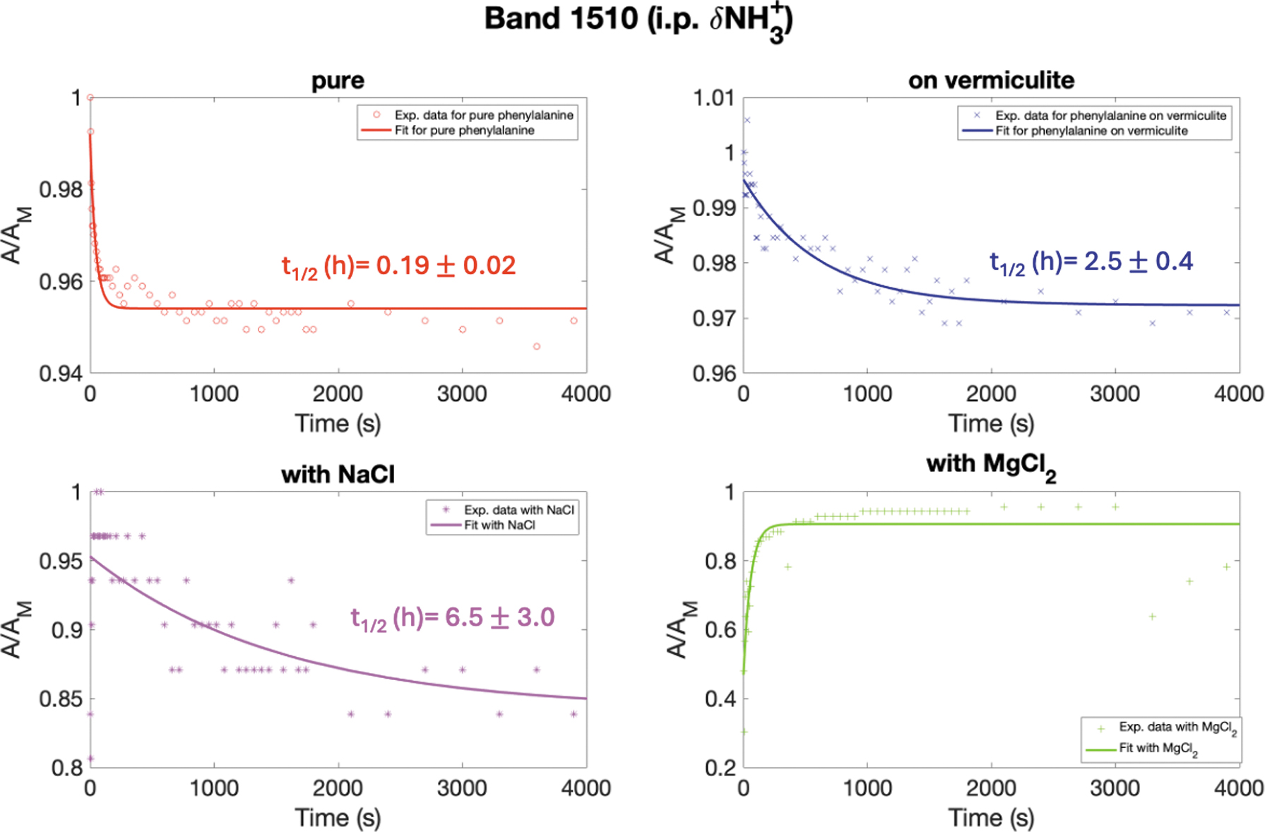
Degradation kinetics curve for the band at 1510 cm−1 (i.p. δ NH3 +) of pure L-phenylalanine (red) and L-phenylalanine adsorbed on vermiculite (blue), in the presence of NaCl (magenta) and in the presence of MgCl2 (green), and the corresponding half-lifetimes of degradation under martian UV flux expressed in hours. i.p. = in plane.
The rest of the bands of the pure L-phenylalanine showed no degradation, which indicates a good stability of the amino acid against UV. Considering that the stability of the amino acid could have complicated the study of the role of vermiculite, it was decided to extend the irradiation time from 1h10 to up to 10h30. However, no further degradation in the pure L-phenylalanine bands was observed. Moreover, the bands detected in the spectra of L-phenylalanine adsorbed on vermiculite also do not show any changes that can be appreciated under the resolution of the instrument, which indicates that they may not be degrading under UV light or at a rate that cannot be detected with our experimental setup. When taking these data into account, it would seem that the L-phenylalanine is stable under UV light, and according to the band 1510 cm−1 for which a degradation could be appreciated, the vermiculite seems to have a photoprotecting nature for L-phenylalanine.
In the presence of NaCl, the band at 1496 cm−1 decreases much more slowly (t 1/2 = 6.5 ± 3.0 h) than in the pure (t 1/2 = 0.19 ± 0.02 h) and in the sample adsorbed onto vermiculite (t 1/2 = 2.5 ± 0.4 h) (Fig. 8), indicating a photoprotective behavior of sodium chloride. In the presence of MgCl2, an increase in the kinetics curve of the band is observed (Fig. 8). This increase could be due to a change in the background continuum from the desorption of water over time caused by UV light. Because water affects the entire spectrum, it can also change the background's position when it evaporates and result in an apparent increase of some bands (Fig. S8). These data indicate that chloride salts protect the amino acid from degradation under UV light, exhibiting a photoprotective behavior toward L-phenylalanine.
Moreover, as can be observed in Fig. 9 in the case of L-phenylalanine adsorbed on vermiculite in the presence of magnesium chloride, new bands in the 1700–1400 cm−1 range formed during UV irradiation. These bands appear at 1657, 1649, and 1639 cm−1 and are situated within the broad water bending band at 1650 cm−1 described above (Fig. 7). They could have appeared due to the decrease in the water content of the sample during the irradiation process where water molecules are cleaved to form radicals. The emergence of new bands in the 1550–1400 cm−1 region is indicative of the formation of new molecules from the interaction of the adsorbed L-phenylalanine on vermiculite and magnesium chloride under UV irradiation, possibly formed through the reactivity of radicals coming from the water adsorbed by the salt, which would form new molecules when interacting with L-phenylalanine, or through catalytic effects of Mg2+. It has been shown that, following the photolysis of a parent molecule, the produced fragments can rearrange or react with their environment to form new molecules (Poch et al., 2014).

IR spectra of L-phenylalanine adsorbed on vermiculite with MgCl2 at different irradiation times. New bands are indicated by their wavenumber.
Vermiculite's influence on undecanoic acid and phenylalanine molecules under UV radiation
Undecanoic acid
Undecanoic acid molecules degraded more when adsorbed onto vermiculite compared to the pure sample under UV radiation. The photocatalytic decomposition of carboxylated molecules has been studied by Shkrob et al. (2010), who proposed that the occurrence of photo-Kolbe reactions at the surface of semiconducting iron (III) oxides initiated by the absorption of UV light was at the origin of the catalytic degradation of the molecules. Moreover, studies have shown that phyllosilicate clay, among which is vermiculite, could behave as a semiconductor material and thus as a photoactive medium in the presence of UV radiation (Martínez-Costa et al., 2018). The degradation observed for the C = O stretching band could, therefore, be due to the decarboxylation of the undecanoic acid molecules.
Phenylalanine
Phenylalanine molecules were less degraded when adsorbed onto vermiculite compared to the pure sample under UV radiation. Different studies have previously shown the photoprotective effect of clay minerals on the evolution of amino acids under UV radiation (Kate et al., 2005; Poch et al., 2015; dos Santos et al., 2016; Ertem et al., 2017). Several mechanisms that explain this phenomenon were proposed such as mechanical shielding but also a stabilizing molecule-mineral interaction that could allow a more efficient energy dissipation (Poch et al., 2015). Moreover, the overall low degradation of L-phenylalanine suggests its high stability. The stability of the molecule is dependent on the substituents bonded to the α-carbon atom. A study by Kate et al. (2005) found that alkyl substituent groups attached to the α-carbon atom contribute toward the stability of the resulting alkyl amine radical that forms after UV-induced decarboxylation and prolong the life of the amino acid. We infer a similar reasoning for aromatic amino acids such as L-phenylalanine, where the highly stable aromatic phenyl group should contribute to the stability of the molecule and, therefore, its photoprotection. Moreover, Poch et al. (2014) results indicate that aromatic molecules are at least 10 times more resistant to martian UV compared to non-aromatic molecules.
Moreover, in Fig. 10 the ToF-SIMS images obtained for L-phenylalanine and undecanoic acid adsorbed on vermiculite are shown. In this figure, areas where L-phenylalanine molecules are agglomerating can be observed, whereas the undecanoic acid is overall homogeneously distributed on the vermiculite. Fornaro et al. (2018b) suggested that the formation of molecular aggregates in which some molecules are directly exposed to radiation while others are covered and more protected could also be a mechanism of photoprotection.
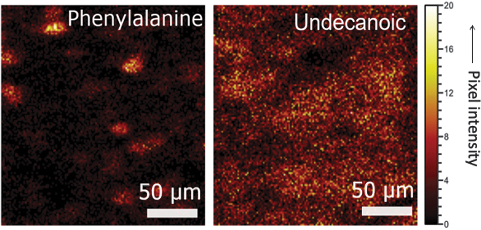
ToF-SIMS image of L-phenylalanine (C9N12O2 +, m/z 166.09) and undecanoic acid adsorbed on vermiculite (C11H21O2 -, m/z 185.15).
This study shows the consequences of UV radiation for organic molecules embedded in the phyllosilicate structure in the first layer of martian soil and directly interacting with UV light. The nature of the molecule and its interaction with vermiculite seem to impact the photodegradation of the molecule. These results are of importance for missions that analyze samples from the first layers of martian soil such as the MSL mission with the Curiosity rover or the Mars 2020 mission with the Perseverance rover.
This is why the future ExoMars mission rover, Rosalind Franklin, will be equipped with a drill capable of collecting samples down to 2 m in depth, where organic molecules adsorbed onto the mineral's structure are not directly interacting with UV light and where phyllosilicates such as vermiculite will act as a protective layer against UV radiation.
Chloride salts' influence on undecanoic acid and phenylalanine molecules under UV radiation
UV degradation kinetics study has revealed that the two inorganic chloride salts can protect undecanoic acid and phenylalanine molecules against UV irradiation. However, the exact mechanism of the protection is not clearly understood. A study by Chang (1982) on the photoprotective properties of inorganic chloride salts on wood speculated that the incorporation of inorganic ions on wood could result in organic-ion complex systems capable of emitting effective light energy by absorbing and reemitting light through some photophysical decay process. Accordingly, less energy will be absorbed by the organic to initiate photoreactions. Moreover, the newly formed complex system could shift the absorbing zone to a shorter wavelength, which could interfere with the photochemical reaction (Chang, 1982). A similar reaction pathway could be inferred for undecanoic acid adsorbed on vermiculite in the presence of MgCl2 and NaCl.
Another possibility is that the organic molecules are physically protected by the recrystallized chloride salts as they form a barrier that protects them from UV light. Indeed, most minerals that precipitate from aqueous solution incorporate fluid inclusion during growth. These fluid inclusions can capture molecules attached to the crystal surface. Cockell and Raven (2004) showed that halite (NaCl) encrustations can act as screens by scattering UV radiation when precipitated as a mass of small crystals that make these environments suitable for photosynthetic processes on Mars or early Earth. Therefore, during sample preparation, undecanoic acid and phenylalanine molecules could have been entrapped as inclusions in magnesium and sodium chloride salts, thus forming a protective cage that prohibits UV light from interacting with the organic compounds.
Moreover, a study by Liu and Kounaves (2021) looked at the degradation of several amino acids (glycine, proline, and tryptophan) under UV irradiation in martian conditions in the presence of chloride (NaCl) and oxychlorine (NaClO3 and NaClO4) salts. While glycine's degradation increased in the presence of the salts, the authors showed that, under ambient martian conditions, salts were protecting proline from degradation. Furthermore, tryptophan, an aromatic amino acid like phenylalanine, did not degrade with or without salt, which is consistent with the stabilizing effect of the aromatic ring described above. The study by Liu and Kounaves (2021) showed that the photocatalyzing effect of chloride salts as described in the present study might depend on the nature of the amino acid, as well as the affinity of the salts to water.
Overall, our study shows that chloride salts could help preserve organic matter from UV radiation. Therefore, environments that contain chloride salts could make good targets for molecular analysis by Mars rovers, and their presence could be a criteria for choosing future sites of investigations on Mars.
Conclusion
In this work, undecanoic acid and L-phenylalanine adsorbed onto vermiculite with and without the presence of chloride salts was studied by way of FTIR spectroscopy to assist the Mars exploration missions to detect and identify organic molecules by showing the changes on the IR spectra of organic molecules due to the different adsorptions. The presence of chloride salts plays a role in the adsorption process of the organic molecules onto the vermiculite matrix. We observed that MgCl2 favors adsorption of the organic species more than NaCl as seen in XRPD. Moreover, the present study also considered the stability of these two organic molecules against UV irradiation, an important degradation factor that may affect the presence of organic molecules on the surface of Mars. The irradiation laboratory simulations performed in this work showed that vermiculite can act as a photocatalyzer for the undecanoic acid and as a photoprotector for L-phenylalanine and that the degradation due to the UV radiation can be abated by the presence of sodium or magnesium chlorides. This study confirms that the straightforward classification of martian minerals as photocatalytic or photoprotective is not possible, since the behavior of minerals under martian conditions may depend on the organic molecules involved and their specific interactions with the mineral surface sites. However, with regard to the present study, it should be noted that chloride salts could preserve the integrity of organic molecules such as amino acids or fatty acids by protecting them from photocatalysis; therefore these materials would be interesting targets for the detection of organic matter by current and future missions performing in situ measurements.
Footnotes
Acknowledgments
ASI/INAF agreement 2023-3-HH.0 and the “ADI 2020” project funded by the IDEX Paris-Saclay, ANR-11-IDEX-0003-02. The authors declare no conflicts of interest.
Supplementary Material
Supplementary Figure S1
Supplementary Figure S2
Supplementary Figure S3
Supplementary Figure S4
Supplementary Figure S5
Supplementary Figure S6
Supplementary Figure S7
Supplementary Figure S8
Abbreviations Used
References
Supplementary Material
Please find the following supplemental material available below.
For Open Access articles published under a Creative Commons License, all supplemental material carries the same license as the article it is associated with.
For non-Open Access articles published, all supplemental material carries a non-exclusive license, and permission requests for re-use of supplemental material or any part of supplemental material shall be sent directly to the copyright owner as specified in the copyright notice associated with the article.
