
Abstract
Background:
Laparoscopic cholecystectomy ranks among the five most common surgical procedures performed in the United States. Studies show the frequency of gallstone spillage during laparoscopic cholecystectomy ranges from 5.7% to 36%, with 0.3% of these cases resulting in abscess formation. To our knowledge, we report the only documented case of a patient who had their gastric remnant fistulized to the anterior abdominal wall from a gallstone abscess found 12 years after routine laparoscopic cholecystectomy.
Case:
We report a case of 39-year-old female who was found to have a gallstone abscess that resulted in a fistula of her remnant stomach to her anterior abdominal wall. Initial workup of her epigastric pain was attributed to her chronic pain and marginal ulcers found on endoscopy. However, further investigation revealed her ongoing symptoms were due to a gallstone abscess. She had prompt resolution of her symptoms after the true etiology of her problem was recognized and treated.
Conclusions:
Although rare, gallstone abscesses can pose a diagnostic challenge for physicians and result in significant morbidity for patients. Clinical examination yields few clues, and without a high clinical suspicion, radiographic imaging is largely nondiagnostic. Below we review the literature on spilled gallstones, recommended treatment, and discuss the workup to allow for prompt and accurate diagnosis.
Introduction
S
The diagnosis of gallstone abscess can often be difficult to discern from other causes of abdominal pain, and its peak incidence of discovery averages 3 years. Vague complaints of fevers, chills, nausea, vomiting, loss of appetite, weight loss, and colicky dull abdominal pain that may be progressively worsening over days to months are classic for gallstone abscess.2,3,5,6 Due to its rarity and the variability in location and characteristic of pain, depending on the site of abscess formation and peritoneal irritation, diagnostic clues that can lead to prompt recognition and intervention are critical for clinicians.
To our knowledge, there have been no reported cases of a gastric remnant fistula due to a gallstone abscess discovered 12 years after laparoscopic cholecystectomy. We report a case of a 39-year-old female who presented with chronic colicky abdominal pain attributed to other conditions on initial evaluation. The clinical and pathologic characteristics of gallstone abscess and literature review is discussed below to aid in accurate diagnosis and intervention.
Case Report
A 39-year-old Caucasian female presented to the emergency room with a 3-day history of colicky dull epigastric pain radiating to her back with decreased oral intake. She complained of subjective fevers and chills, with intermittent nonbilious emesis. One month prior, outpatient upper endoscopy performed for similar symptoms was diagnostic for a small marginal ulcer. Treatment was initiated and did not completely alleviate her symptoms. Her past medical history is significant for morbid obesity, hypothyroidism, fibromyalgia, polycystic ovary syndrome, chronic pain syndrome, post-traumatic stress disorder, anxiety, and depression. Her surgical history included laparoscopic cholecystectomy, performed for symptomatic cholelithiasis and laparoscopic Roux-en-Y gastric bypass. The 2001 operative report from the laparoscopic cholecystectomy was reviewed. There were no reported complications, and the gallbladder was removed without an endoscopic retrieval bag through the epigastric trocar site. In 2006, she underwent elective Roux-en-Y gastric bypass for morbid obesity for which she reported an unremarkable postoperative course and lack of continued follow-up.
On physical evaluation, she was afebrile with normal vital signs. Her abdomen was soft and nondistended, but she had epigastric tenderness and associated guarding. Her laboratory results were unremarkable. Ultrasonography was nondiagnostic due to body habitus, but a hypoechoic area in this region was visualized and consistent with abscess. A computed tomography (CT) scan of the abdomen and pelvis showed a 3.2 cm×3.3 cm cystic mass with a thickened wall and surrounding inflammatory changes suggestive of an abscess just below the left hepatic lobe (Figs. 1 and 2). She was then admitted to the hospital with the diagnosis of intra-abdominal abscess of unclear etiology, possibly from a perforated marginal ulcer, and started on antibiotics. Subsequent attempts at CT guided percutaneous catheter placement were unsuccessful due to severe wall thickening. However, 2 cc of purulent material was aspirated and sent for culture and came back positive for Escherichia coli.

Axial computed tomography (CT) image demonstrating fistula tract (arrow) from gastric remnant stomach to gallstone abscess.
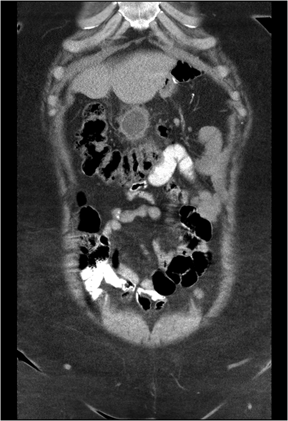
Coronal CT image demonstrating 3.2 cm×3.3 cm gallstone abscess with surrounding inflammatory changes just below the left lobe of the liver.
She then underwent surgery for abscess drainage. A fistula tract from the anterior abdominal wall to the distal portion of the thickened inflamed gastric remnant contained a large palpable abscess cavity. The fistula tract was removed in its entirety, and the remnant stomach was visually inspected to be grossly normal prior to closing the small defect in the stomach. The pathology report showed a 10 cm×8 cm×3.5 cm fistula tract containing a 3 cm×2.5 cm tannish, brown, cholesterol gallstone. Her postoperative course was unremarkable, and she was discharged home with 4 weeks of oral antibiotics, with resolution of her symptoms reported at outpatient follow-up.
Discussion
The incidence of gallbladder perforation is around 18.3% with subsequent spillage of gallstones in 40% of cases, most of which are unretrieved. 3 The most common complications of spilled gallstones are abscess formation, followed by fistula formation, intestinal obstruction, pleural empyema, and broncholithiasis. 3 Spillage occurs 75% of the time during dissection off the liver bed and 25% of the time during extraction, with the most significant factor being the surgeon's skill. 7 Abscess formation as a result of spilled gallstones has been reported to range from 4 days to 10 years, with peak incidence of symptoms at 4 months. 6 The mean duration from the time of procedure and development of symptoms is 2.1±0.6 years, while the duration from onset of symptoms until diagnosis is 2.9±1.0 years. 2
Traditionally, dropped stones were considered harmless and believed to reabsorb gradually into the peritoneum. Bacteria inside gallstones have long been thought to be dormant, but recent studies have shown the opposite to be true. 8 One study cultured gallstones obtained during laparoscopic cholecystectomy and found 81% grew bacteria, the most common being gram negative organisms. 9 The infectious process originating from gallstones is independent of contamination of the peritoneal cavity with infected bile.3,9
Gallstone abscess is diagnosed by imaging in 40% of cases. 2 Radiographic diagnosis of gallstone abscesses can be challenging depending on the type of gallstone and the unusual locations in which they may present. 2 CT scan with contrast is the most sensitive for identifying and localizing an abscess cavity; some gallstones or stone fragments may be visualized within the cavity. 2
Prevention of gallstone abscess should include strategies such as intraoperative aspiration of bile to reduce wall tension, careful dissection from the hepatic fossa, and the use of specimen retrieval bags. If perforation occurs, every effort should be taken to retrieve any lost stones, followed by copious irrigation to minimize bile contamination. The use of perioperative antibiotics covering biliary tract flora in any instance of spilled gallstones has also been recommended.3,4 Management strategies for gallstone abscess include the use of antibiotics and adequate drainage of the abscess and removal of the gallstone—either percutaneous or open. 10 Patients should be started on broad spectrum antibiotics with de-escalation following final culture and sensitivity. 4
Conclusion
With a growing trend toward minimally invasive procedures, almost all laparoscopic surgeons will encounter gallbladder perforation and spillage. Although rare, the complication of gallstone abscess can pose serious problems to patients, months to years following surgery. In order to treat our patients in the best possible way, we must employ prevention strategies to avoid complications, be suspicious of unusual presentations of abdominal pain and abscesses after cholecystectomy, and have a low threshold for initiation of treatment.
Footnotes
Disclosure Statement
No competing financial interests exist.