
Abstract
Background:
Carotid intima media thickness (CIMT) is used as a surrogate measure of coronary artery disease. The aim of this research was to evaluate the effects of laparoscopic sleeve gastrectomy (LSG) and laparoscopic gastric plication (LGP), and its associated weight loss, on CIMT.
Materials and Methods:
This prospectively designed study enrolled 48 consecutive obese subjects who underwent either LSG or LGP between January 2013 and June 2014. Patient demographic data (age, sex, height, and weight), body–index (BMI), and metabolic parameters (lipid profile) were measured before and 3 and 6 months after surgery. The degree of liver steatosis was also determined ultrasonographically at the same time.
Results:
LSG was performed in 25 patients (LSG group) and LGP in 23 patients (LGP group). Both groups were similar in terms of age, sex, BMI, and CIMT preoperatively. In both groups, biochemical parameters improved significantly at 3 and 6 months after surgery. Mean CIMT decreased significantly in both groups.
Conclusion:
In addition to the improvement in lipid profiles of obese patients after either LGP or LSG, CIMT decreases after bariatric surgery. This decrease is proportional to the decrease in BMI and cholesterol levels. The risk of future cardiovascular events in obese subjects undergoing bariatric surgery may be decreased.
Introduction
C
Obesity has been strongly associated with hypercholesterolemia. The increased weight has detrimental effect on blood cholesterol and lipid levels, even in young ages. 8 For this reason, weight loss of obese patient has a positive effect on decreasing plasma cholesterol levels. The correlation between elevated low-density lipoprotein (LDL) cholesterol and total cholesterol and CIMT has been proven. 9 Patients with increased CIMT along with raised total cholesterol and LDL cholesterol levels develop early coronary and cerebrovascular diseases. Although previous studies have thoroughly investigated the correlation between lipid lowering medications and CIMT, the reported findings are diverse and nonconclusive.10,11 Nevertheless, other studies have stated that cardiovascular diseases drop off as CIMT progression is slowed down with the lowering of cholesterol levels. 12 Hepatosteatosis is another issue that we come across with obese patients. We do not know exactly if obese patients lose weight after obesity surgery whether the degree of steatosis would decrease or not.
Objectives
The aim of this research is to evaluate the short-term outcomes of bariatric surgery (laparoscopic sleeve gastrectomy [LSG] and laparoscopic gastric plication [LGP]) in terms of weight loss, improvement in lipid profile, and investigate its effect on CIMT and liver steatosis.
Materials and Methods
This study is a prospectively designed study that was conducted on 48 obese subjects who underwent LSG or LGP at the Bagcilar Training and Research Hospital, Istanbul, Turkey, between January 2013 and June 2014. All procedures performed in this study were in accordance with the ethical standards of the institutional and/or national research committee and with the 1964 Declaration of Helsinki and its later amendments or comparable ethical standards. All participants of the study were evaluated by a multidisciplinary team consisting of general surgeon, endocrinologists, dietician, and psychiatrist. The prospective study protocol was approved by the local ethics committee. Informed consent was obtained from all individual participants included in the study. All the operations were performed laparoscopically by one surgeon. Demographic data (age, sex, height and weight, body–mass index [BMI], mass [kg]/height [m2]) of the patients, lipid profile, CIMT, and liver steatosis were recorded preoperatively and 3 and 6 months after surgery. Excessive weight loss (%EWL), which was defined as the percentage of weight lost in a period of time, was calculated at 3 and 6 months after surgery.
Subjects were divided into two groups (LGP group vs. LSG group) according to the procedure performed. Operative indications for LGP were BMI <50, absence of metabolic syndrome, and consent to the procedure. In contrast, operative indications for LSG were BMI ≥40 or BMI between 35 and 40 with the metabolic syndrome like uncontrolled diabetes or osteoarthritis. An exclusion criterion for the study was not willing to be included in the study.
Biochemical analysis
Blood samples were collected after overnight fasting of 12 h. Total cholesterol, high-density lipoprotein (HDL), LDL, and triglycerides were determined by using the Cobas® 6000 autoanalyzer (Roche Diagnostics). Lipid profile of all participants was determined preoperatively and after 3 and 6 months of the operation.
CIMT measurement and hepatic steatosis evaluation
Ultrasonographic evaluation of the patients and hepatic steatosis evaluation were done by a single radiologist using the Esaote MyLab™60. B-mode ultrasound of both the common carotid arteries, 2 cm proximal to the bifurcation, was done in three planes (transverse, anterolateral, and posterolateral) with a linear probe. This ensured standardization of all the measurements (Fig. 1). Ultrasonographic assessment of hepatic steatosis was made by considering the following factors: (a) increase in hepatic echogenicity, (b) decrease in visualization of the vascular walls, and (c) visualization of the diaphragm. Hepatic steatosis was graded as mild, moderate, or severe.
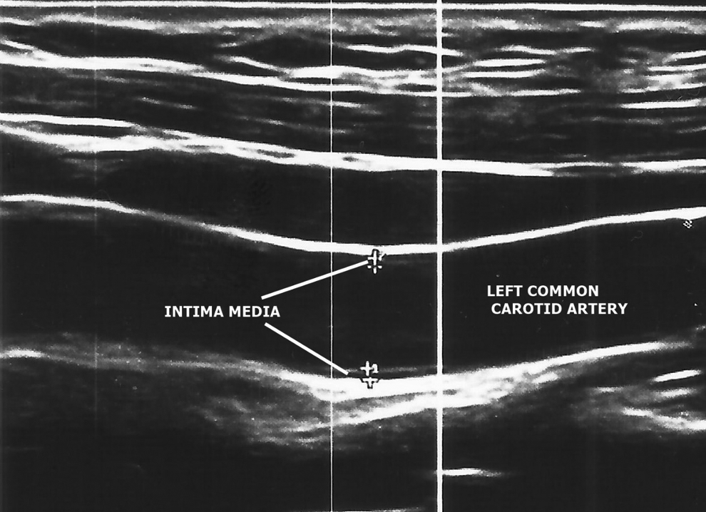
Measurement of the left carotid intima media thickness on B-mode ultrasound. The measurement was 0.87 mm.
Statistical analysis
Statistical analysis was performed by using the NCSS (Number Cruncher Statistical System) 2007 Statistical Software package program. Continuous variables are given as mean ± standard deviation (SD) and categorical variables as numbers and percentages. Paired t-test and chi-square tests were used for comparison of continuous and categorical variables. Analysis of variance and the Newman–Keuls multiple comparison test were used for intergroup analysis. For nonparametric paired variables, the Mann–Whitney U test was used. Paired nominal data were evaluated by using the McNemar's test. All p-values less than 0.05 were accepted as statistically significant.
Results
The study enrolled a total of 68 subjects, of whom 20 dropped out leaving only 48 subjects with complete follow-up records at the time of this writing. Because they did not come to the hospital for control, we had to extract them from the study. There were 25 subjects in the LSG group and 23 in the LGP group. Both groups were similar in terms of demographics (age, gender, and BMI). The LSG group consisted of 7 (28%) males and 18 (72%) females, whereas there were 10 (43.48%) males and 13 (56.52%) females in the LGP group. Mean ± SD age was 42.96 ± 7.87 for the LSP group and 38.3 ± 9.88 for the LGP group. Preoperative BMI was 44.84 ± 3.63 for the LSP group and 45.39 ± 3.69 for the LGP group (Table 1).
BMI, body–mass index; CIMT, carotid intima-media thickness; HDL, high-density lipoprotein; LDL, low-density lipoprotein; LGP, laparoscopic gastric plication; LSG, laparoscopic sleeve gastrectomy.
Mean baseline CIMT was comparable in both groups, 0.62 ± 0.07 (LGP group) and 0.69 ± 0.09 (LSG group) (p = 0.099). Hepatic steatosis grading was also similar in both groups (p = 0.68). Biochemical parameters (HDL, LDL, total cholesterol, and triglyceride) were similar in both groups.
In the third month after surgery, a statistically significant decrease in CIMT was observed in both LSG and LGP groups (p = 0.0001). Paired analysis did not reveal any statistical significance (p > 0.05). When postoperative 6-month CIMT values were compared with baseline or postoperative 3-month CIMT values, a significant decrease was noted in Table 2 (p = 0.0001). Intergroup analysis that was performed by using the Newman–Keuls multiple comparison tests did not reveal any statistically significant correlation between BMI and CIMT.
%EWL, excessive weight loss.
Mean preoperative BMI, which was 44.84 ± 3.63 in the LSG group, decreased to 33.35 ± 3.83 at 6 months after surgery (p = 0.0001). This significant decrease in mean BMI was similar in subjects who had LGP: preoperative 45.39 ± 3.69 and 6 months postoperative 34.37 ± 2.81 (p = 0.0001). Intergroup comparative analysis of preoperative and 6-month postoperative BMI values did not show any statistical significance (p = 0.972).
The calculated %EWL was 38.03 ± 14.48 kg at 3 months after surgery and 59.43 ± 17.14 6 months after surgery for the LSG group and 38.76 ± 10.78 at 3 months and 54.33 ± 10.37 at 6 months for the LGP group.
A statistically significant decrease in hepatic steatosis grade was observed among subjects in the LSG group at both 3 and 6 months postoperatively (p = 0.001). For the LGP group, hepatic steatosis grade did not decrease significantly (p = 0.17) at 3 months postoperatively but at 6 months (p = 0.001) postoperatively. When both groups were compared, patients from the LSG group were found to have lower grades (p = 0.034) at 3 months postoperatively. No differences were found at 6 months postoperatively (Table 3).
The serum lipid profile showed a statistically significant increase in HDL levels in both groups, LSG group (p = 0.002); LGP group (p = 0.0001). LDL levels did not decrease significantly in both groups (p = 0.604 for LSG group and p = 0.484 for LGP group). Serum total cholesterol levels decreased significantly (p = 0.014) at 3 and 6 months postoperatively when compared to baseline levels in the LSG group. In this group, the difference was more evident when the baseline serum total cholesterol levels were compared to the 6-month postoperative findings (p = 0.006). For the LGP group, the changes in serum total cholesterol levels at 3 and 6 months postoperatively were not significant (p = 0.778). Serum triglyceride levels decreased significantly at 3 and 6 months postoperatively in the LSG group (p = 0.002). Similarly, a significant decrease was observed in the LGP group at the same time points (p = 0.001). When both groups were compared, the lipid profiles were similar at the described time points (p = 0.258 and p = 0.431) (Table 4).
Discussion
In this study, the effect of two different bariatric surgery procedures (LSG and LGP) on CIMT was investigated. Pignoli et al. were the first to measure CIMT in 1986 and this was embraced by numerous investigators who performed extensive studies on the utility of this CIMT. Today, CIMT is widely used and regarded as a reliable marker for arteriosclerosis. 13 Lorenz et al. in their 2007 systematic review of 8 studies, which included 37,000 patients, showed that a 0.1 mm increase in CIMT increases the risk of heart attack (myocardial infarction [MI]) by 10–15% and stroke by 10–13% in patients with cardiovascular disease. 14 This review substantiated the utility of CIMT as a marker for the assessment of MI and stroke risk. There are multiple factors that can affect CIMT values. Among these are age, gender, race, lipid profile, and obesity. A study by Besir et al. 15 on 2,298 Turkish patients reported age as the only independent predictor of CIMT. When the findings from a study by Besir et al. were applied to our study, subjects from the LSG group should have had a CIMT value of 0.49 instead of our reported higher mean CIMT of 0.68 and 0.47 for the LGP group in contrast to the reported mean CIMT of 0.61. The ethnic similarities observed in both studies show that obesity may contribute to higher CIMT values. For both adult and pediatric populations, a positive correlation between BMI and CIMT has been previously reported.16–18 Kotsis et al. 19 studied 3,173 obese subjects and found a positive correlation between both demographic (BMI, male gender) and metabolic (serum cholesterol and blood glucose levels) parameters. In agreement with these studies, findings from our study were consistent with a positive correlation between BMI and CIMT values at 3 and 6 months after surgery.
In obese subjects, other factors that contribute to the increased CIMT values are serum lipid profile and glucose levels. A positive correlation between BMI and lipid profile parameters (LDL, triglyceride, and total cholesterol levels) has been reported previously. 20 In our study, HDL levels increased significantly when compared to baseline levels irrespective of the bariatric surgery procedure performed (p = 0.002 for LSG group and p = 0.0001 for LGP group). Although LDL is the most important part of the lipid profile for atherosclerosis and decrease of LDL levels was expected in both groups, this was not observed in our study. This may be due to the eating habits of our obese population. Total cholesterol levels decreased significantly in the LSG group (p = 0.014), however, the change was not significant in the LGP group (p = 0.778). For serum triglyceride levels, the decrease was significant in both groups (p = 0.002 for LSG group and p = 0.001 for LGP group). Liver steatosis has been included in this study for the correlation of change of steatosis with decrease of blood cholesterols and weight loss. The results showed that in addition to the decrease in lipid profiles and CIMT values, a decrease in the severity of hepatic steatosis was observed in the patients who underwent LSG. Follow-up ultrasound assessment confirmed a lesser degree of hepatic steatosis compared to preoperative results. This finding is similar to what has been reported in the literature previously.21,22
To the best of our knowledge, this study is the first study to objectively compare two different bariatric procedures and investigate their effect on CIMT by including parameters (age, lipid profile, and BMI) that are known to affect CIMT. Study limitations include a smaller sample size and consideration of only two bariatric procedures. Also, the short follow-up period did not allow us to fully evaluate the long-term effects of both procedures on CIMT and its associated parameters.
Conclusion
In conclusion, we were able to show that CIMT, a reliable marker of arteriosclerotic disease, which can be measured noninvasively, decreases after bariatric surgery. In addition to the well-known merits of bariatric surgery, subjects undergoing bariatric surgery may benefit from an improvement in lipid profiles and a decrease in the risk of cardiovascular events in the future.
Footnotes
Author Disclosure Statement
No competing financial interests exist.