
Abstract
Abstract
Ectopic breast tissue is defined as glands of breast tissue located outside of the normal anatomic breasts. Historically, ectopic breast tissue has been thought to arise from a remnant of the embryonic mammary ridge along the “milk line” or the midaxillary line from the axilla to the groin, including the vulvar region. Extramammary tissue displays the same pathologic and physiologic changes as normal breast tissue and is often discovered in multiparous women as the result of swelling from lactational activity. We present a case report of a gravid patient with lactating vulvar mass and a brief historical perspective of vulvar ectopic breast tissue.
Introduction
Extramammary tissue displays the same pathologic and physiologic changes as normal breast tissue and is often discovered in multiparous women as the result of swelling from lactational activity. 2 We present a case report of a lactating vulvar mass in a patient with a significant history of familial breast cancer and a brief historical perspective of ectopic vulvar tissue.
Case Report
A 31-year-old African American female gravida 5 para 2022 at 35 6 weeks dated by her last menstrual period and an 11-week ultrasound presented to the labor and delivery unit with complaints of “vaginal cyst pain” that had been gradually increasing over the past few weeks. The patient had a history of multiple sclerosis, gestational diabetes, anemia, migraine headaches, obesity with a body mass index of 37 kg/m2, and a significant family history of breast cancer. On physical examination, the patient appeared well developed and well nourished. Her skin was unremarkable, and her breasts were normal bilaterally without masses or lesions. A nonpigmented, 2–3-cm mobile, soft tissue mass was noted on the inferior portion of the right labia majora. Batholin's and Skene's glands appeared to be within normal limits, and the introitus was otherwise unremarkable.
The decision was made to perform a bedside excisional biopsy of the vulvar mass using local analgesia. The procedure was performed without complications. The lesion removed was a solid, fatty-appearing, encapsulated nodule measuring 2.6×1.9×1.3 cm. Examination of the gross tumor specimen revealed a polypoid fragment of tan-pink wrinkled tissue. Microscopic evaluation revealed a tumor composed of benign ectopic breast tissue with lactational changes and no evidence of dysplasia or malignancy.
Discussion
Ectopic breast tissue is hormonally responsive and often shows pathologic changes. In fact, one of the most referenced cases of accessory breast tissue is from 1827 and involves Therese Ventre of Marseilles, France, of whom the story is told that she had an accessory breast on the lateral thigh that produced milk when she became pregnant (Fig. 1). 3 Additional rare cases of breastfeeding or pumping ectopic breast tissue have been reported.4,5
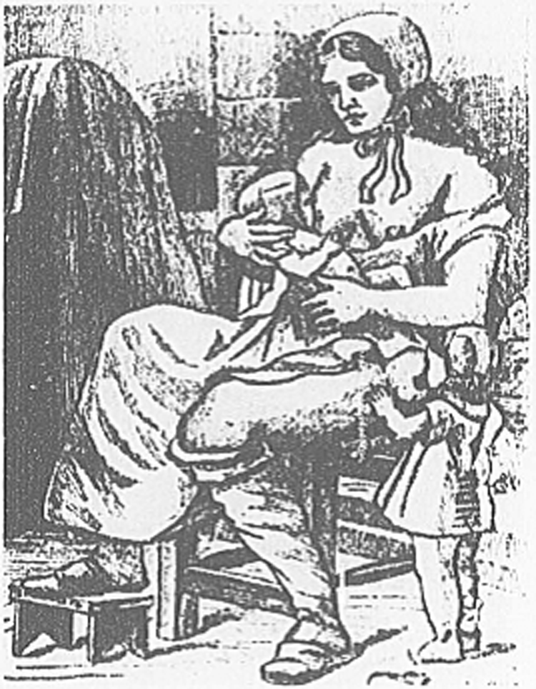
Therese Ventre of Marseilles, France, representing lactating accessory breasts. 3
Historically, multiple breasts were associated with fertility. Goddesses of fertility such as the Phoenician goddess Astarte, the Incan goddess Mama Allpa, and Artemus of Ephesus have been depicted with polymastia or supernumerary breast tissue. 6 A recent artistic representation of Cybele, an ancient Phrygian goddess of fertility, by Mihail Chemiakin not only shows polymastia, but also supernumerary breasts along the milk line or mammary ridge as well (Fig. 2).7,8
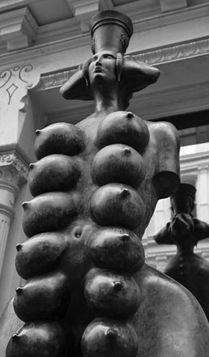
Sculpture of Cybele, the Goddess of Fertility, by Mihail Chemiakin. Photograph by Joe Goldberg. 7
Traditionally, it was presumed that breast tissue developed during the embryonic period during the fourth and fifth week of gestation. Breasts, or mammary glands, are believed to be modified sweat glands that develop as a thickening of the ectoderm called the mammary ridge or milk line (Fig. 3). Multiple glands develop along ridges that extend bilaterally from the midaxillae to the inguinal region. Normally, only one pair of mammary glands persists in the pectoral region to become the normal anatomic breasts. Ectopic breast tissue is thought to be a result of an embryonic abnormality due to a remnant of embryonic mammary tissue and can occur anywhere along the milk line.

Mammary milk lines. 8
A recent theory has also been suggested to describe the specific etiology of vulvar ectopic breast tissue. This theory suggests that mammary-like glands exist in the anogenital region that are distinct and separate from remnants of the mammary ridge. 9 Supporters of this view suggest that there is no convincing evidence for extension of mammary tissue beyond the pectoral region, but that this tissue is still estrogen and progesterone receptor positive, and therefore lesions of these glands appear very similar to the mammary glands. Supporters of this view suggest that breast pathologists specialize in these lesions.
Ectopic breast tissue in the vulva was first reported in 1875
8
:
The patient, a woman, age 30, who was suckling her child, had a pedunculated tumor, the size of a large goose's egg, attached to the lower and inner part of the left labium majora. It was covered over with skin and its pedicle was the size of a man's thumb. In front, at its upper part, there was an eroded ovoid patch from which milk-like fluid escaped.
After careful examination after removal, the mass was considered to be consistent with a “true supernumerary mamma of the vulva.” 8
Then in 1905, a case of bilateral vulvar nipples was reported 10 and in 1923 a case of lactational vulvar tissue was reported. 11 In 1917 a review of 11,000 cases of aberrant mammary tissue revealed only two cases of vulvar involvement. 12 In 1954, two more cases of ectopic breast fibroadenoma of the vulva were reported, 2 and by 1967, over 20 cases of ectopic vulvar breast tissue had been reported. 13 According to a recent review of the literature, in total, about 50 cases of benign breast tissue in the vulva have now been reported, with over 20 cases being malignant lesions. 2
Mammary tissue in the vulva also displays all of the characteristics of normal breast tissue including lactation, growth during puberty, fibrocystic disease, growth of fibroadenomas, phyllodes tumors, milk cysts, extramammary Paget's disease, and any of the various histological malignancies. 2 Lactating breast tissue in the vulva generally develops in later gestations and may persists after the period of lactation. Since 1926, when the first case of vulvar lactating breast tissue was reported, a few additional cases have been reported.2,14–18 Of the reported vulvar ectopic breast tissue, nearly one-third of these cases have had a primary cancer.19,20
Conclusions
When clinicians are evaluating a patient during the peripartum period and are confronted with a vulvar mass, or any abnormal mass along the mammary ridge line, it is imperative to consider ectopic breast lesions in the differential diagnosis. Like the normal anatomic breast, ectopic breast tissue is hormonally sensitive and can hypertrophy during pregnancy or lactation or with exogenous hormone usage. Because ectopic breast tissue in the vulva can undergo malignant transformation, excision of this tissue is generally recommended. As the majority of information addressing ectopic breast tissue of vulva comes primarily from case reports and historical perspectives, additional research is needed to further define the most appropriate clinical evaluation and to more accurately define the embryonic origins of mammary tissue.
Footnotes
Disclosure Statement
No competing financial interests exist.