
Abstract
Abstract
Background:
Breast anthropometric morphology affects various factors with diverse physiognomy, making accurate measurements very difficult. The aim of this study was to measure the female breast using anthropometry and to use this method on normal subjects to examine breast asymmetry and consider the influence of age, height, weight, body mass index (BMI), parity, delivery mode, and breastfeeding in premenopausal Korean women.
Subjects and Methods:
In total, 17 parameters of breast were measured with participants in a standing position. Breast volume was also assessed.
Results:
The mean values of the right and left breast volumes were calculated as 386.0±342.5 mL and 393.3±347.2 mL, respectively. With aging, the height of women decreased, but the weight, BMI, upper chest, middle chest, lower chest, waist, and hip widths, nipple–nipple length, and ptosis increased with statistical significance. No asymmetric differences were observed between each breast, except for nipple–inframammary fold length in 20–30-year-old women and upper arm length in 41–50-year-old women. In our study, the breast volume increased with age as a result of weight gain, but the delivery mode and breastfeeding did not affect anthropometric breast measurements.
Conclusions:
In conclusion, age, weight, and BMI are important factors in determining breast anthropometry in our study. The results of the present study will help in the comparison of the anthropometric breast values of Korean women with those of women in other countries and may also be useful in the understanding of breast physiologic change-related obstetrical factors and epidemiologic factors.
Introduction
O
Breasts are secondary sexual characteristics of the female gender and have many anatomic variations with respect to volume, width, length, projection, density, composition, shape, and placement on the chest wall. In addition, changes in the shape of the body and the extremities can affect the shape or location of the breasts. Breast shape is affected by physiological changes associated with puberty, ovulation, gestation, lactation, and aging. The shape of the breast in an adult woman is conical for those who are nulliparous and may become ptotic after menopause. The size, stiffness, and nodularity of the breast in an adult woman may change with weight, menstrual period, gestation, and lactation. 1
The goal of the present study was to measure the descriptive indices of the breast, to determine the average values of breast parameters and the affecting variable factors, and to calculate the volume of the breasts in 250 premenopausal Korean women between 20 and 50 years of age. An additional goal was to obtain data on breast ptosis, asymmetry rates, and related epidemiologic and obstetrical factors. The values obtained for the breasts of premenopausal women in this study may be used as ideal augmentation and reduction values in aesthetic breast surgery and are important for comparison with the anthropometric parameters obtained from different races.
Subjects and Methods
Two hundred fifty premenopausal Korean female volunteers 20–50 years of age were included in this study, which was conducted in May 2011–June 2012. The present study was reviewed and approved by the Institutional Review Board of the Catholic Medical Center (protocol number HC11OISI0024). All participants signed an informed consent form.
All subjects with a history of breast surgery or disease, chest deformity, or any health problems were excluded. The effects of age, weight, height, and body mass index (BMI) were assessed using partial correlation coefficients to eliminate the effects of the other potentially confounding variables, including breastfeeding and parity.
The measurements were obtained with the participants in the anatomic position during the follicular phase of the menstrual cycle: body erect, arms at the sides and palms forward. 2
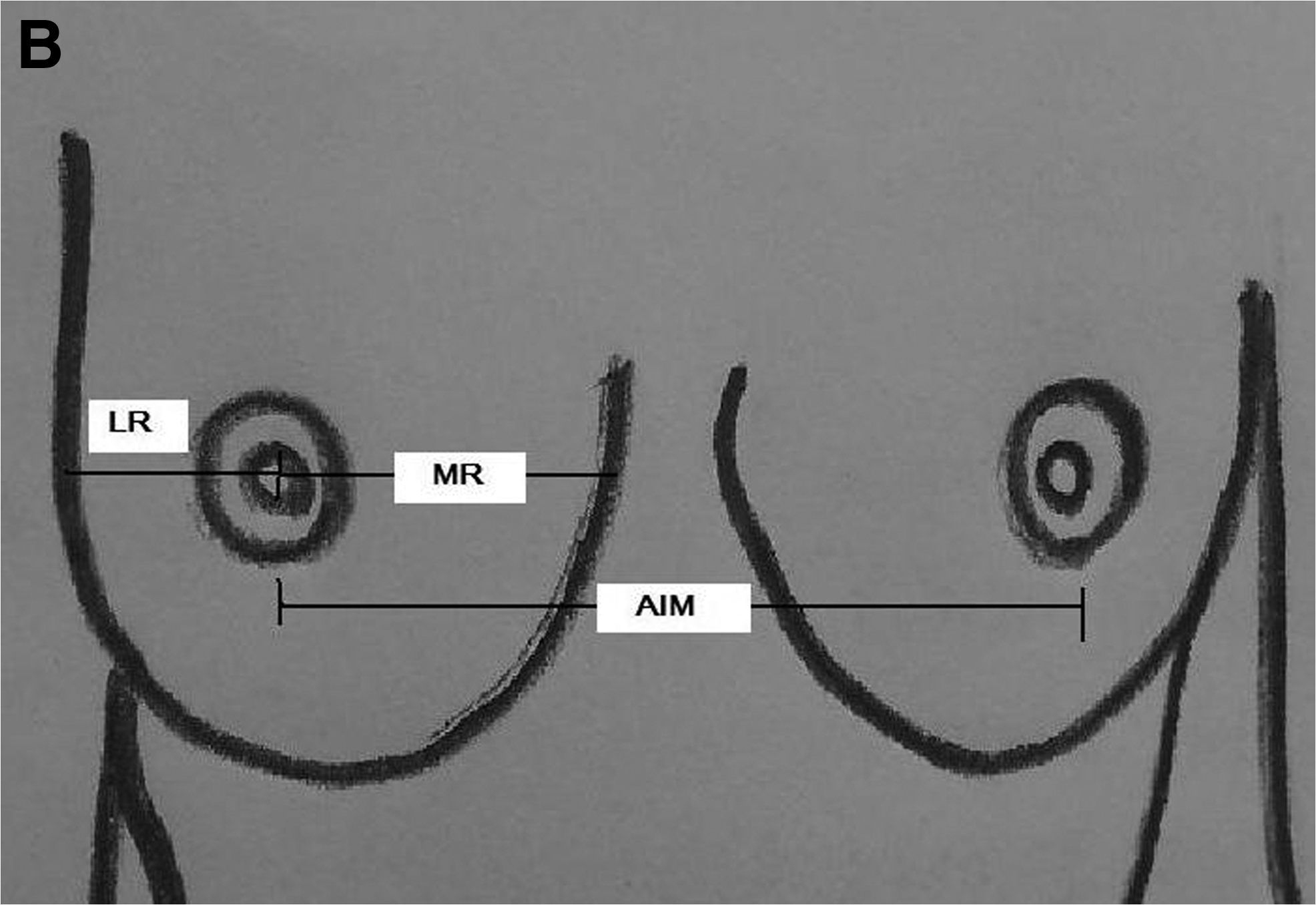
Our team (M.J.K. and S.J.K.) measured the following 17 parameters: body weight, height, upper chest width (CC1), middle chest width (CC2), lower chest width (CC3), shoulder width (SC), waist width (LC), hip width (BC), clavicle–nipple length (CNL), sternal notch–nipple length (SNL), nipple–nipple length (AIM), upper arm length (HL), medial mammary radius (MR), lateral mammary radius (LR), nipple–inframammary fold length (IR), mammary projection (MP), and stage of breast ptosis (Fig. 1).

According to the anatomic method, breast volume (mammary volume [MV]) was calculated, according to the following formula defined by Qiao et al. 3 and inserting the measured MR, LR, and IR values for each participant: MV=1/3×3.14×MP 2 ×(MR+LR+IR−MP).
The breast ptosis was evaluated according to the classification of Kirwan 4 : Stage A, nipple position 2 cm above the inframammary crease (IMC); Stage B, nipple position 1 cm above the IMC; Stage C, nipple position even with IMC; Stage D, nipple position 1 cm below the IMC; Stage E, nipple position 2 cm below the IMC; and Stage F, nipple greater than 2 cm below the IMC.
The measurements were expressed as mean±standard deviation values. Normal distribution was tested with the one-sample Kolmogorov–Smirnov test, and the differences among age groups were tested with the analysis of variance test, followed by the post hoc Scheffé's test. Fisher's exact test was used to compare the breast ptosis, according to age, and Student's t test was applied to compare the right and left breasts and to study the breast anthropometric measurements according to breastfeeding, as the variables were not normally distributed. The association between variables was assessed using a Spearman correlation analysis. The level of statistical significance was determined as p<0.05.
Results
The study was performed on 250 Korean female volunteers; 71 volunteers were 20–30 years old, 65 volunteers were 31–40 years old, and 114 volunteers were 41–50 years old. Eighty-seven volunteers were nulliparous (included all of 71 women in the 20–30-year-old group), and the remaining 163 subjects had between one to five children. Of these 163 subjects, 133 had breastfed their children. In the 163 women in the delivery history group, 116 (71.2%) had undergone normal spontaneous delivery, and 47 (28.8%) had undergone cesarean section. All 250 women were in a premenopausal state, and the mean age was 37.1±8.7 years, the mean height was 159.9±4.9 cm, the mean weight was 56.3±7.4 kg, the mean BMI was 22.0±2.6 kg/m2, and the mean breast volume was 386.0±342.5 mL for the right breast and 393.3±347.2 mL for the left breast.
The mean values of the body and breast measurements are presented in Table 1 according to age. The weight, BMI, CC1, CC2, CC3, LC, BC, SC, and AIM all statistically increased with age. Regarding age, the AIM was 18.5±1.7 cm in 20–30-year-old women and 19.4±1.7 cm in 41–50-year-old women. This indicates that the breasts become more laterally positioned with increasing age.
The p value for the difference between 20–30 years old versus 31–40 years old versus 41–50 years old was calculated by the analysis of variance test, followed by the post hoc Scheffé test.
20–30 years old versus 31–40 years old, b20–30 years old versus 41–50 years old, c31–40 years old versus 41–50 years old.
AIM, nipple–nipple length; BMI, body mass index; CC1, upper chest width; CC2, middle chest width; CC3, lower chest width; LC, waist width; BC, hip width; SC, shoulder width.
Ptosis is defined as the case where the nipple position is 1 cm below the IMC (Stages D–F) in our study. Of the 250 participants, ptosis was observed in 11.3% of the 20–30-year-old group, 26.2% of the 31–40-year-old group, and 49.1% of the 41–50-year-old group (p<0.0001) (Table 2). Of the 250 participants, ptosis was observed in 12.6% of the nulliparous group and in 42.9% of the parous group (p<0.0001). Of the 163 parous women, ptosis was observed in 43.6% of the breastfeeding group and 40% of the non-breastfeeding group (p=0.083).
The p value of the difference between 20–30 years old versus 31–40 years old versus 41–50 years old was calculated by Fisher's exact test.
By the classification of Kirwan 4 : Stage A, nipple position 2 cm above the inframammary crease (IMC); Stage B, nipple position 1 cm above the IMC; Stage C, nipple position even with IMC; Stage D, nipple position 1 cm below the IMC; Stage E, nipple position 2 cm below the IMC; and Stage F, nipple greater than 2 cm below the IMC.
Comparing the right and left breast differences according to age, there was no significant difference between the CNL, SNL, MR, LR, MP, and MV values of the right and left breasts (p>0.05). The left IR (6.2±1.3 cm) was significantly greater than that of the right IR (5.9±1.3 cm) (p=0.047) in the 20–30-year-old group, and the right HL (28.8±1.6 cm) was significantly greater than that of the left HL (28.3±1.5 cm) (p=0.019) in the 41–50-year-old group (Table 3).
The p value for the difference between right and left breasts calculated by Student's t test.
The p value for the difference between 20–30 years old versus 31–40 years old versus 41–50 years old for the right side calculated by analysis of variance, followed by post hoc Scheffé test.
The p value for the difference between 20–30 years old versus 31–40 years old versus 41–50 years old for the left side calculated by analysis of variance, followed by post hoc Scheffé test.
Statistically significant difference (p<0.05).
20–30 years old versus 31–40 years old, b20–30 years old versus 41–50 years old, c31–40 years old versus 41–50 years old.
CNL, clavicle–nipple length; HL, upper arm length; IR, nipple–inframammary fold length; LR, lateral mammary radius; MP, mammary projection; MR, medial mammary radius; MV, mammary volume; SNL, sternal notch–nipple length.
The volume of each breast was calculated (Table 3). In the 250 women, the left breast volume was greater than the right breast volume on average without statistical significance, and there were significant differences between the groups according to age (296.3±258.6 mL for the right breast and 303.3±270.6 mL for the left breast in the 20–30-year-old group and 493.6±391.3 mL for the right breast and 500.7±400.4 mL for the left breast in the 41–50-year-old group; p<0.0001).
A positive correlation was found between the mean breast volume and the age and weight. Adjusted for weight, the correlations of age and both breast volumes were 24.2% in the right breast volume and 18.8% in the left breast volume with statistical significance in a regression analysis (p<0.0001) (Table 4).
r2 means that predictor factors from X to Y.
Adjusted for weight.
For multivariable analysis, R2=0.242, p<0.0001.
For multivariable analysis, R2=0.188, p<0.0001.
MV, mammary volume.
One hundred thirty-three (81.6%) women had breastfed in the group of 163 parous women of the total of 250 volunteers; no significant difference was observed between the MR, LR, MP, and MV values, according to breastfeeding (Table 5). A comparison of the mean values of the right and left breast measurements is given in Table 5. No statistical significance was observed in ptosis (Stages D–F), according to breastfeeding in parous women (p=0.083).
Data are mean±standard deviation values. The p values were calculated by Student's t test.
AIM, nipple–nipple length; BMI, body mass index; IR, nipple–inframammary fold length; MR, medial mammary radius; LR, lateral mammary radius; Lt, left; MP, mammary projection; MV, mammary volume; Rt, right.
Discussion
A wide variation has been observed in the color, shape, size, and projection of the breast morphology, as well as in the nipple–areola complex, due to race, aging process, hormonal changes (pregnancy and menopause), and variation of weight.3,5–7 The size and shape of the breast also vary depending on the fat tissue content; obesity is therefore an important factor. 8
An understanding of the morphology and physiology of the breast and the many endocrine interrelationships is essential to the study of the pathophysiology of the breast and in the management of breast disorders.
Although anthropometric measurements of the breasts are important, breast volume measurements have not been carried out on a routine basis because there is still no commonly accepted standard method. Six main methods of breast volume measurement can be used: (1) Archimedes Principle (displacement of water); (2) anthropometry (anatomic) measurement; (3) imaging (mammography, magnetic resonance imaging, computed tomography, and ultrasound); (4) the Grossman–Roudner device method; (5) casting; and (6) biostereometrics (three-dimensional surface scanning). 9 Breast volume assessment by the Archimedes Principle should not be used despite its acceptability, as patients had difficulty in performing the test adequately. Anatomical measurement was easy to do and well tolerated by the patients; however, selection of the appropriate formula is still a problem, and doing calculations is impractical. The magnetic resonance imaging and biostereometrics methods are ideal in terms of accuracy and reproducibility, but they are complex and too expensive to be performed as a routine investigation. 9 Casting and water displacement methods have not become routine practices owing to low patient comfort, complications in their application, and relatively low levels of accuracy. 9 We carried out breast measurements using a linear method of anthropometric (anatomic) measurement. 3 We suggest that further prospective studies of accurate of breast volume measurement using a standardized method.
Although the height decreased with age, the weight, BMI, shoulder, and the waist and hip circumference all increased with age in the premenopausal Korean women. In particular, the AIM increased with age; this means that there is inferior migration of the nipple, and the vertical components of the breast with increasing age and lateral ptosis are consistent with the effects of gravity on the aging breast with the loss of elasticity. Weight correlates significantly with many of our morphometric measurements, consistent with previous findings. 10
The age and parity factors are significantly correlated with breast ptosis (Stages D–F) (49.1% of the 41–50-year-old group and 42.9% of the parous group, p<0.0001), but the breastfeeding might not affect breast ptosis (43.6% of the breastfeeding group and 40% of the non-breastfeeding group among 163 parous women, p=0.083) in our study. Rinker et al. 11 reported that the risk of breast ptosis increased with each pregnancy, but breastfeeding did not seem to worsen these effects, and these results were consistent with our study.
The amount of fatty and connective tissue of the breast decreases with age, menopausal involution of the breast results in a reduction of both the number of ducts and lobules, and the lobules shrink and collapse, in which then the breasts represents ptosis. 8 Breast ptosis is an important factor in women regarding the aspect of cosmetics; the study of the affecting and relating factors to breast ptosis enables surgeons to plan and manipulate reconstructive or aesthetic breast surgeries more easily.
The mean SNL value was 18.59 cm in the study of Westreich, 5 20.63 cm in the study of Penn, 12 and 19.052±0.096 cm in the study of Qiao et al. 3 In our study, the values for the right and left breasts were 20.0±1.8 cm and 19.5±1.7 cm, respectively, in the 20–30-year-old group.
The mean IR values were 8.3 cm and 6.836±0.086 cm in the studies of Avşar et al. 13 and Qiao et al., 3 respectively, with respective mean breast volumes of 407.2 mL and 325.369±12.66 mL. In our study, the mean IR were 5.9±1.3 cm (right) and 6.2±1.3 cm (left), with mean breast volume of 296.3±258.6 mL (right) and 303.0±270.6 mL (left), in the 20–30-year-old group.
Ramsey et al. 14 chose a volume of 400–700 cm3 to correspond to small breasts, 700–1100 cm3 to correspond to medium-sized breasts, and a volume of >1,000 cm3 to correspond to large breasts. In our study, the brest volume of Korean women corresponded to a small breast, and several differences between races were observed.
Although there was no significant difference between right and left breast volume, the left breast volume was larger than the right breast volume at all ages of our study groups, and these results concur with those of Westreich. 5 Breast cancer occurs more frequently in the left breast, and the left breast is typically slightly larger than the right breast. 13 Our results showed that the IR and breast volume of the left breast were greater than those of the right breast, and the HL of the right arm was greater than that of the left arm, possibly because of the predominance of right-handed people in Korea. Therefore, further studies of the association of being a right-handed person and breast anthropometric measurements are needed. Female breasts are often not completely symmetrical, and breast asymmetry results from many factors that can be roughly classified into congenital and acquired causes.
Vandeput and Nelissen 15 reported that breast volume was independent of weight and chest diameter. However, in the study of Westreich, 5 breast volume was found to increase by 20 mL for each kilogram of weight gain above the ideal weight. Our study, likewise, found a positive correlation between breast volume and both age and weight (p<0.001). Considering that while age increases, weight and BMI also increase in premenopausal Korean women, in our study weight is therefore the most important factor in affecting breast volume. In general, the breast reduces in volume after menopause. However, menopausal women usually have more fat than premenopausal women, and older women are also typically heavier than younger women. 16 Therefore, with age, breast ptosis is more common, and the breast volume is larger in premenopausal women, and the various factors, including age, hormones, weight, and BMI, are related to the breast anthropometric changes.
Few studies related to breastfeeding and breast anthropometric measurements have been conducted until now. No relationship was observed between breastfeeding and breast anthropometric measurements in parous women in our study, but the number of women who had not breastfed was limited in our study (n=30), and all of 20–30-year-old women were nulliparous. Therefore, we need further studies of the affecting factors of delivery, parity, and breastfeeding with anthropometric measurements.
Conclusions
These results of such anthropometric studies will help to suggest standard data of Korean women and will help to understand the relationship among the multiple factors of age, weight, obstetrical factors, and breast change.
Footnotes
Acknowledgments
The statistical consultation was supported by Catholic Research Coordinating Center of the Korean Health 21 R & D Project (A070001), Ministry of Health &Welfare, Republic of Korea.
Disclosure Statement
No competing financial interests exist.