
Abstract

I
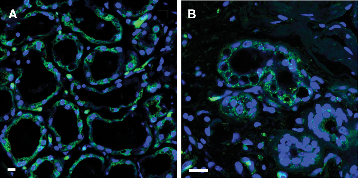
Mammary epithelial cells in the human lactating breast expressing markers known also for mesenchymal cells. CD44-green
Although the exact nature, origin, and properties of BSCs are still not fully established, state-of-art investigations using a plethora of techniques combining ex vivo breastmilk cell molecular and gene expression analyses, single cell analyses, and microscopic examinations coupled with immunohistochemical staining of resting and lactating mammary tissues as well as in vitro and in vivo studies, have revealed that the cellular composition of breastmilk is dynamic and heterogeneous.1,2 It largely reflects the status and cellular heterogeneity of the lactating mammary epithelium when both the mother and infant are healthy during established lactation, with the majority of cells being of epithelial origin among the less than 2.5% of immune cells that protect the mammary gland and infant from infection, which originate from the maternal circulation.1,2 Breastmilk epithelial cells constitute a cellular hierarchy comprising the mature milk-synthesizing lactocytes, few myoepithelial cells, progenitor cells with limited expansion and differentiation potential, and BSCs with multilineage potential, being capable of expanding in culture and differentiating into multiple cell lineages.1,2 Evidence has supported the origin of the majority of BSCs from the lactating epithelium, whereas rarer cells expressing markers of the hematopoietic stem cell lineage have also been found in breastmilk samples, potentially originating from the maternal circulation.1,2 These cells, however, are not MSCs and their origin and properties remain to be established.
Therefore, no convincing evidence currently exists supporting the presence of MSCs in breastmilk. Rather, it has been shown that breastmilk and the lactating breast contain different populations of epithelial cells, some of which express markers also known to be expressed by MSCs, such as CD44, CD105, vimentin, and others (Fig. 1). 1 In the lactating epithelium, these markers may have a functional significance. It has been hypothesized that they may represent cells undergoing epithelial-to-mesenchymal transition (EMT), which have been shown to coexpress mesenchymal and epithelial markers. 1 Importantly, the demonstration that epithelial cells in the lactating breast express these markers provided strong evidence that they cannot be mesenchymal (Fig. 1). 1 In the context of the normal mammary gland, EMT has been reported in both the human and mouse, and has been hypothesized to facilitate the normal expansion of the gland and interaction between the lactating epithelium and stroma. 1
Taken together, current evidence of the heterogeneity of breastmilk cell populations does not support the presence of MSCs in breastmilk. Further analyses are required to decipher the role and properties of EMT and the different cell types in breastmilk and the lactating mammary gland.
Footnotes
Disclosure Statement
No competing financial interests exist.