
Abstract
Abstract
Aim:
The aims of this study were to investigate the effects of calcium at the same concentration as that found in human milk on the viability, proliferation, and adhesion of MCF-7 human breast ductal carcinoma cells by exposing them to calcium at the same frequency as in breastfeeding.
Materials and Methods:
High-concentration calcium was applied for 30 minutes every 4 hours for 24, 48, and 72 hours. Cell proliferation and viability were measured using a hemocytometer and the MTT cell viability assay. The effects of calcium treatment were evaluated by a comparison among a multiple-, single-dose calcium treatment, and a control group.
Results:
We show that calcium at the same concentration as that in milk caused a decrease in the number of cells but did not affect cell viability.
Conclusions:
The results of this study suggest that calcium caused a lowering of the number of cells from the luminal surface of the breast by triggering proliferation under the condition of fluidity. Calcium and fluidity together serve to eliminate breast cancer stem cells during the lactation period. Effects of the other components of milk can be analyzed by the new method developed in this study.
Introduction
B
Invasive ductal carcinoma of the breast originates from the luminal epithelium. Cancer cells may contain estrogen receptors and exhibit rapid proliferation. 7 Rapidly proliferating cells detach for mitosis and reattach to nearby cells after mitosis. The membranes and the channels of the cell work actively to divide. 8 Therefore, anticancer drugs are more toxic to rapidly proliferating cells. 9
During the lactation period, mothers breastfeed their babies about six times a day. Milk remains inside the breast ducts during and after breastfeeding and is present until it dries. 10 Therefore, channel epithelial cells are exposed to milk daily for about 30 minutes every 4 hours. Moreover, it is believed that cancer cells with rapid proliferative capacity cannot continue to exist in the breast tissue during the lactation period due to the repeated exposure to high calcium concentrations and milk fluidity.
The fluidity event serves as a natural barrier in the human body. Many diseases, ranging from infections to cancers, occur when fluidity is reduced in the digestive and urinary systems. In this context, it is known that constipation leads to an increase in the incidence of colon cancer.11–13
In human breast milk, calcium is found at a concentration of 30 mg/100 mL (7.5 mM). The levels of electrolytes in milk, such as calcium, remain at a constant concentration from the early to late stages of breastfeeding. 5 Calcium, a vital electrolyte, is required for survival, and plays an important role in the cell death mechanisms of apoptosis and necrosis. In Jaffe's study, it was shown that cells exposed to high levels of calcium with great frequency were characterized by broken gap junctions; the cells could gain oncogenic potential through proto-oncogene activation. 8
We hypothesized that weakened gap junctions in conjunction with fluidity cause an increased capacity for proliferation and the elimination of breast cancer stem cells through the calcium in milk.6,14 Therefore, in this study, we aimed to investigate the effects of a medium with a similar calcium concentration as that found in human milk with similar exposure frequency as that occurring during breastfeeding on viability, proliferation, and adhesion on the MCF-7 human breast ductal carcinoma cell line under culture conditions.
Materials and Methods
Chemicals and reagents
Calcium and other chemicals used for cell culture, including the medium, serum, trypsin–ethylene diamine tetraacetic acid (EDTA), and antibiotic mixture, were purchased from Sigma Chemical Company (St. Louis, MO). 3-[4.5-Dimethylthiazol-2-yl] −2.5-diphenyl-tetrazolium bromide (MTT) was purchased from Roche Applied Science (Indianapolis, IN).
Cell culture
In this study, the human breast ductal carcinoma MCF-7 cell line was used. The cells were grown in high-glucose Dulbecco's modified Eagle medium (DMEM) supplemented with 10% fetal calf serum (FCS) and 1% antibiotics (penicillin–streptomycin–amphotericin B mixture) in a 5% CO2 humidified atmosphere at 37°C. When the cells became 70% confluent, they were passaged using 0.25% trypsin–EDTA solution. Proliferated cells were subcultured twice a week for 3 weeks.
Design of a cell culture breastfeeding model
The study comprised three groups. The first group was the control, in which the cells were incubated in normal medium supplemented with FCS and antibiotics for 30 minutes once every 4 hours, as shown in Figure 1. Calcium was applied once in the second group at the beginning of the experiment. In the third group, calcium was applied for 30 minutes every 4 hours (Fig. 1). Calcium was used at a concentration of 0.3 mg/mL (7.5 mM) in DMEM in the second and the third groups. This concentration is similar to the calcium concentration found in human milk. After 30 minutes treatment with high-concentration calcium-containing medium in the third group, the medium was carefully replaced with normal medium containing FCS and antibiotics. This procedure was performed every 4 hours in the third group. This experiment continued for 24, 48, and 72 hours, and was replicated three times.
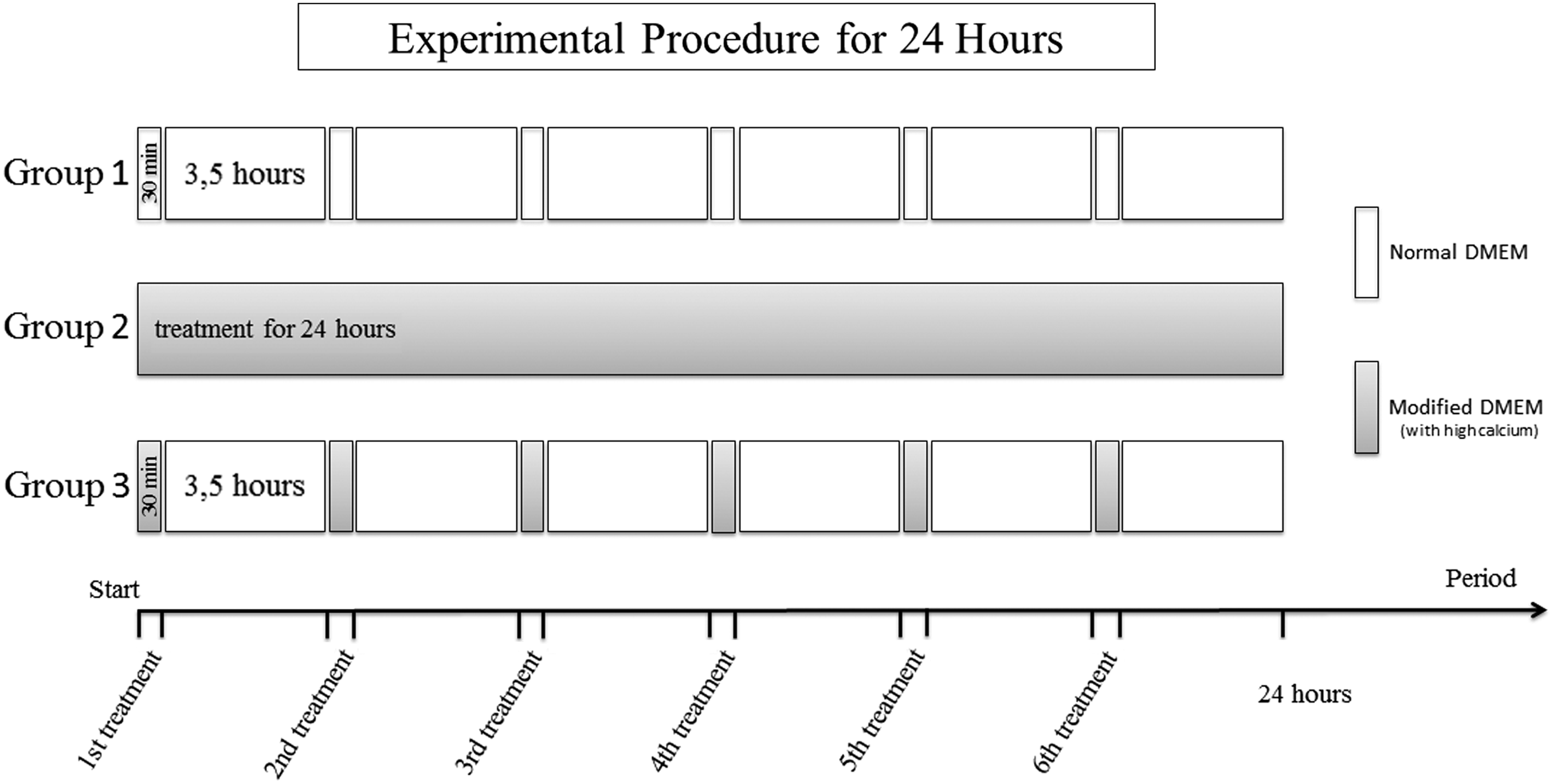
Experimental schema of calcium application. This procedure was continued for up to 3 days. Normal medium was applied in the control group for 30 minutes every 4 hours throughout the experiment. Medium containing high-concentration calcium was used for a single application in the calcium group at the beginning of the experiment. Medium containing high-concentration calcium was also administered in the repeated calcium application group for 30 minutes every 4 hours throughout the experiment. DMEM, Dulbecco's modified Eagle medium.
Determination of the cell proliferation rate using a Neubauer hemocytometer
Semiconfluent cells were harvested using trypsin–EDTA, and the number of cells was determined using a Neubauer hemocytometer slide. Cells were seeded at 20 × 104 per well in a 6-well plate. After 1 day, cell adhesion was determined by visual inspection, and our breastfeeding model experimental procedure commenced. After 24, 48, or 72 hours, the cells were collected, and the number of cells in each group was determined using a Neubauer slide. The number of cells in the groups were compared with the control group, and percentages of groups were calculated by comparison with the control which was accepted as 100%. Percentage of group = (average cell count of group/average cell count of the control) × 100.
Measurement of cell viability by MTT assay
MCF-7 cell viability was determined using the MTT assay. MTT is a water soluble tetrazolium salt, which is converted to insoluble purple formazan by cleavage of the tetrazolium ring by succinate dehydrogenase within the mitochondria. The formazan product is impermeable to the cell membrane and therefore accumulates in healthy cells. The MTT assay has been tested for its validity in various cell lines. 15 The colorimetric MTT test is useful to quantify the activation level of cells, independent of proliferation. 16
After the cells were collected from the 6-well plate at the end of the experiment, 100 μL of the cell suspension from each group was seeded into a 96-well plate. After seeding, 10 μL of the MTT solution was added to each well. The cells were then incubated under humidified air with 5% CO2 at 37°C. At the end of incubation, 100 μL of dimethyl sulfoxide was added to each well containing the formazan solution and allowed to incubate overnight. Formazan absorbance was measured at 570 nm using a microplate enzyme-linked immunosorbent assay reader (Multiskan FC, Thermo Fisher Scientific, Finland). The absorbance of groups was shown in Figure 2. The percentage of viable cells was calculated in comparison with that of control, and is shown in Figure 3. Control was considered 100%.
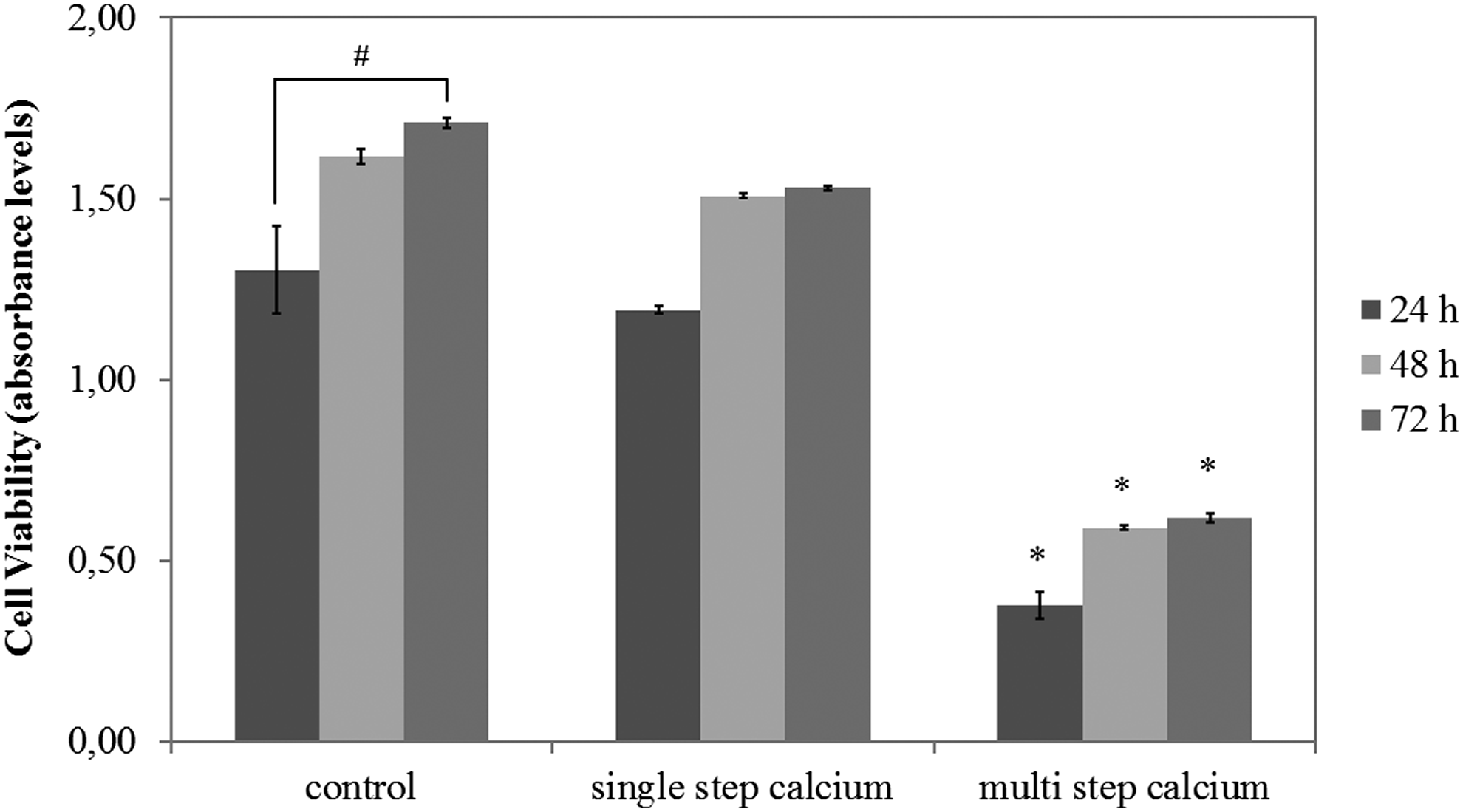
MCF-7 cell viability after 24, 48, and 72 hours of culture determined using the MTT assay. Formazan was decreased in the repeated calcium application group but not in the single calcium application group, *p < 0.05. In the control group, formazan increased significantly, #p < 0.05.
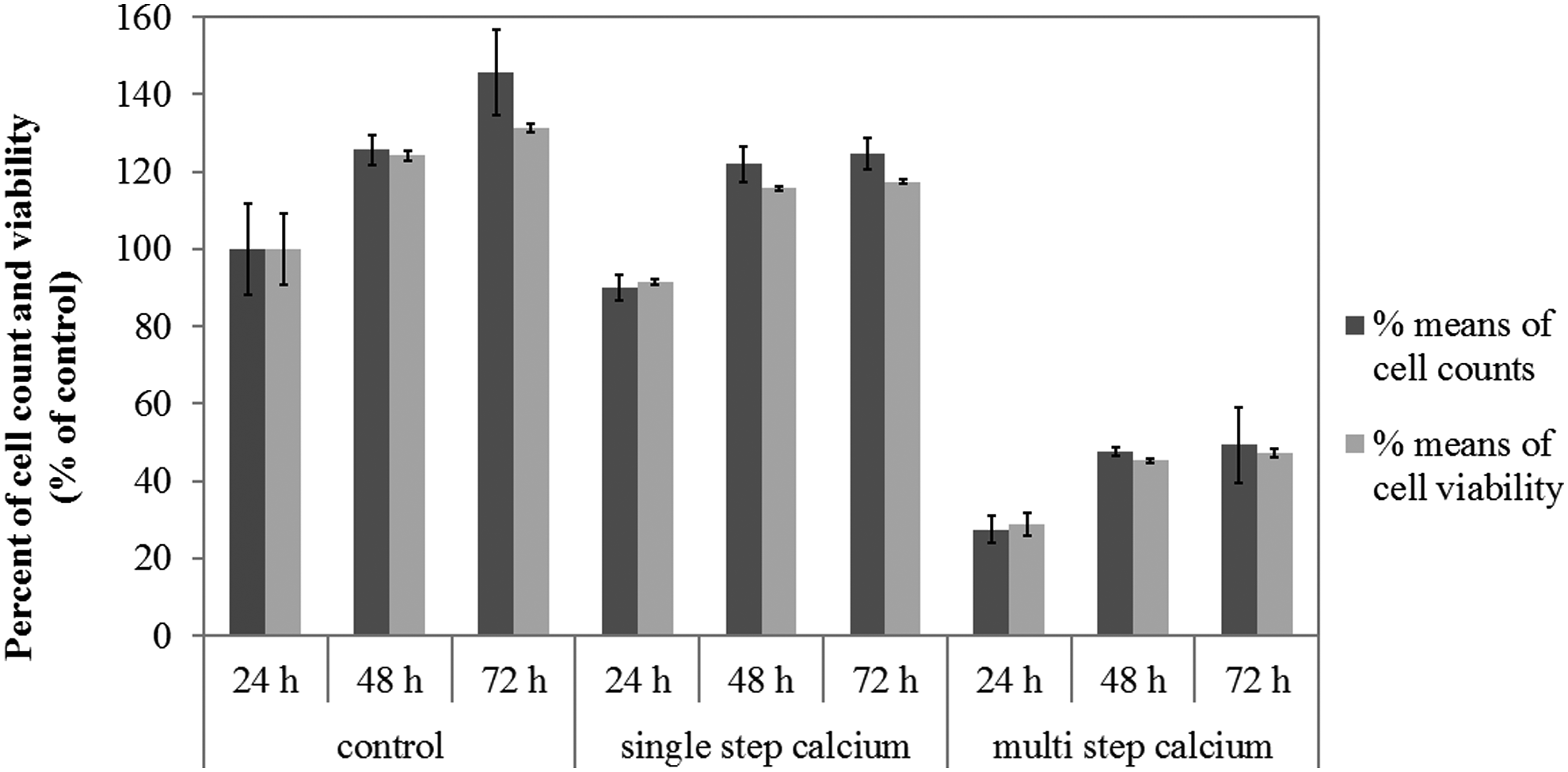
Comparison of the percentage of number of cells and viability in each group. The data indicate that there was no decrease in cell viability.
Statistical analysis
All experiments were performed three times. All data are presented as the mean ± standard error of the mean of at least three experiments. For multigroup comparisons, an analysis of variance followed by Tukey's test was performed. p < 0.05 was considered statistically significant.
Results
Index of cell proliferation
Compared with the control group, no significant positive or negative effects related to proliferation were observed in MCF-7 breast cancer cells treated with a single dose of high-concentration calcium (0.3 mg/mL) (Fig. 4A). This result was consistent over time. On the contrary, the cell counts in the group treated with multiple doses of high-concentration calcium (0.3 mg/mL) significantly decreased (Fig. 4A, and 4B). The number of cells in the control group increased steadily day by day. Conversely, cell proliferation decreased in the calcium-treated groups; cell counts of the calcium-treated groups slowly increased after 48 hours (Fig. 4B). Although the single-dose calcium treatment caused cell loss 10 percent, multiple-doses calcium treatment increased the amount of cell loss to 63% at the end of 24h (Fig. 3).
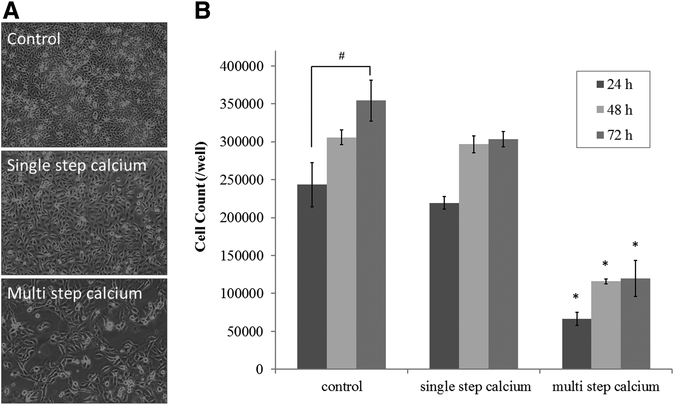
Effects of calcium on the MCF-7 breast cancer cell line.
Cell viability
The MTT assay absorbance values, which measure the mitochondrial activity of living cells, decreased in correlation with the cell concentration of the groups (Fig. 2). Figure 3 shows that neither the single-dose nor multiple-doses of high-concentration calcium treatment significantly affected cell viability.
Discussion
This study was performed to investigate the protective effect of breastfeeding against breast cancer. In this study, a breastfeeding model was created. Specifically, the effects of high levels of calcium, similar to a mother's milk, and the effects of fluidity, a physiological defense mechanism against breast cancer, were investigated. The findings of our study showed that exposure to high-concentration calcium at frequent intervals reduced the adhesion ability of cells, and that the cells were removed from the environment upon renewal of the medium.
The literature contains a large number of meta-analyses indicating that breastfeeding can reduce the risk of breast cancer.2,17–21 In addition, it has been reported that long-term breastfeeding further enhances the protective activity against breast cancer. 1 Many studies have shown that the migration and proliferative ability of breast cancer cells were improved by inducing calcium-sensitive receptors.22,23 The observed decrease in number of cells suggests that proliferation was inhibited. However, calcium causes detachment of the cells that induce proliferation in adherent cells. In our model, detached cells were removed upon renewal of the medium 30 minutes after high-concentration calcium application, resulting in low number of cells. In our study, 7.5 mM calcium was used as the high concentration. This concentration did not inhibit the proliferation of MCF-7 cells. Previous studies have reported that the concentration of extracellular calcium that causes toxicity varies depending on the cell type.24–26
Previous studies showed that a calcium-rich diet and intake of drugs with calcium decrease the risk of developing colon cancer.27,28 Some studies have shown that dietary calcium increases the risk of developing prostate cancer, another type of cancer with luminal characteristics.29,30 Much evidence for the preventive effects of calcium against colon cancer exists; however, its effects on prostate cancer are controversial.
McCullough et al. reported an association between high calcium intake and reduced breast cancer risk in postmenopausal women. 31 However, Chlebowski et al. showed that calcium and vitamin D supplementation did not reduce the incidence of invasive breast cancer in postmenopausal women. 32 Fluidity is an important natural defense mechanism for displacing products and metabolites. Many different types of cells are present in fluids removed from the body, such as milk, menstrual blood, and urine. The population of cells in these fluids is heterogeneous. Many studies have shown that stem cells are also found in body fluids, including milk.33–35
Cancer cells and stem cells are similar with regard to antigenic determinants on the membrane surface.36,37 Some researchers have identified breast cancer cells as stem-cell-like cancer cells.38–40 In addition, Reya et al. reported that carcinogenic changes might occur through activation of various signaling cascades in stem cells. 41 The concept that breast cancer can originate from stem cells suggests that the elimination of stem cells may prevent future carcinogenesis. Our study showed that calcium and fluidity of the milk contribute to this elimination process.
If it is considered that the fluidity in the mammary ducts continues for more than a year during breastfeeding, this long retention period may also be important for the prevention of breast cancer formation. 1 Previous studies have shown that women who breastfed their babies long term had a lower rate of breast cancer than women who did not. 18 Intestinal tissue cells, which are affected by toxic substances in feces, can become cancerous in patients with low intestinal motility. This condition suggests that cancer cells proliferate due to a lack of displacement from the environment for various reasons, causing tumor formation. 12 Therefore, the fluidity in luminal tissues is an important factor for prevention of cancer development.
Conclusions
This study has illuminated some of the mechanisms associated with the protective effects of breastfeeding on breast cancer. This cell culture model, first tested in this study, may be a method that can be used for future research into breast cancer during the lactation period. To clarify the effects of breastfeeding on breast cancer, the investigation of the effects of other components in milk may provide important data for the exploration of pathogenesis.
Footnotes
Disclosure Statement
No competing financial interests exist.