
Abstract
Introduction:
Breastfeeding is a fundamental biological function in mammals, allowing the progeny to develop in a physiological way. A physical and emotional dialog between mothers and offspring during breastfeeding has been described as part of the attachment relationship, and a synchronicity between maternal and neonatal brains can be hypothesized. This study aimed to assess if neonatal and maternal cortical areas activated during breastfeeding are functionally synchronized since the second day of life.
Materials and Methods:
Twenty mothers and their term newborns were enrolled. Cortical activation during breastfeeding was identified by multichannel near-infrared spectroscopy, which detects changes in haemoglobin concentration from multiple cortical regions. Functional activity was simultaneously detected (hyperscanning) in mothers and newborns’ frontal and motor/primary somatosensory cortical areas during the first 5 minutes of breastfeeding. Cluster analysis and Student’s t test were used to detect oxygenated haemoglobin increase, as cortical activation estimate. Wavelet transform coherence (WTC) analysis was used to identify a possible synchronization between maternal and neonatal activated cortical regions.
Results:
Mothers showed an activation of the central motor/primary somatosensory cortex, above the sagittal fissure. In newborns, the bilateral frontal cortex was activated. WTC analysis revealed two different cyclical synchronizations between mothers and infants’ activated cortical regions.
Conclusions:
Such evidence may reflect a very early common sharing of experiences, possibly associated with reciprocal dynamic motor adjustments, hormonal coregulation, and somatic stimulations and sensations. The observed cyclical neural synchronization, between the mother and her newborn’s cortex during breastfeeding, may play an important role in promoting their bonding.
Introduction
Breastfeeding constitutes a fundamental characteristic of mammals, 1 allowing offspring to grow and develop in a physiological way. 2 The breastfeeding interaction with the mother satisfies both the nutritional and relational needs of the newborn. 3
Therefore, breastfeeding is a relational experience and can be considered part of the attachment relationship. 4 Attachment bonds are another crucial characteristic of mammals, which are rapidly formed 5 and involve multiple maternal and neonatal functional dimensions. Mother–infant reciprocal behaviors are associated with specific brain activations, affiliative hormones secretion (e.g., oxytocin), and neurovegetative responses, connecting a mother and her own newborn’s physiology, behavior, and (inter)subjective experience.6,7 The biobehavioral synchrony between caregiver and infant is considered a key feature of human attachment, 5 encompassing physiological coordination, heart-rate rhythms coupling, and hormonal release coregulation between partners of the attachment dyad.8–10
The neurophysiology of human breastfeeding has been studied too, both in mothers and in newborns. Electroencephalography (EEG) assessment in one nursing woman revealed electroencephalographic signs of “relaxation behavior” (groups of synchronic, 6–10 Hz, 100–150 μV EEG waves) 15–80 seconds after the start of suckling. 11 In the breastfeeding newborn, an EEG response was found in the posterior cortical areas, 12 whereas when using multichannel near-infrared spectroscopy (fNIRS), the response was found in the motor, somatosensory, and right parietal cortex. 13 We wondered if such maternal and neonatal brain activities are also synchronized since the first days of life. 5
Hyperscanning is a functional neuroimaging method that can identify the presence of a coherence in brains’ neural activities of two or more people engaged in a relationship.
14
Coherence in brain activities, or interpersonal brain synchronization (IBS), was studied in parent–infant dyads too. Using fNIRS, it was found that maternal affectionate touch increases the neural synchrony between mother and her 4
The aim of this observational study was to assess whether maternal and neonatal cortical areas, activated during breastfeeding, are functionally synchronized already on the second day of life. We hypothesized that such synchronization is present and may be part of the earliest relational exchange taking place during breastfeeding.
Materials and Methods
Participants
Twenty mother–newborn dyads were enrolled. Mean maternal age was 32.15 ± 3.51 years (range: 27–37 years) and mean educational level was 13.7 ± 3.9 years (range: 8–18 years). Exclusion criteria for mothers were history of neurological or neurosensory disorder, psychosis, depressive or bipolar disorder, substance abuse or addiction, and medications that could interfere with a functional neuroimaging assessment (e.g., analgesics and sedatives). Possible postpartum depression symptoms were excluded using the Edinburgh Postnatal Depression Scale. 18
Newborns were all healthy, term, and vaginally delivered. There were 12 females and 8 males. Their mean gestational age was 39.2 ± 0.9 weeks (range: 38–41 weeks), and their mean birth weight was 3566 ± 298 g (range: 2950–4080 g). All babies were assessed at 2 days of postnatal age. No infant had taken any medication that could interfere with a functional neuroimaging assessment (e.g., analgesics and sedatives).
The study was conducted according to the guidelines of the Declaration of Helsinki, as revised in 2013, and was approved by the Institutional Review Board of the Institute for Maternal and Child Health IRCCS “Burlo Garofolo” (Trieste, Italy). Both parents signed a written informed consent, after full technical and procedural explanation.
Multichannel near-infrared spectroscopy
FNIRS is a functional neuroimaging technique detecting and localizing the activation of the cerebral cortex by continuously monitoring changes in cortical haemoglobin concentration. Optical fibers (optodes), emitting and detecting near-infrared light, are used to measure changes in the concentration of oxygenated haemoglobin (HbO2) and deoxygenated haemoglobin. 19 An increase in HbO2 concentration is considered to reflect cortical activation on the basis of neurovascular coupling. 20 Each pair of adjacent emitter and detector optodes defines a single channel. fNIRS systems have considerably improved spatial resolution, compared with one- or two-channel NIRS devices. 21 The technique is better detailed elsewhere.22,23
In this study, we used the Hitachi ETG-4000 OT device (Hitachi Medical Corporation, Tokyo, Japan). This device can simultaneously record cortical activity from 48 channels, which can be arranged in different patterns to cover specific areas of the cerebral cortex. It does not allow monitoring functional activity from the overall cerebral cortex; thus, the researcher has to choose the specific areas on which to focus, according to the study aim. It emits near-infrared light at two wavelengths, 695 and 830 nm, and the reflected light is sampled at a frequency of 10 Hz. Changes in HbO2 concentration in response to a stimulation or experience are estimated as mM·mm units, i.e., the product of the hemoglobin concentration changes, expressed in millimolar, and the optical path length, expressed in millimeters.
To assess and localize the dyad’s cortical activity, 24 detecting channels were placed on mother scalp and 24 channels on newborn scalp, by rubber (mother) or silicone rubber (newborn) fiber holders, for a total of 48 detecting channels. Adjacent emitters and detectors were placed 3 cm apart. It was chosen to position fiber holders on participants’ scalp according to previous literature15,24 and referring to the international 10–20 EEG placement system. 25 On mothers, the optodes were arranged in a 4 × 4 pattern (16 optodes; eight emitters and eight detectors) and positioned over the frontal and motor/primary somatosensory cortex. Specifically, the anterior row of channels (Fig. 1a) was put on the virtual line joining Fp1 and Fp2 points, placing channel 23 on Fpz.
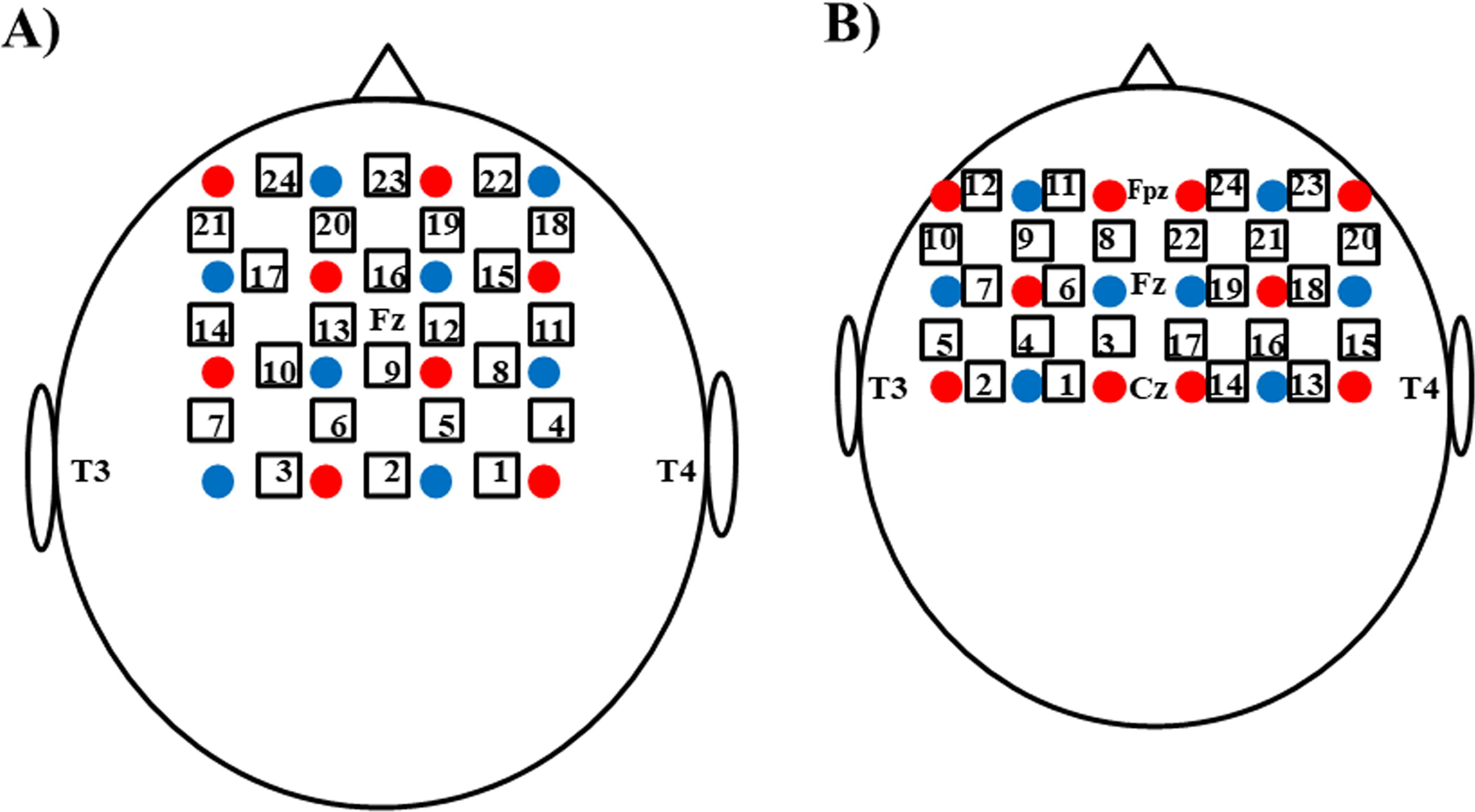
Schematic representation of optical fibers positioning on left and right side of
On newborns, the optodes were arranged in two 3 × 3 patterns (18 optodes; ten emitters and eight detectors), positioned over the left and right side of the frontal and motor/primary somatosensory cortex. Specifically, the anterior row of channels of both fiber holders (Fig. 1b) was put on the virtual line joining Fp1 and Fp2 points, keeping in contact the holders over the sagittal fissure.
Procedure
The dyads’ cortical activity assessment during breastfeeding was performed on newborn’s second day of life. Only babies showing effective breastfeeding were selected. fNIRS monitoring was done in the nursery, at mother’s bedside, when newborns showed signs of hunger. The room was dimly lit and as quiet as possible. The biological nurturing approach was adopted by all the dyads for reciprocal positioning of mother and baby during breastfeeding (Fig. 2). 26

Picture showing the real setting for fNIRS data collection during breastfeeding (written informed consent for image publication obtained from both parents).
After noticing neonatal hunger signs, the mother adopted a relaxed semi-reclined position in her bed. The mother placed the baby prone between her chest and abdomen just below the breast. Infants maintained their airways free and their bodies in full contact with mothers at all times. An investigator positioned the fiber holder on mother and newborn’s head, keeping them in place by elastic bands. After checking the adequacy of the signal, fNIRS monitoring of maternal and neonatal cortical activation during breastfeeding was started. A 10-second baseline was collected before newborn attachment to the nipple. After attachment, fNIRS monitoring was performed during the first 5 minutes of breastfeeding. At the end of fNIRS recording, the fiber holders were removed from the mother’s and newborn’s scalps and breastfeeding continued freely.
Data analysis
First, to identify the dyad’s cortical areas simultaneously activated during breastfeeding, HbO2 concentration changes detected by fNIRS were statistically analyzed. In rodents, it was found that, when using fNIRS, an increase in HbO2 reflects a change in the same direction of regional cerebral blood flow, which in turn reflects cortical activity.27,28 To remove interferences on the haemoglobin signal related to physiological noise (slow cerebral blood flow fluctuations, heartbeat, and breathing), a bandpass filtering of the near-infrared signal between 0.02 and 1 Hz in mothers and between 0.02 and 0.5 Hz in newborns was applied. 29 To prevent motion artifacts, rapid changes in HbO2 concentration (signal variations > 0.1 mM·mm over two consecutive samples) 30 were identified in the recorded data by the software already included in the fNIRS device. We also visually checked the signals recorded in each channel of all participants to detect low signal-to-noise ratio due to suboptimal transmission of near-infrared light. Data from channels with a signal interfered by motion artifacts or suboptimal transmission of near-infrared light were replaced, in the statistical analysis, with the mean of the entire dataset, 23 as an estimate of central tendency, separately calculated for each experimental phase (baseline and breastfeeding).
An exploratory statistical analysis was conducted to identify the possible presence and extension of specific functional regions of interest (ROIs) inside the cortical areas monitored in mothers and newborns during breastfeeding. For this purpose, mean HbO2 changes, detected in single channels during the first 5 minutes of breastfeeding, were analyzed by agglomerative hierarchical cluster analysis, performed separately in mothers and newborns.
To test whether the identified ROIs (clusters of channels) in mothers and newborns were significantly activated, we applied one-tailed paired Student’s t test. For each mother and baby’s ROI, the mean of relative HbO2 changes during baseline (10 seconds) and the first 5 minutes of breastfeeding, after the newborn’s attachment to the nipple, were calculated for each channel belonging to a cluster (i.e., an ROI) and then averaged among ROI’s clustered channels. To identify ROI’s activation, HbO2 mean changes during baseline and during the first 5 minutes of breastfeeding were compared by one-tailed paired t tests, as there was only one direction of interest to test the statistical significance (HbO2 increase). 31
In case no ROI revealed a functional activation during breastfeeding, we focused our exploratory analysis on the overall cortical area monitored by fNIRS to identify single activated channels. Accordingly, HbO2 mean changes during baseline and during the first 5 minutes of breastfeeding were compared, channel by channel, by one-tailed paired t tests. In this latter case, a false discovery rate (FDR) approach was used to control type I error in multiple testing situations (p = 0.05). 32 Analyses to identify cortical activation during breastfeeding were performed separately for mothers and newborns and conducted using SPSS version 22.0 for Windows (Armonk, NY, IBM Corp.).
The possible synchronization between the simultaneously activated cortical ROIs and/or channels in mothers and newborns (IBS) during breastfeeding was assessed by wavelet transform coherence (WTC) analysis.33,34 By this data processing approach, the cross-correlation between two time series can be measured, as a function of signal frequency and time. 35 Monte Carlo simulation methods are applied to estimate the statistical significance of the wavelet coherence, by computing wavelet coherence for 300 randomly sampled surrogate datasets and comparing them with the actual dataset coherence. 34 The wavelet coherence toolbox for MatLab 34 was used, which included a subsequent contribution for applications with fNIRS data, 33 available from: https://www.alivelearn.net/?p=1561 (last accessed: 05/08/2024). WTC data processing generates a 2D coherence map, which shows the cross-correlation intensity of the two time series included in the analysis on a graduated color scale, varying from blue (low intensity) to yellow color (high intensity). The 5% statistical significance level against red noise of the coherence between the time series is expressed by a thick contour enclosing frequency-synchronized signals over time on the coherence map.
Results
Mother and newborn’s cortical activation during breastfeeding
Ten newborns were breastfed from the right breast and 10 newborns from the left breast. In breastfeeding mothers, agglomerative hierarchical cluster analysis performed on mean HbO2 changes, detected in single channels during the first 5 minutes of breastfeeding, gave the output reported in Table 1 (agglomeration schedule). Looking at the squared Euclidean distance coefficients among clustered channel pairs (see Table 1),we decided to consider only the first seven stages of channel clustering to identify a functionally activated ROI in mothers. Channels 13-15-16-18-19-22-23-24 were initially included, covering the sagittal fissure, right frontal cortex, and bilateral prefrontal cortex (see Fig. 1a). The functional activation of this ROI during breastfeeding was assessed comparing, by one-tailed t test, the HbO2 mean changes during baseline and during the first 5 minutes of breastfeeding, both averaged among the eight clustered channels, but no significant increase was found (t(19) = 0.530; p = 0.30). The same data processing approach was performed on each ROI emerging from stage six to one of the clustering process (see Table 1), but no significant activation emerged in any case. Therefore, an exploratory analysis of the overall monitored cortical area in mothers was performed to identify single activated channels, by one-tailed t test comparison between baseline and breastfeeding experience, channel-by-channel, with FDR control for multiple testing (p < FDR 0.05). We observed one significant functional activation in channel 2 (t(19) = –3.379; p = 0.0015), approximately located on central motor/primary somatosensory cortex, above the sagittal fissure. Such activation is illustrated in Figure 3.
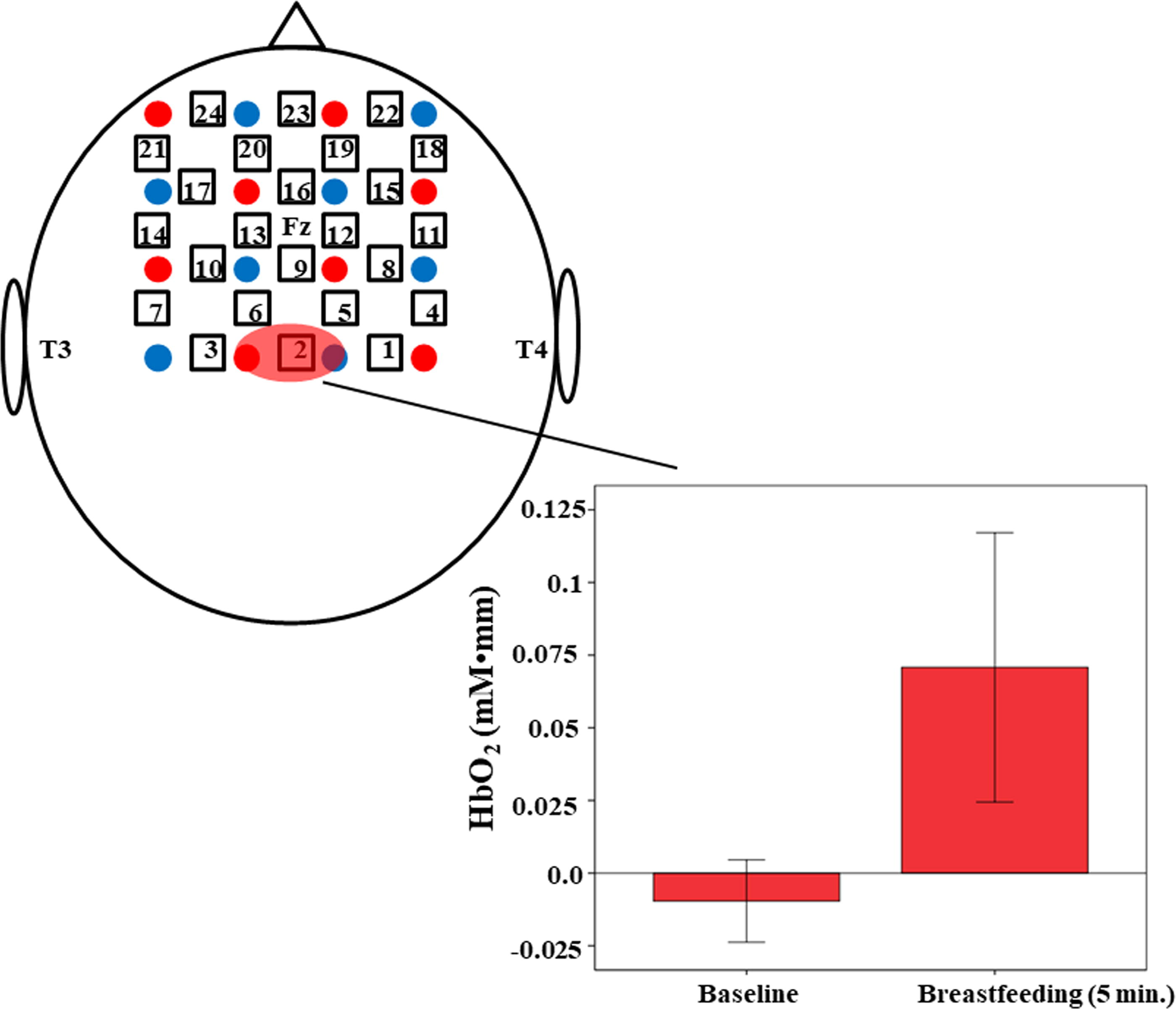
Bar chart of mean oxygenated haemoglobin (HbO2) variation over the baseline and the first 5 minutes of breastfeeding in channel 2 of mothers (approximately located on central motor/primary somatosensory cortex), which showed a significant activation compared with baseline (*p < FDR 0.05). Channel’s positioning is showed on a simplified representation of the head. Whiskers represent ± 2 standard error variability around the mean of HbO2 variation, which is reported on the y axis in mM·mm unit (see text). FDR, false discovery rate.
Cluster Analysis’ Agglomeration Schedules, Limited to the First 9 out of 23 Stages a
The first seven clustering stages were selected to identify the ROI in both groups.
In breastfed infants, agglomerative hierarchical cluster analysis performed on mean HbO2 changes detected in single channels gave the output reported in Table 1. Looking at the squared Euclidean distance coefficients among clustered channel pairs (see Table 1), we decided to consider only the first seven stages of channel clustering to identify a functionally activated ROI in newborns. Channels 6-9-10-11-12-18-19-21-22-23-24 were initially included, covering the newborn’s anterior frontal cortex, bilaterally (see Fig. 1b). The functional activation of this ROI during breastfeeding was assessed comparing, by one-tailed t test, the HbO2 mean changes during baseline and during the first 5 minutes of breastfeeding, both averaged among the 11 clustered channels, but no significant increase was found (t(19) = −1.407; p = 0.09). No statistical significances were observed also when the same data processing approach was performed on each ROI emerging from stage six to four of the clustering process (see Table 1). However, the ROI emerging from stage three showed to be activated by breastfeeding (t(19) = −1.894; p = 0.037). It included channels 9-11-21-22-23-24 and covered, bilaterally, the anterior newborn’s frontal cortex, with a wider extension on the right side. Such activation is illustrated in Figure 4.

Bar chart of mean oxygenated haemoglobin (HbO2) variation over the baseline and the first 5 minutes of breastfeeding in the bilateral anterior frontal cortex of newborns (more extended on the right side), which showed a significant activation compared with baseline (*p < FDR 0.05). Channel’s positioning is showed on a simplified representation of the head. Whiskers represent ± 2 standard error variability around the mean of HbO2 variation, which is reported on the y axis in mM·mm unit (see text).
Mother–newborn IBS during breastfeeding
Basing on Monte Carlo simulations, WTC analysis including the averaged time series of mothers’ channel 2 (central motor/primary somatosensory cortex) and newborns’ activated ROI (bilateral anterior frontal cortex) revealed two significant cyclic functional synchronization between such activated cortical regions (5% statistical significance level against red noise, see Fig. 5). The first one occurred at a signal sampling frequency between 0.8 and 4.0 seconds, appeared about 40 seconds after breastfeeding started and lasted until the end of the 5 minutes of fNIRS monitoring. The second one occurred at a lower signal sampling frequency and was composed of two synchronization cycles: an earlier cycle was already present when breastfeeding began, occurred at a sampling frequency of about 30 seconds and ended about 130 seconds after the beginning of breastfeeding. A following cycle started about 260 seconds after the beginning of breastfeeding, occurred at a sampling frequency between 13 and 26 seconds and was still present at the end of fNIRS monitoring. Further features of the WTC output are described in detail in Figure 5. Supplementary Figures S1–S20 show the results of WTC analysis performed, separately, in each one of the 20 dyads (see Supplemental files).
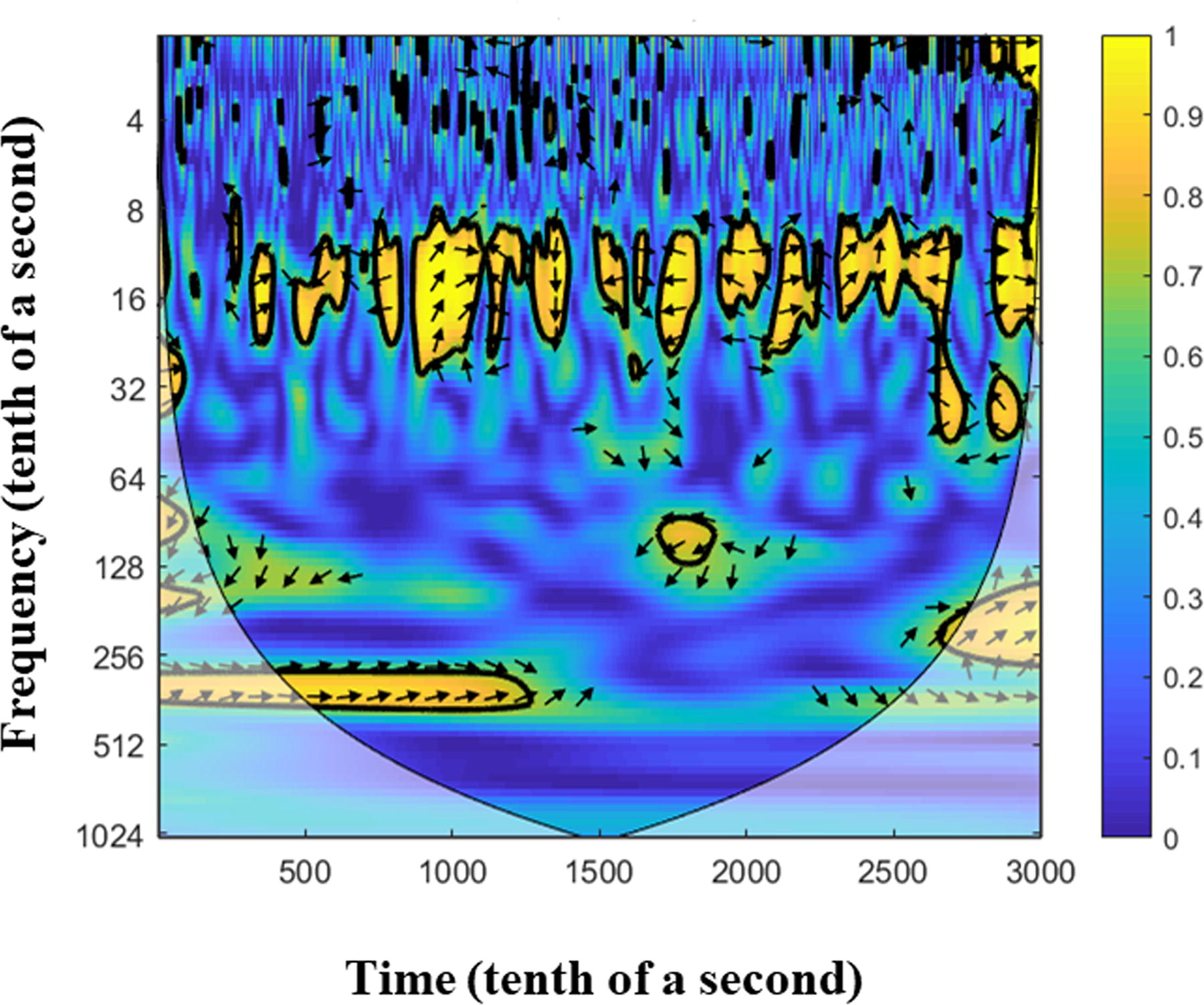
Interpersonal brain synchronization (IBS), analyzed by wavelet transform coherence (WTC), between average time series of oxygenated haemoglobin signal from mother’s central motor/primary somatosensory cortex (channel 2) and baby’s bilateral anterior frontal cortex, detected over the first 5 minutes of breastfeeding. WTC value is represented on a color scale (see the right bar). The 5% statistical significance against red noise, resulting from Monte Carlo simulations, is showed as a ticked contour surrounding frequency synchronized signals. Two cyclic functional synchronizations can be observed, a first one in the frequency band between 0.8 and 4.0 seconds (0.25–1.25 Hz) and a second one in the frequency band between 13.0 and 30.0 seconds (0.03–0.08 Hz). By arrows, further features of the IBS are specified. Arrows pointing to the right indicate an in-phase cross-correlation (signals move in the same direction), whereas arrows pointing to the left indicate an antiphase cross-correlation (signals move in opposite directions). Moreover, arrows pointing down, right-down, or left-up all indicate that the first variable (newborns in this study) is leading the synchronization, whereas arrows pointing up, right-up, or left-down all indicate that the second variable (mothers in this study) is leading the synchronization. Concerning the IBS at the faster signal sampling frequency, arrows direction and their inclination angle changed throughout the cyclic periods of synchronization. There were both in-phase coherence between the dyad’s cortical activation and antiphase coherence. Moreover, the IBS could be led by the mother or by the newborn. Concerning the IBS at the slower signal sampling frequency, arrows direction indicates an in-phase IBS coherence in both cycles, whereas arrows angle inclination indicates an in-phase IBS lead by the newborn in the second cycle.
Discussion
The aim of this observational study was to assess whether there is a functional synchronization between the maternal and neonatal cortical areas activated during the first 5 minutes of breastfeeding on the newborns’ second day of life. When looking at mothers, we observed an activation of the central motor/primary somatosensory cortex, above the sagittal fissure. EEG previously revealed signs of “relaxation behavior” mainly in central–parietal and parietal–temporal regions, with a prevalence in the right hemisphere, 15–80 seconds after the beginning of breastfeeding. 11 However, in our study, we monitored only mothers’ frontal and motor/primary somatosensory cortex. The observed maternal activation during breastfeeding seems to partially overlap one of the cerebral regions in which a “relaxation behavior” was previously detected by EEG. 11
Such activation may be associated with the somatic sensations experienced by mothers during breastfeeding. The localization of the maternal cortical activity (e.g., the motor/primary somatosensory cortex surrounding the sagittal fissure) may be compatible with areas involved in the processing of motor command to and of somatosensory information from the trunk/abdomen.36,37 Mothers described breastfeeding as a close, physical experience, creating unity between them and their newborns. 38 Thus, the central motor/primary somatosensory cortex may have a role in the processing of the breastfeeding experience referred by mothers.
When observing newborns, we found a simultaneous activation of the anterior frontal cortex, extended on the right side. A previous study described an EEG response in the posterior cortical areas of newborns during breastfeeding, with a slight predominance on the right side. 12 Although we did not assess the posterior cortical areas, a predominance of the right side was observed. Previous studies with fNIRS found that, since their first days of life, newborns are frontally activated by mother’s milk odor 26 and infant-directed speech, 39 more intensely on the right side in the latter case. Therefore, the newborn’s frontal cortex seems to play a pivotal role in processing different aspects of very early relational experiences.
After identifying the dyad cortical areas significantly activated during breastfeeding, the possible synchronization between these functional activations of mother and newborn’s cerebral cortex was assessed by WTC analysis. We found that mother’s motor/primary somatosensory cortex and newborn’s frontal cortex are cyclically synchronized. A cyclical synchronization was observed in a sampling frequency of the HbO2 signal ranging between 0.8 and 4.0 seconds throughout the first 5 minutes of such primitive experience. Cycles of synchronization showed both in-phase coherence (e.g., HbO2 variation signals move in the same direction) and antiphase coherence (e.g., HbO2 variation signals move in opposite directions). Similarly, the leading of IBS changed throughout the cycles, oscillating between mother’s leading, newborn’s leading, or no leading. Another cyclical synchronization was observed in a slower sampling frequency of the HbO2 signal, ranging between 13.0 and 30.0 seconds. As it was already present when breastfeeding started, it seems to reflect a more general mother–newborn synchronization associated with their wider experience of being in reciprocal contact. It can be speculated that a so slow periodic synchronization of the HbO2 signal may be mediated by an affiliative hormonal response, e.g., by oxytocin pulsatility and the related neural networks. 40
There is little doubt that breastfeeding is a form of attachment between mothers and infants. Attachment has been also defined as an interactive regulation of biobehavioral synchronicity between organisms. 6 In line with this definition and as already showed in older infants, 16 we observed a synchronous activation of mothers’ and their neonates’ brains. Such synchronization may reflect a common sharing and exchange of experiences in the dyad, associated with reciprocal dynamic motor adjustments and coregulation, somatic and olfactory sensations, hormonal synchronicity, and attachment-related emotions during breastfeeding. It can be also speculated that the observed cyclical neural synchronization may play an important role in promoting the earliest bonding between mother and newborn.
Some limitations of the study need to be acknowledged. As we did not have any simultaneous magnetic resonance imaging data, the described cortical localizations can only be approximate. We assessed mothers and newborns only for 5 minutes. What happened beyond this period was not explored. However, the choice to restrict fNIRS monitoring only for 5 minutes was done to minimize disturbance to the breastfeeding dyads. Moreover, there were no video recordings and analysis of breastfeeding behavior. This might have helped to better identify potential sources of the shifts in phase coherence between mothers and newborns. Finally, our sample size was small and our results need to be confirmed in larger samples.
Conclusions
Our study shows that, already on the second day of life, a dialog is present between the cortical functional activations of the mother–newborn dyad during the first 5 minutes of breastfeeding. Such evidence may reveal a common sharing of experiences, possibly associated with reciprocal dynamic motor adjustments and coregulation, somatic and olfactory sensations, hormonal synchronicity, and attachment emotions. The cyclical neural synchronization observed, with the associated shared subjective experiences, may play an important role in promoting the bonding between a mother and her newborn, since the first days of life. However, further research is needed to better understand and deepen these preliminary observational results.
Footnotes
Acknowledgments
The authors would like to thank all the mothers and their babies who participated to this study. The authors also wish to thank Ms. Maria Carmela Telesca for her fundamental support in the literature review.
Authors’ Contributions
S.B.: conceptualization (equal); methodology (lead); investigation (equal); formal analysis (lead); writing—original draft (lead); project administration (lead). El.C.: methodology (supporting); investigation (equal); writing—original draft (supporting). J.B.: conceptualization (equal); writing—review and editing (lead). En.C.: investigation (equal); formal analysis (supporting); writing—review and editing (supporting). C.P.: investigation (equal); formal analysis (supporting); writing—review and editing (supporting). F.M.: investigation (equal); writing—review and editing (supporting). L.T.: conceptualization (equal); writing—original draft (supporting); supervision (lead).
Data Sharing Statement
Disclosure Statement
The authors declare no competing interests.
Funding Information
This research was funded by the Ministry of Health, Rome, Italy, in collaboration with the Institute for Maternal and Child Health IRCCS “Burlo Garofolo,” Trieste, Italy (grant number
References
Supplementary Material
Please find the following supplemental material available below.
For Open Access articles published under a Creative Commons License, all supplemental material carries the same license as the article it is associated with.
For non-Open Access articles published, all supplemental material carries a non-exclusive license, and permission requests for re-use of supplemental material or any part of supplemental material shall be sent directly to the copyright owner as specified in the copyright notice associated with the article.