
Abstract
Exposure time and addition of sucrose to the vitrification medium as well as the solid-surface vitrification (SSV) on the morphology of bovine preantral follicles were evaluated. Ovarian tissue was exposed for 1, 5, or 10 min to 4.0 M ethylene glycol with or without the addition of 0.5 M sucrose. Subsequently, the tissue was washed out from cryoprotectants or vitrified by the SSV method. Independently of the presence of sucrose, exposure to vitrification solution for 10 min did reduce the percentages of normal follicles when compared with control. However, the highest rates of normal follicles were attained when tissue was previously exposed to the vitrification solution, with sucrose added or not, for 10 min. Although the SSV is a promising procedure to be applied in ovarian tissue, an optimal vitrification solution for bovine ovarian tissue needs to be developed.
Ovaries from 8 adult cows were used. From each ovarian pair, 13 pieces of 3 × 3 × 1 mm were collected (Fig. 1a). As controls, 1 fragment was fixed for histology and the remaining fragments were either exposed or exposed and cryopreserved in the presence of a serum-free vitrification solution containing 4.0 M EG combined or not with 0.5 M sucrose. Exposure to cryoprotectant solution was performed for 1, 5, or 10 min. After this, the ovarian fragments were submitted to cryoprotectant removal or vitrification. For vitrification, a previously described SSV method was used. 4 For the warming, samples were exposed to air for 1 min at ∼25°C and subsequently submitted to cryoprotectant removal by washing the ovarian fragments in serum-free minimum essential medium (MEM) at 37°C for 3, 5, and 7 min. All reagents were obtained from Sigma-Aldrich. As a conventional screening of cryopreservation protocols, the ovarian pieces were fixed for histology and the preantral follicles were classified as morphologically normal or degenerated. 8 Data were analyzed using one-way analysis of variance and Tukey's post hoc test (P < 0.05).
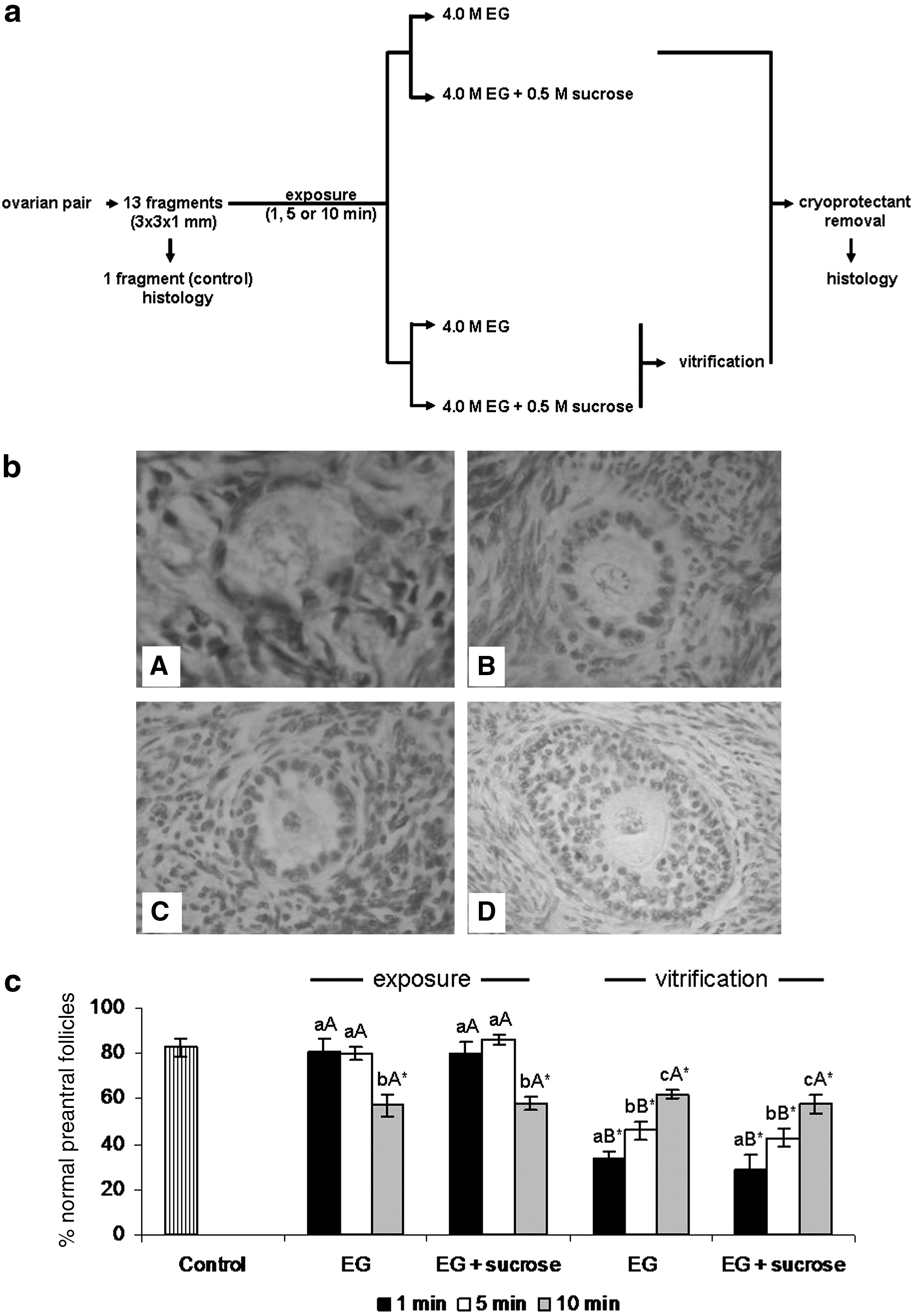
At least 150 bovine preantral follicles were evaluated per treatment. Figure 1b illustrates morphologically normal preantral follicles from control (Fig. 1b-A), after exposure (Fig. 1b-B), as well as degenerated (Fig. 1b-C) and normal (1b-D) vitrified preantral follicles. The mean percentages of morphologically normal preantral follicles immediately after collection are presented in Fig. 1c. It was observed that 83% ± 4% of the follicles in the control tissues were morphologically normal. Exposure to EG, for 1 and 5 min, combined (80% ± 3% and 80% ± 5%) or not (81% ± 6% and 86% ± 2%) with sucrose, did not reduce significantly the percentages of morphologically normal preantral follicles when compared with control. However, exposure to EG for 10 min, with (58% ± 3%) or without (57% ± 5%) sucrose, resulted in a significant reduction in the percentages of normal preantral follicles. Vitrification resulted in a significant decrease in the percentages of normal preantral follicles independently of the exposure time and presence of sucrose. The highest percentages of normal preantral follicles were observed when vitrified ovarian tissue was previously exposed for 10 min to the cryoprotectant, with sucrose added (58% ± 4%) or not (62% ± 2%).
We have shown the ineffectiveness of the addition of 0.5 M sucrose to the vitrification medium on the morphology of bovine preantral follicles. Although sucrose is known for its cryoprotective effect, 4 probably the concentration of 0.5 M was not sufficient to improve vitrification, or it is simply ineffective to preserve bovine ovarian tissue. The study by Cetin and Bastan 9 has shown the importance of 1.0 M sucrose inclusion to the vitrification medium of bovine oocytes. However, in a recent study, it has been shown that 0.5 M sucrose was deleterious to preantral follicles vitrified enclosed in the bovine ovarian tissue. 7 When Gandolfi et al. 1 performed the vitrification of bovine ovarian tissue, the sugar of choice was trehalose, resulting in even lower percentages of normal preantral follicles when compared with the present study. Usually, bovine ovarian tissue has been exposed to the vitrification solution for 1 min 7 or 5 min. 1 However, to our knowledge, this is the first time that the exposure period of bovine ovarian tissue to a vitrification solution has been tested. We have shown that a period of 1 or 5 min was not sufficient to protect the bovine ovarian tissue against the cryodamage. Indeed, the composition of our vitrification solution is different from those used by the former authors. However, when we have vitrified caprine ovarian tissue with the same solution after 5 min exposure, most of the preantral follicles were properly preserved. This may suggest species-specific differences or simply that bovine ovarian tissue might be more fibrous than the caprine ovarian tissue. Although the long exposure time of 10 min was more toxic to the ovarian tissue, it confers a better protection to the tissue during vitrification. Since the development of the SSV method for oocytes, 10 this procedure has been repeated with success in caprine 4 and human 6 ovarian tissue. The application of the SSV method resulted in the morphological preservation of around 58% preantral follicles. Even though this rate is far from optimal values, it was higher than the rates (maximum of 29%) observed previously when a tissue was loaded in plastic straws. 1 Different from carrier systems such as straws and cryovials, the SSV method (i) allows a fast cooling–warming of the tissue, (ii) applies a small volume of cryoprotectant, and (iii) eliminates the isolating layer (plastic wall of vials) between LN2 and vitrification solution.11,12 Therefore, the SSV method appears as an efficient option to vitrify bovine ovarian tissue. Further development of efficient vitrification solutions for bovine ovarian tissue, probably sugar-free, requires the application of more accurate assays such as biochemical, functional, and developmental analyses.
Footnotes
Acknowledgment
R.R. Santos was supported by CNPq (project 483439/2009-6), Brazil.
Author Disclosure Statement
There is no conflict of interest.