
Abstract
Recovery of microRNAs (miRs) from tissue samples of many kinds has become an area of increasing interest and importance in recent years. We isolated and then amplified and quantitated miRs from human peripheral blood samples stored in the University of Colorado Denver Skin Cancer Biorepository to determine whether miR recovery was possible and consistent over time in storage. Forty-five blood samples from patients with different stages of malignant melanoma were collected in PAXgene Blood RNA tubes and then stored at −80°C prior to RNA preparation. The samples examined had been stored from 4 weeks to 3 years. Total RNA was prepared, followed by miR isolation, amplification, and quantitation using real-time polymerase chain reaction. A widely expressed miR, miR-221, was used as a standard for comparison across samples and storage time. miR-221 was recovered from all samples, with no differences observed with longer storage time. These studies show that miRs can be recovered and quantified from human blood samples stored for up to 3 years.
Introduction
At the University of Colorado Denver, we have a skin cancer biorepository in operation since 2005. During this time we collected 300 tissue samples and >1500 samples of whole blood, plasma, serum, and white blood cells from patients with skin cancers of various kinds. Blood cells were collected and stored using the PAXgene Blood RNA system. 4 From previous studies, such as those described above, we knew that total RNA was well preserved using this system, but it was less clear whether miRs can also be preserved, isolated, amplified, and quantitated. Further, there are little data on how long samples can be stored and then used for miR collection, measurement, and molecular analysis.3,5 The aim of the present study was to elucidate whether we could extract total RNA from stored blood samples collected from patients with malignant melanoma while simultaneously isolating and amplifying miRs. In the present article, we outline the methods and demonstrate that miRs can be isolated and amplified in samples stored for at least 3 years.
Materials and Methods
Peripheral blood from patients with malignant melanoma was collected directly into PAXgene Blood RNA tubes (PreAnalytiX; Qiagen/BD) in accordance with institutional review board approval and patient consent. These tubes contain a reagent that stabilizes intracellular RNA. A summary of patient data can be found in the Table 1. The stage of melanoma shown on subsequent figures is the stage of each patient at the time the blood was collected. The staging system is that according to AJCC guidelines. 6
Patient blood samples were collected using our standardized laboratory procedure. First, 2.5 mL of peripheral blood was collected from each patient and the tubes were inverted 3–4 times. Second, the samples were immediately transported to the laboratory at room temperature where they remained at 25°C for an additional 2 h to ensure complete lysis of blood cells. Lastly, the tubes were stored at −20°C for 1–2 days and then directly placed in a −80°C freezer until processed (Table 2). RNA stored at these temperatures is stable for at least 50 months. 4 Total RNA greater than 18 nucleotides, including miRs, was isolated using the PAXgene Blood miRNA kit from samples stored at varying lengths of time. 4 The samples were centrifuged, washed, and digested with proteinase K and then homogenized using shredder columns and treated with isopropanol. Total RNA was quantified by a UV spectrophotometer (Nanodrop 1000; ThermoFisher). Absorption was measured at 260 and 280 wavelengths. One hundred nanograms of RNA was then reverse transcribed into polyadenylated cDNA using the miScript Reverse Transcription Kit (Qiagen). miRs-16, -155, -181a, and -221 were quantified using real-time polymerase chain reaction (PCR) (CFX96; BioRad) and SYBR green chemistry (miScript SYBR Green PCR Kit; Qiagen). The 2−ddCt method for relative quantitation was used to calculate miR levels, 7 using the small RNA molecule, U6, as the internal housekeeping gene. A normal (nonmelanoma patient) sample was used as control. See Table 3 for the following primer sequences: RNU6B (U6) (Qiagen); miR-16, miR-155, miR-181a, and miR-221 (Integrated DNA Technologies, Inc.). Statistical analysis was performed using GraphPad Prism 5.01 (GraphPad Software, Inc.). Each data point represents a patient sample. Error bars represent the standard error of the mean. An unpaired t-test (2-tailed) analysis was performed and a P value of <0.05 was considered significant. Time of storage was defined as the length of initial blood collection to the time of preparation of RNA (Table 2).
Freezing profile for 2.5 mL blood collected in PAXgene Blood RNA Tube. The evaluation shows a prefreeze rate of −0.82°C/min, with a postfreeze rate of −2°C/min and an end temperature at −80°C.
Freezing rate of 2.5 mL blood collected in PAXgene Blood RNA Tube, measured at 5-min intervals from room temperature (25°C) to −20°C and from −20°C to −80°C.
Results
Blood samples from 45 patients stored for varying lengths of time and with various stages of malignant melanoma were studied. The main objective was to determine whether we could recover amplifiable miRs from these samples. We examined multiple miRs but concentrated our study on miR-221, which is known to be widely expressed in multiple tissues.8–11 Figure 1 shows total extracted RNA (ng) grouped by stage of disease (I, II, III, and IV). There were no significant differences between the average total extracted RNA among the different stages. Although the smallest P value was calculated between stages II and III (P = 0.0795), this was not considered significant. The total RNA extracted for all stages ranged from 870 to 9485 ng, with a mean of 3617 ng. Four miRs were then amplified and quantitated as described in the Materials and Methods section. Details of the findings are shown in Fig. 2a–d. miR-221 could be recovered and amplified from blood cells of all patients with malignant melanoma, with increasing expression of miR-221 by stage of melanoma. Figure 2a shows the relative miR-221 level in each stage. A significant difference was seen between stages I and III (0.01), stages I and IV (0.03), stages II and III (0.001), and stages II and IV (0.004). The increase of miR-221 with increasing stage may indicate that this is biologically significant for melanoma and may also simply reflect differences in numbers of blood cells. Before any conclusions can be drawn, a larger study of cases and controls will need to be conducted. miRs-16, -155, and -181a were also amplified from stages I and IV melanoma patients (Fig. 2b–d). The levels of these miRs were not significantly different among early- and late-stage melanoma patients (miR-16, P = 0.95; miR-155, P = 0.98; and miR-181a, P = 0.82). To demonstrate that we could detect mRNA in these same samples, levels of p27 (normalized to actin) were amplified and quantitated successfully using quantitative RT-PCR (Fig. 3). We then examined the total RNA yield in these 45 samples by duration of storage (Fig. 4a). The average storage time, as defined in the Materials and Methods section, at −80°C of these samples was 2.25 years, with a range from 0 to 3 years. There was a minor difference in total RNA yield between 2 and 3 years of storage, but this was not thought to be significant. We also examined the expression of miR-221 by duration of storage (Fig. 4b). There were no significant differences calculated among samples based on length of storage (P > 0.1).
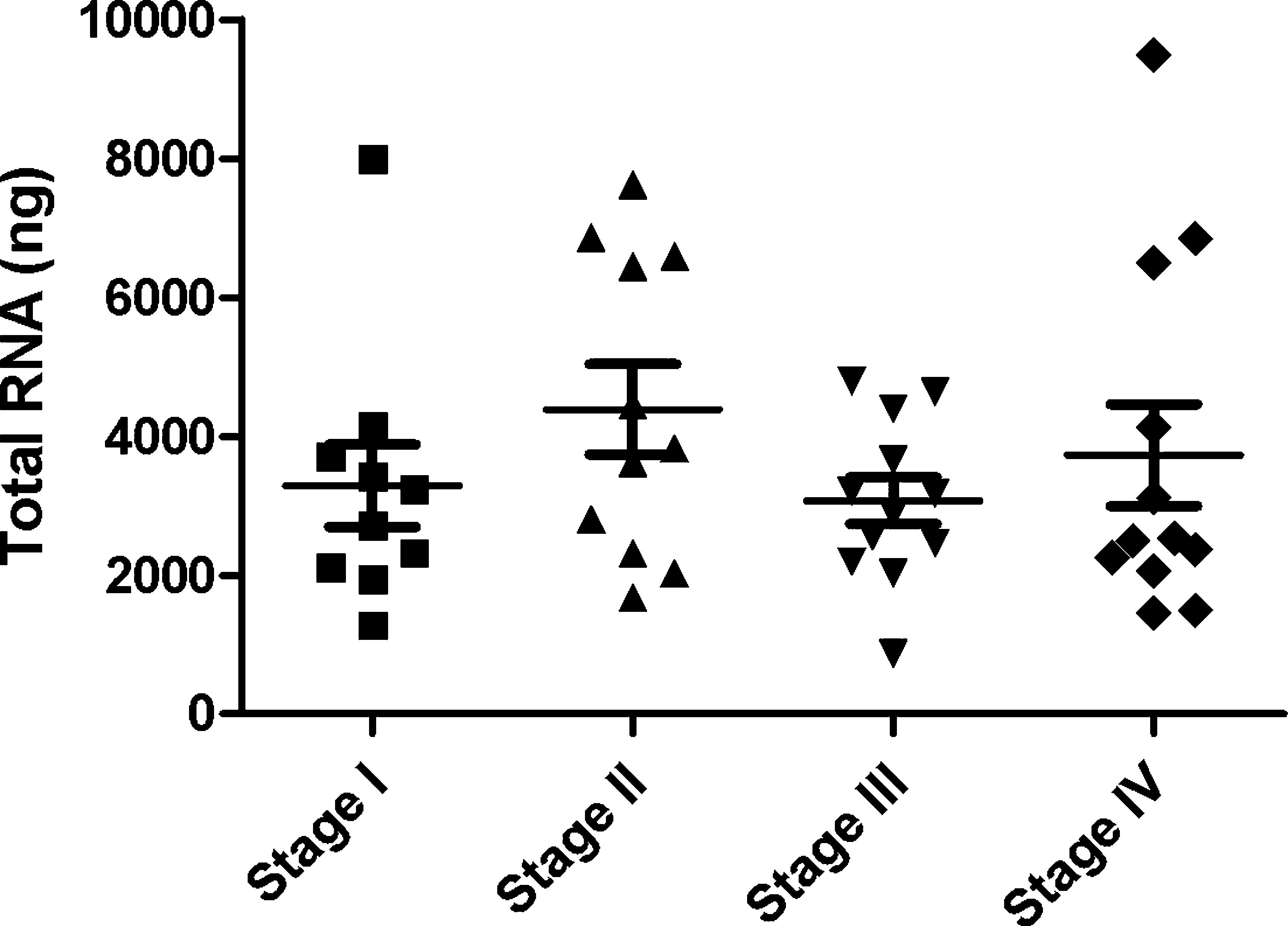
Total RNA from 45 blood samples from patients with stages I–IV malignant melanoma stored for varying lengths of time. Each point is the yield from a single patient sample, with the means for each stage shown by the thin horizontal bar. The standard errors are also shown. The mean yield was 3617 ng for all samples, with no differences in total amounts of RNA by stage of disease.
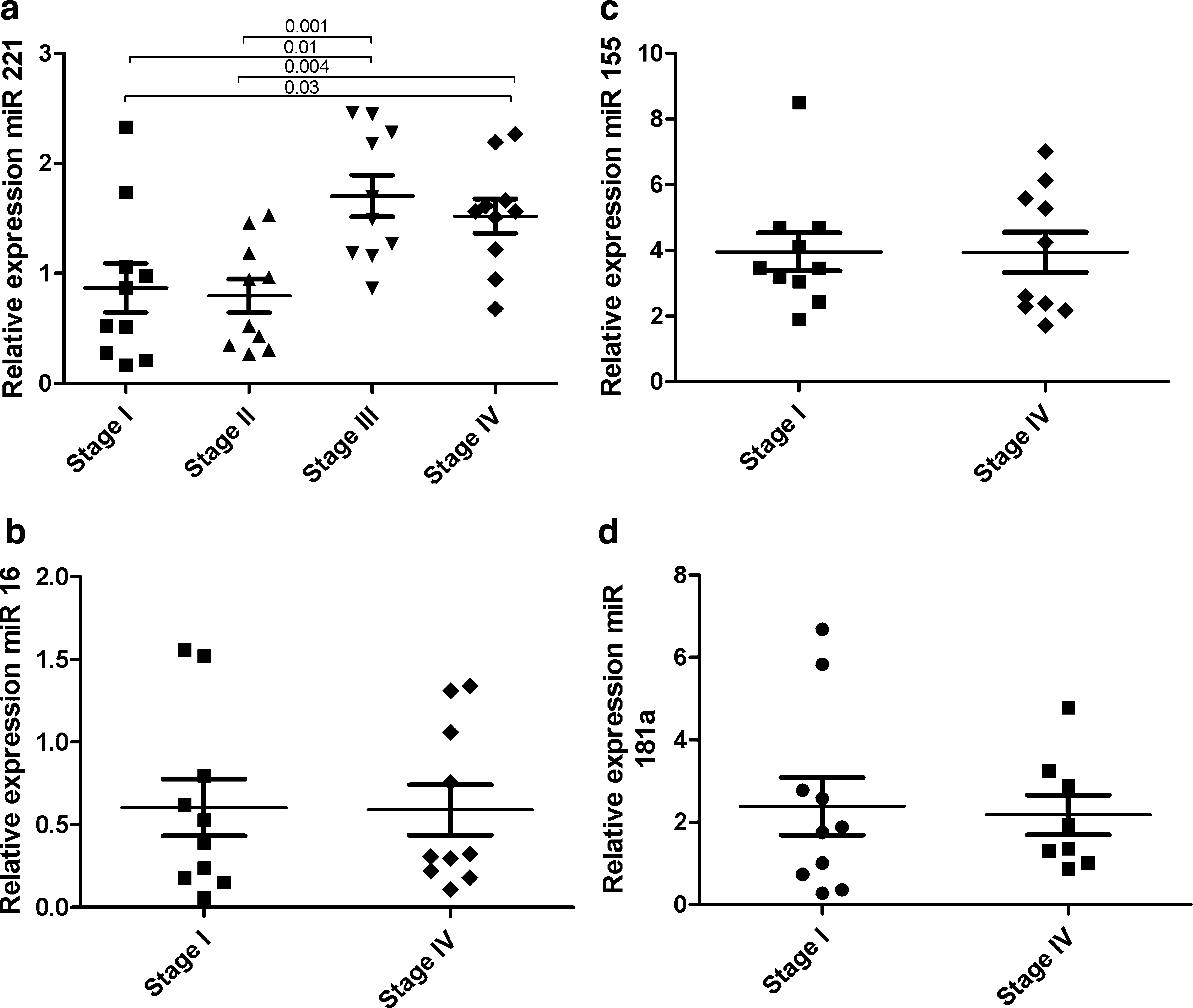
Relative expression of 4 different miRs relative to U6 in blood samples shown by stage of melanoma. Each point represents a single patient sample and the horizontal bars are mean values and standard error of the mean.

A small representation of stage I and stage III melanoma patients were analyzed for p27 mRNA levels. Amplification and quantitation was performed as described in the methods section using actin as the internal control gene.

Discussion
miRs are a recently discovered class of small RNAs that have important regulatory properties in almost all cellular and biological systems. miRs are frequently encoded in larger RNAs, which are processed to yield small-molecular-weight (19–25 nt) molecules whose major function appears to be regulation of gene translation by binding to the 3′UTR of mRNAs. A single miR may be involved in the regulation and coordination of multiple genes. Alterations in expression, binding, and function have been described in many human diseases. Recovery of miRs from both frozen and nonfrozen tissues stored for long periods has been well documented.5,12–14 There are, however, little data about miR recovery from stored blood samples. Kruhoffer et al. 3 examined recovery of total RNA and miR from blood cells stored in the same PAX system as used here. Their recovery of total RNA (mean: 5900 ng) was similar to that in the present study (mean: 3617 ng). They also demonstrated that recovery of total RNA was stable for at least 2.5 years. They did not report on the viability of miR recovery over time. In contrast to our study, they added an extra step to concentrate and obtain adequate amounts of miRs for locked nucleic acid-based oligonucleotide microarrays. Using the array method, they detected 42 miRs in blood samples and validated expression profiles using quantitative PCR for 3 of the miRs.
In the present study, we have demonstrated that miRs can be recovered, amplified, and quantitated without concentration directly from blood RNA stored in the PAXgene RNA Blood system for as long as 3 years. Further, we have shown that miR recovery is independent of total RNA recovery. In our study, we concentrated on a well-known and widely expressed miR, miR-221. We were able to show that there were differences in expression of this miR in different stages of melanoma, indicating that our ability to amplify this miR was likely a specific event and, in the future, may have importance in understanding the development and staging of melanoma. More importantly, for now, this suggests that other miRs expressed by blood cells can be quantitated in other human diseases for diagnostic, prognostic, and therapeutic purposes. This may have particular importance for hematologic neoplasms.
These studies are ongoing and we are presently examining the recovery of multiple miRs from blood samples stored over longer periods of time and under various conditions. We hope that these studies will be of value to biorepositories and investigators from various disciplines.
Footnotes
Acknowledgment
This work was supported by the Moore Family Foundation.
Author Disclosure Statement
No competing financial interests exist.