
Abstract
In the past decade, it has increasingly been reported that epigallocatechin-3-gallate (EGCG), a major catechin derivative extracted from Green tea, has various bioactivities, including a cell-protective action on mammalian cells and tissues. In this study, we have tested a commercial preservation solution containing EGCG (Theliokeep®) in both two- and three-dimensional cultures of human periosteal sheets, which have been used as an osteogenic grafting material for periodontal regenerative therapy. When periosteal sheets were 3D-cultured on collagen mesh, cell viability was maintained for 2 days using the hypothermic EGCG preservation solution. Replenishment of EGCG solution with 2-day intervals prevented the time-dependent decline in cell viability at 3 days and later. As observed in nonpreserved control cultures, most cells were positive for proliferating cell-nuclear antigen (PCNA) in the cultures preserved at 4°C in the EGCG solution, whereas PCNA-negative cells were increased in the cultures preserved at 4°C in the MesenPRO medium. In periosteal sheets 2D-cultured in plastic dishes, the EGCG solution occasionally was associated with vacuole formation in the cytoplasm, but cells could again expand in the culture medium at 37°C. As observed in the nonpreserved periosteal sheets control, the osteogenic induction upregulated alkaline phosphatase in those cells and tissues preserved in the EGCG solution. The EGCG solution protected cells from the cold shock-induced membrane phospholipid peroxidation. Our data suggest that the EGCG solution acts as an antioxidant to protect periosteal cells from cold shock and preserves cells under chilled conditions. The limited period of preservation time could be expanded by repeating replenishment of the EGCG solution or by optimizing the formula to be more favorable for human periosteal sheets without sacrificing cell viability. This methodology of preserving human cultured periosteal sheets with EGCG would be expected to support and spread the clinical use of regenerative therapy with autologous periosteal sheets.
Introduction
In the past 5 years, we have investigated a therapeutic methodology using cultured autologous periosteal sheets for periodontal regeneration and successfully treated more than 70 patients.7,8 Briefly, this methodology utilizes small periosteum tissue segments collected from patients' periodontal tissue that is explanted in culture to form sizable cell multi-layered sheets. 9 These cell multi-layered sheets are then implanted into the site of a periodontal osseous defect. However, the major disadvantage of this methodology is its time-consuming process. Usually, it will take 6 weeks until implantable cell cultured sheets are prepared. This may pose a challenge in arranging therapeutic schedules among patients, dentists, and technicians in cell-processing centers.
To solve this problem, periosteal sheets in the process of preparation should be safely preserved for relatively short periods of time without further culturing. In a previous study, 10 we demonstrated that the central regions of periosteal sheets, which are excised from the peripheral regions, could be cryopreserved with 10% dimethylsulfoxide-containing fetal bovine serum at −75°C, when the periosteal cells enter the early growing phase. After thawing, cells could again be explanted from these central regions and utilized to form periosteal sheets, as observed in native periosteum tissue segments. However, this methodology requires additional cultivation after thawing, and therefore, hospital clinics could not use periosteal sheets cryopreserved and transported from the cell-processing centers to these clinics for regenerative therapy unless the hospitals possess a cell-culture facility.
If periosteal sheets could be preserved under chilled conditions, and if re-cultivation of preserved periosteal sheets is not required prior to clinical use, this practical problem regarding the scheduling of patient treatment would be solved. In this article, we report the performance of the commercial preservation solution containing EGCG tested with our periosteal sheet cultures.
Materials and Methods
Isolation and culture of human periosteal sheets
Human periosteum tissue segments were aseptically dissected from the periodontal tissues of the healthy periodontal buccal tissue side of the retromolar area of the mandible in non-smoking volunteers.9–11 Small tissue segments (1×1 mm) were initially placed in 40-mm culture dishes and cultured in Medium 199 (Invitrogen, Carlsbad, CA) supplemented with 10% fetal bovine serum (FBS) (Invitrogen), 25 μg/mL ascorbic acid 2-phosphate and antibiotics or MesenPRO-RS™ (Invitrogen) supplemented with 2% FBS and 25 μg/mL ascorbic acid 2-phosphate in humidified 5% CO2, 95% air at 37°C. When uniform cell outgrowth was observed around the cultured periosteum tissue segments (∼9 days), these periosteum segments were cut and transferred onto atelocollagen mesh made of bovine epidermis (Integran®; Koken, Tokyo, Japan). For osteogenic induction, human periosteal sheets cultured on plastic dishes were treated with 3% KE-200, a commercial osteogenic-inducing agent (DS Pharma, Osaka, Japan) that contained dexamethasone, β-glycerophosphate, and L-ascorbic acid.9–11
In some experiments, we used dispersed periosteal cells. These cells were trypsinized from periosteal sheets cultured in Medium 199+10% FBS for 3–4 weeks, and then subcultured in the same medium.
All subjects enrolled in this study positively responded to an Informed Consent which was approved by the ethical committee for human subject use at Niigata University Medical and Dental Hospital in accordance with the Helsinki Declaration of 1975 (revised in 2000) and this protocol was approved on June 22, 2006.
Preservation of cultured periosteal sheets
Periosteal sheets were cultured for a total 20–24 days, then subconfluent cultures of dispersed periosteal cells were washed twice with HBSS and then incubated at 4°C with the preservation solution (Theliokeep®; Bio-Verde, Kyoto, Japan). Unless otherwise described, this solution was not replaced with fresh solution during the preservation at 4°C. At the end of incubation in the preservation solution, periosteal sheets were washed twice with PBS and incubated with Medium 199+10% FBS in a CO2 incubator for 2 h prior to the WST-8 assay (2 h) (Dojin, Kumamoto, Japan) for determination of cell number.
To confirm that EGCG, among the bioactive contents of the preservation solution, played an important and major role in protecting cells from cold shock, EGCG (Sigma, St. Louis, MO) was added to Medium 199+1% FBS at a concentration of 50 μg/mL and applied to the dispersed periosteal cells as indicated above.
Histological examination
Periosteal sheets cultured on plastic plates were harvested, fixed, and embedded in paraffin, while periosteal cells cultured on collagen mesh were directly fixed and embedded in paraffin, as described previously. 9 The blocks were sectioned sagittally at a thickness of 6 μm for staining with hematoxylin and eosin (HE), or Mason-Trichrome (MT).
For immunohistochemical staining,10,11 the sections were subjected to antigen-retrieval, blocking of endogenous peroxidase, and blocking of nonspecific binding; the sections were probed with mouse monoclonal anti-PCNA (1:100) (Santa Cruz Biotechnology, Santa Cruz, CA), or rabbit monoclonal anti-CD146 antibody (1:100) (Abcam), followed by probing with ImmPRESS® anti-mouse or rabbit IgG (Vector). Immunoreactive proteins were visualized by DAB substrate solution (Kirkegaard & Perry Laboratories, Inc., Gaithersburg, MD).
Staining for ALP activity of fixed periosteal sheets was performed as described previously.9–11
Detection of apoptotic cells
Apoptotic DNA fragmentation was detected in situ by 3′-end labeling with the Apoptosis Detection kit, (Wako, Osaka, Japan). This kit is based on the TUNEL procedure. Briefly, the de-paraffinized sections were permeabilized with the protein digestion enzyme, and DNA strand breaks were labeled with fluorescein-12-dUTP. The sections were then probed with HRP-conjugated anti-fluorescein antibody and visualized with diaminobenzidine (DAB). Counter-stain was performed faintly using hematoxylin.
Detection of membrane phospholipid peroxidation
Dispersed periosteal cells were washed twice with HBSS and treated with 5 μg/mL diphenyl-1-pyrenylphosphine (DPPP) (Dojin) in HBSS for 15 min in a CO2 incubator. 12 Cells were again washed twice with HBSS and subjected to hypothermic preservation, as described above. At the end of the incubation period, cells were enzymatically detached, smeared on slide glasses, and observed under a fluorescent microscope (Nikon, Tokyo, Japan) with the wavelengths of DAPI (excitation, 340–380 nm; dichroic mirror, 400 nm; emission, 435–485 nm).
Results
Periosteal sheet preparation using collagen mesh
In a parallel study (Uematsu et al., manuscript submitted), we have found that human periosteal sheets can be tightly secured on collagen mesh (Integran®) and easily form cell multi-layers in the medium formulated for supporting stem cells, MesenPRO-RS™. In terms of thickness, the resulting periosteal sheets resembled native periosteum tissue. Therefore, we have chosen the collagen mesh as a scaffolding material to prepare a 3D-reconstructed model of periosteal sheet in this study.
The time-course growth (proliferation and penetration) of periosteal cells explanted from the original tissue segments cultured on collagen mesh is shown in sagittal sections in Figure 1. In the initial period (∼5 days after transfer) (Fig. 1A), cells slowly expanded from the original periosteum tissue segments and grew on and just below the surface. In the following period (∼10 days), cells started vigorous penetration into the deeper regions of the collagen mesh (Fig. 1B) and eventually reached the bottom surface (15∼20 days) (Figs. 1C and 1D). The WST-8 assay demonstrated the time-course increase in cell number in periosteal sheets (Fig. 1E). The maximal number of cells reached the steady-state level at 13 days and could be sustained at this level for at least an additional 9 days. Based on these initial findings, periosteal sheets cultured on collagen mesh for 20–26 days were subjected to the following experiments.

Time-course proliferation and penetration of periosteal cells explanted from the original periosteum tissue segments cultured on collagen mesh.
Effects of hypothermic preservation on cell viability and proliferation
Periosteal sheets cultured on collagen mesh (for 20–26 days), which were fully saturated with periosteal cells, were preserved in the EGCG solution at 4°C. The time-course loss of cells in preserved periosteal sheet cultures on collagen mesh is shown in sagittal sections in Figures 2A–2D. At Day 1 of preservation, cells were present on and just below the bottom surface (Fig. 2A). However, cells gradually disappeared from the side of the bottom surface with time (Figs. 2B–2D). On the other hand, when periosteal sheets were preserved in the MesenPRO medium at 4°C, most cells were dead within 2 days (Fig. 2E). The WST-8 assay demonstrated a decrease in cell numbers in a quantitative manner (Fig. 2F). The cell numbers were sustained by the EGCG solution at initial levels for 2–3 days, but declined with time in the subsequent 2–3 days, and reached a plateau level within 5–7 days. Interestingly, refreshment of the EGCG solution at 2-day intervals prevented the steep decline and successfully sustained cell viability.
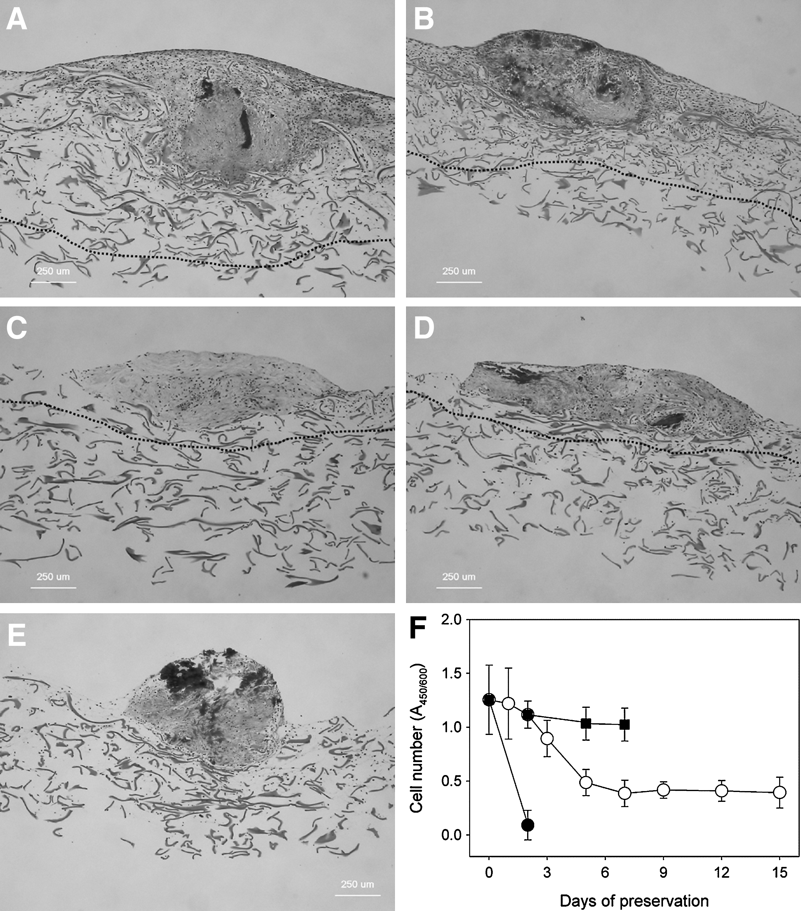
Time-course loss of cells in preserved periosteal sheet cultures on collagen mesh. As described in the legend of Figure 1, periosteal sheets cultured on collagen mesh for 20–26 days were subjected to preservation with the EGCG solution at 4°C.
To identify whether the reduced cell number was due to apoptotic cell death, we have immunohistochemically examined expression of PCNA and DNA fragmentation. Immunohistochemical findings of proliferating and apoptotic cells in periosteal sheets are shown in Figure 3. Periosteal sheets cultured on collagen mesh in the MesenPRO medium for 24 days were preserved at 4°C in the EGCG solution for 10 days or in the MesenPRO medium for 2 days. In the control of nonpreserved periosteal sheets that were cultured in the MesenPRO medium for 29 days, PCNA-positive cells (more than 80%) were widely distributed in the distal region of the explanted cell multi-layer (Fig. 3A), while apoptotic cells were not found in this region (Fig. 3D). Similar findings were obtained from periosteal sheets preserved in the EGCG solution (Figs. 3B and 3E). In contrast, the number of PCNA-positive cells was substantially decreased in the periosteal sheets preserved in the MesenPRO medium (Fig. 3C). However, apoptotic cells were rarely detected in this region (Fig. 3F). The positive control for apoptotic cells was prepared by treating the section with DNase. Almost all cells appeared positive under the same experimental conditions (Fig. 3G).

Immunohistochemical findings of proliferating and apoptotic cells in periosteal sheets. Periosteal sheets cultured on collagen mesh in the MesenPRO medium for a total of 24 days were preserved at 4°C in the EGCG solution for 10 days
Effects of hypothermic preservation on cell morphology and membrane phospholipids
Here, the scaffolding material was switched from collagen mesh to a transparent plastic culture dish, and possible changes in cell morphology were examined microscopically. Cell outgrowth from periosteal sheets preserved in the EGCG solution is shown in Figure 4. Cell expansion from periosteal sheets, which were 2D-cultured in Medium 199+10% FBS for 21 days and subsequently preserved at 4°C in the EGCG solution for 2 days was demonstrated. When preserved periosteal sheets were re-cultured in Medium 199+10% FBS at 37°C, the outline of the periosteal sheet was traced as shown in Figure 4A. At 24 h, periosteal cells grew across the line (Fig. 4B). Induction by cold shock of vacuole formation in periodontal cells is shown in Figure 5. In several periosteal sheets cultured for 21 days in plastic dishes, vacuoles were formed in the cytoplasm of some, but not all, cells during hypothermic preservation with the EGCG solution for 2 (Fig. 5A), 4 (Fig. 5B), and 7 days (Fig. 5C). In addition, those cells could not be excluded or rescued by an additional 96-h re-cultivation with Medium 199+10% FBS (Fig. 5D). As a control, periosteal sheets were directly preserved in Medium 199+10% FBS at 4°C for 8 h. At this early time point, almost all cells in the peripheral region became round (Fig. 5E) and detached with further incubation at approximately 24 h (data not shown).

Cell outgrowth from periosteal sheets preserved in the EGCG solution. Periosteal sheets cultured for 21 days in plastic dishes in Medium 199+10% FBS were preserved in the EGCG solution at 4°C for 2 days
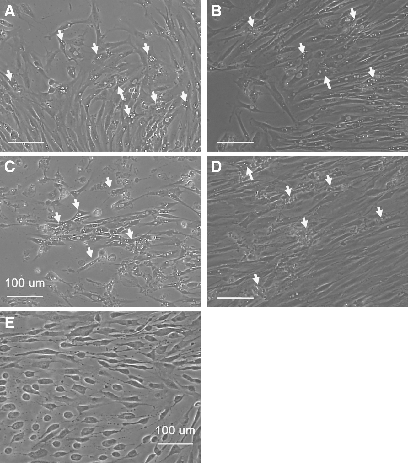
Induction by cold shock of vacuole formation in periosteal cells. Periosteal sheets cultured on plastic culture dishes in Medium 199+10% FBS for 21 days were preserved in the solution containing EGCG at 4°C for 2
To reveal the mechanism of EGCG action, we have examined the initial morphological changes of periosteal cells, which were enzymatically dispersed from periosteal sheets in sparse cultures. Protective effects of EGCG solution on the morphological changes in dispersed periodontal cells and membrane phospholipid peroxidation caused by cold shock is shown in Figure 6. As shown in Figure 6A, several cells became circular by hypothermic preservation with Medium 199+10% FBS for 20 h. In the EGCG solution at 4°C (Fig. 6C), cell outlines became clearer than that of normal cultures maintained at 37°C (Fig. 6E), but cells were not converted to a round shape. To obtain supporting data for the major involvement of EGCG in this action, Medium 199 was supplemented with 1% FBS and 50 μg/mL EGCG and used for hypothermic preservation. As expected, cells were well preserved without formation of round cells (Fig. 6F). To examine membrane phospholipid peroxidation, cells pre-labeled with DPPP were detached and smeared on glass slides for microscopic examination (Figs. 6B and 6D). Under chilled conditions, many bright spots were distributed in cells preserved in Medium 199+10% FBS at 4°C for 20 h (Fig. 6B), but not in cells preserved in the EGCG solution (Fig. 6D).

Protective effects of the EGCG solution on the morphological changes in dispersed periosteal cells and membrane phospholipid oxidation caused by cold shock.
Finally, to examine the effects of hypothermic preservation on osteoblastic differentiation, we have treated periosteal sheets, which were preserved in the EGCG solution for 2 or 7 days, with the osteogenesis-inducing agents (3% KE-200). ALP expression in preserved periosteal sheets is shown in Figure 7. Preserved periosteal sheets were capable of upregulating ALP in response to the osteoinduction (for 8 days) both after 2- (Fig. 7A) and 7-day preservations (Fig. 7B), as much as the control of nonpreserved cultures (Fig. 7C).

ALP expression in preserved periosteal sheets. Periosteal sheets, which were cultured with Medium 199+10% FBS for 21 days, were preserved with the EGCG solution at 4°C for 2
Discussion
In a previous study, 10 we attempted to preserve periosteal sheets at −75°C, and found that the periosteal sheets consisting of proliferating cells could be successfully cryopreserved. After thawing, cells were immediately explanted to form complete periosteal sheets with cell multi-layers. However, this methodology required re-cultivation in order to release dimethylsulfoxide contained in the preservation solution as cryoprotectants from cells and to re-gain cellular activity prior to clinical use. Practically, since the peripheral regions were removed for efficient retrieval after thawing, it would take 3–4 weeks to form an implantable size section of periosteal sheets in culture after thawing. Therefore, we believe cryopreservation methodology is not suitable for relatively short-term preservation of periosteal sheets. For example, in the case of coordinating and scheduling therapy between hospital clinics and cell-processing centers, short-term preservation of periosteal sheets would be advantageous.
To solve this problem, we have explored and attempted to develop a hypothermic preservation methodology. In a series of experiments, we have found that the preservation solution containing EGCG could be used for the purpose of preserving periosteal sheets for relatively short periods of time. Essentially as reported by other investigators,1–3,6,13 in our study, the EGCG solution appeared to effectively protect cells from membrane phospholipid peroxidation caused by cold shock. 14 Since the phospholipid oxidation product acts as a cytotoxic agent, 15 we suggest that the EGCG solution could effectively protect viable cells from the cold shock-induced cell death.
While our results demonstrate the value of short-term preservation with EGCG under refrigerated conditions in preserving cells and tissues, this methodology still contains some disadvantages. The first one is that the viability of periosteal cells can be maintained only for 2–3 days. Second, vacuole formation cannot be completely prevented in the cytoplasm of some cells. However, we did not observe substantial growth arrest or apoptotic cell death in the re-cultivation process. It has often been reported that EGCG causes growth arrest through upregulation of p21WAF1 or other components involved in regulation of the cell cycle,4,5,16 and these initial changes are followed by apoptotic cell death. The reason why EGCG did not cause apoptotic cell death in our periosteal cells might be related to their nature; that is, human periosteal cells contained in periosteal sheets prepared by explant cultures constitutively express p21 WAF1 at higher levels, which is not deleterious to cell viability (Kawase et al., unpublished observation). Therefore, we speculate periosteal cells may not further upregulate p21WAF1 in response to applied EGCG. However, further investigation should be performed to clarify this mechanism of action, and thereby to optimize the hypothermic preservation of periosteal sheets at the ordinary temperature as reported elsewhere.1–3,6
Compared to our previous cryopreservation methodology, one more advantage of this methodology is that the preservation solution does not contain FBS. We routinely expand periosteal sheets with medium supplemented with 10% FBS as approved by our University Hospital's Institutional Review Board (IRB), but we recognize that the current medium should be replaced with xeno-free medium for preparation of greater clinically effective and reliable “grafting materials” as soon as possible. Therefore, we are now vigorously screening and investigating the applicability of serum-free (or low serum-containing) stem-cell media. When an appropriate medium is found, in combination with the EGCG preservation solution, it will provide not only the flexibility in scheduling patient therapy but more reliability in ongoing periodontal regenerative therapy using autologous periosteal sheets.
Conclusions
Our data suggest that the preservation solution containing EGCG, Theliokeep®, is useful for a short-term refrigerated preservation of human periosteal sheets. The opportunity to preserve human periosteal sheets by this technique would help clinicians in overcoming challenges posed in transporting periosteal sheets from the clinical laboratory or cell-processing facility to the treatment center or clinic and also allow timelier scheduling of patients. Further optimization of the preserving solution containing EGCG (e.g., addition of other supplemental factors) would be expected to improve the performance of this solution in preserving the periosteal sheet cultures for clinical use.
Footnotes
Author Disclosure Statement
The authors declare that they have no conflicting financial interests.
This project was funded and supported by a Grant-in-Aid for Scientific Research from the Ministry of Education, Sports, Science, and Technology, Japan, and by Practical Application research from the Japan Science and Technology Agency.