
Abstract
Red blood cell (RBC) units are administered routinely into patients expressing a wide range of acute and chronic conditions (e.g., anemia, traumatic bleeding, chronic diseases, and surgery). The modern blood banking system has been designed to answer this need and assure a continuous, high quality blood supply to patients. However, RBCs units can be stored under hypothermic conditions for only up to 42 days, which leads to periodic shortages. Cryopreservation can solve these shortages, but current freezing methods employ high glycerol concentrations, which need to be removed and the cells washed prior to transfusion, resulting in a long (more than 1 hour) and cumbersome washing step. Thus, frozen RBCs have limited use in acute and trauma situations. In addition, transportation of frozen samples is complicated and costly. Freeze drying (lyophilization) of RBCs has been suggested as a solution for these problems, since it will allow for a low weight sample to be stored at room temperature, but reaching this goal is not a simple task. We studied the effect of different solutions (IMT2 and IMT3) containing trehalose and antioxidants or trehalose and human serum albumin, respectively, on freezing/thawing and freeze drying of RBCs. In addition, we evaluated the effect of cells concentrations and cooling rates on the post thaw and post rehydration recoveries of the RBCs. Finally, we developed a new radio frequency (RF) lyophilization device for a more rapid and homogeneous sublimation process of the frozen RBCs samples. Recovery and free Hb were measured as well as oxygen association/dissociation and cell's deformability. We found that IMT3 (0.3 M trehalose and 10% HSA) solution that was directionally frozen at a rapid interface velocity of 1 mm/sec (resulting in a cooling rate of 150°C/min) yielded the best results (better than IMT2 solution and slow interface velocity). Freeze thawing gave 100% survival, while freeze drying followed by rehydration with 20% dextran-40kDa solution resulted in 75% survival. However, recovery following freeze drying was possible only when 20% Dextran-40 solution was used as the rehydration medium. The rehydrated cells were not stable upon an eight-fold dilution. The RF lyophilization system increased the sublimation rate more than twice compared to conventional drying and maintained a high survival rate of the RBCs after partial drying.
Introduction
Degradation of the RBC under these storage conditions continues, and has been demonstrated through alterations in cell content of ATP, 2,3 DPG, and by alteration in RBC membrane deformability properties.2–4 These changes can be demonstrated as early as 2 weeks after collection, making the use of "old" units undesirable in emergency cases such as severe bleeding.
Cryopreservation allows for long-term storage of RBCs units. However, only units collected from rare type donors are mainly cryopreserved today, accounting for less than 1% of the collected blood units. There are two accepted cryopreservation methods; both employ the use of glycerol as a cryoprotectant. One method uses 20% (v/v) glycerol and the unit is stored in liquid nitrogen (LN) tanks, and the other uses 40% (v/v) glycerol and storage is done in −80°C mechanical freezers. 5 The last method is the most commonly used today.
The use of cryopreserved RBCs units is limited due to the cumbersome handling required for freezing and the complicated, time-consuming process required for thawing and deglycerolizaiton. Both these processes entail a significant increase in unit cost. The thawing and deglycerolization process renders the use of cryopreserved units impractical in battlefield and trauma scenarios. In addition, transportation of cryopreserved units is also costly, more difficult, and prone to hazards.
It has been shown that cryopreserved units can be stored successfully for even up to 37 years, although currently the FDA approves the use of units stored up to 10 years. 6 In addition, cryopreservation maintains the RBC quality at the time of freezing as it is currently the only method that puts the cells in a state with little or no active metabolism, whereas at hypothermic storage there is still a degree of metabolism as seen with associated RBCs storage lesions. 2
It is these two difficulties, the short shelf life of RBCs stored in hypothermia and the complexity of the logistics involved in handling cryopreserved RBC units, that leads to the need for a simple method for storing and handling blood at high quality for long periods of time. Lyophilization of RBCs has been highly desirable for decades, dating back to the early work of Meryman. 7 More recently, Goodrich et al. reported some success in this regard, work that was expanded and extended by work at Crowe's laboratory.8,9 Success by all those workers was limited, so we have set out to re-visit this field and seek solutions to the problems encountered by these previous groups of investigators.
In order to freeze dry successfully, several conditions need to be met: (1) The cell membrane has to be stabilized in order to minimize damage which occurs during the different lyophilization steps: (a) During freezing mechanical damage caused by ice crystal formation, (the increase in size of ice crystals during slow freezing will increase mechanical damage and osmotic stress due to the increased concentration of solutes) and lipid phase transition as lipids in the cell membrane turn from a liquid form into a gel form;10,11 (b) during the drying process, damage is induced by free radicals, peroxides, browning reactions, cross-linking of proteins, and membrane fusion;12–14 (c) during rehydration there is a lyotropic phase transition as the membrane changes its form, from gel into liquid. 15 (2) The matrix/solution the cells are being lyophilized in needs to have a high glass transition temperature (Tg) in order to support and protect the biological materials for long periods of time at elevated temperatures.
We have developed a directional freezing (DF) method (MTG freezing device), in which controlling ice propagation through the sample is achieved by regulating the velocity of movement through a pre-determined thermal gradient. The DF method permits a precise and uniform cooling rate, which has allowed for successful freezing in the absence of liquid CPAs, of high volume samples and of organs.16–19
In addition to this technology, we have developed lyophilization solutions based on sugars, antioxidants, and proteins (named IMT1, IMT2, and IMT3). Using these solutions, we have been able to lyophilize sheep leukocytes and show that their DNA remained intact after 3 years storage at room temperature. Further, human hematopoietic stem cells that were lyophilized and rehydrated with water were viable and maintained their capacity to grow and differentiate in culture into the different blood lineages. 18 This was the first report to show cells that have undergone complete lyophilization and following rehydration have maintained not only their viability but also their functionality. 18 These findings were confirmed recently by Buchanan et al. 20
The present study is focused on showing the feasibility of freeze drying RBCs in small volumes (1 mL samples) and low hematocrit (Hct) levels of 0.4%. We have evaluated the effect of initial cell concentrations, freezing solution, and DF parameters on recovery rate and other in vitro parameters following freeze thawing and freeze drying.
Materials and Methods
Chemicals were purchased from Sigma-Aldrich (St. Louis, MO) unless otherwise indicated.
Hematology analysis and hemolysis measurement
Total hemoglobin (Hb) and complete blood counts (CBC) were measured using an automatic blood counter (Pentra 60, ABX Diagnostics, France). The hematocrit (Hct) was measured using a MicroHematocrit capillary tube centrifuged at 1000 g for 4–7 min (MPW Medical Instruments, Poland). Plasma Hb and total Hb was measured using the HemoCue device (Ängelholm, Sweden) according to the manufacturer instruction.
The percentage of the free Hb was calculated as follows:
Recovery was therefore defined as:
Percent numerical recovery was defined as:
Morphology assessment
A light microscope Eclips 50i (Nikon, Tokyo, Japan) equipped with a digital camera DXM1200F (Nikon, Tokyo, Japan) used for photographing RBCs samples. Image-J software was used for processing.
Freezing solutions
IMT2 freezing solution preparation
0.1 M trehalose and 0.945 mg/mL Epigallocatechin gallate (EGCG) (>98% purity; Zhejiang Yixin Pharmaceutical, Lanxi, China) dissolved into Ca+2- and Mg+2-free PBS (Biological Industries ltd, Beit Haemek, Israel).
IMT3 freezing solution preparation
0.3 M trehalose, 10% (w/v) human serum albumin (HSA) dissolved into Ca+2- and Mg+2-free PBS (Biological Industries Ltd).
Cell preparation for freezing
pRBC units were ordered from the Israeli blood bank (Magen David Adom). Following a complete blood count (CBC) using the Pentra 60 counter (ABX-Horiba, Montpelier, France) and free Hb determination, RBCs were diluted to yield the indicated cell concentration or Hct with the indicated freshly prepared freezing solution. The cell suspension was divided into glass tubes (MTG-516 freezing apparatuses, Manara, Israel). Sample volume was 1 mL per tube in all experiments performed.
Freezing methods
Freezing was performed in MTG-1314 and MTG-516 apparatuses, (Core Dynamics, Nes Ziona, Israel). The MTG apparatuses are based on the technology of directional freezing (DF), named multi-thermal gradient (MTG), which enables freezing at an accurate cooling rate and the control of ice crystal morphology during the freezing process. 21
Directional freezing with the MTG apparatuses was performed in the following manner:
150°C/min DF (MTG 516–glass tubes)
The freezing machine temperatures were set to: +5°C to −120°C to −130°C; the velocity at which the samples were pushed through the freezing machine was set to 1 mm/sec, resulting with a measured (using thermocouples) cooling rate of 150°C/min.
3.6°C/min DF (MTG 1314–glass tubes)
The freezing machine temperatures were set to: +4°C–block A and a gradient of −10°C to −70°C, the velocity at which the samples were pushed through the temperature gradient was set to 0.2 mm/sec, resulting in a cooling rate of 3.6°C/min. The following freezing samples were kept in liquid nitrogen (LN) until use.
Thawing
Thawing was performed in a 37°C water bath; after thawing, free Hb and cell concentration were determined.
Drying and rehydration
Lyophilization was performed with a commercial lyophilizer, Virtis Encore (VirTis, NY). Drying was performed by transferring frozen samples into pre-cooled shelves (-55°C) for 24 h at a vacuum of 5 mTorr.
Dried samples were rehydrated immediately following lyophilization with double distilled water (DDW), DMEM or Dextran 40 rehydration solution (20% (w/v) Dextran-40 and 10 mM HEPES in DDW) as indicated, warmed to 37°C with a volume of 0.9 mL.
Cell stability assessment following drying and rehydration with Dextran-40 rehydration solution
Following rehydration, cells were diluted in three consecutive cycles, in each cycle cells were diluted in a 1:1 v/v ratio, followed by 5 min incubation prior to the next dilution cycle. This resulted with an 8-fold dilution.
Trehalose to RBCs ratio calculation
In order to determine the trehalose per RBCs ratio, we assumed the weight of RBCs at 40% Hct, and 4% Hct within 1 mL are equal to 400 and 40 mg (1 μL=1 mg). Trehalose weight was calculated according to trehalose concentration within the sample and by its molecular weight; 0.1 M trehalose in a 1 mL sample is equal to 37.8 mg.
Hb saturation assay
The Hb saturation curve and the P50 were determined by the Hemox-Analyser (TCS Scientific Corp, New Hope, PA). Twenty μL of pRBC were added to 5 mL of buffer (135 mmol/L NaC1, 30 mmol/L TES, 5 mmol/L KC1, and NaOH adjusted to pH 7.4±0.02) (TCS buffer; TCS Scientific Corp.), 10 μL of antifoam solution, and 20 μL of 20% BSA. Nitrogen (100%) was bubbled through the sample at a constant rate that resulted in complete deoxygenation within 20 min, followed by reoxygenation with air for 15 min. The analyzer measured the oxygen tension with a standard Clark O2 electrode (model 5331 Oxygen Probe; Yellow Springs Instrument Co., Yellow Springs, OH).
Rheology analysis—Cell membrane deformability
Analysis of the red cell deformability was conducted using the RheoScan-D ektacytometer (Reho Meditech, Seoul, Korea). 22 Briefly, 6 μL pRBC (700 g for 2 min) are loaded into a disposable test kit (K02) and analyzed per device instruction. The device exerts differential pressure on the red cells and measures the associated light diffraction, from which the cell shape can be determined. The deformability is given in units of elongation index (EI) that represent the relative length to width ratio of the cells. For comparing different RBC samples, results are presented at a pressure of 3 Pa.
The system was evaluated with RBCs that were subjected to deliberate decrease of deformability. Two methods for decreasing deformability were employed:
A. Incubating RBCs with increasing concentrations of glularaldehyde B. Incubating RBCs at 49°C for increasing durations.
Deformability analysis by both methods resulted in decreased EI either as a function of glularaldehyde concentration or longer exposure to high temperature, in agreement with previously published results. 3
Drying using the RF system (Chuno®)
The RF system is connected to a conventional lyophilizer Virtis Advantage Plus. The RF unit is composed of an RF chamber, RF power unit, and RF controller. The condenser temperature during the operation was −82°C. Samples were put inside a glass jar that was placed in the RF chamber. The jar was connected via a vacuum line to the commercial lyophilizer. The RF power parameter is set via the RF controller and the pressure parameter is set in the commercial lyophilizer (see Fig. 6).
Statistical analysis
Experiments were performed with duplicates or triplicates for each treatment and performed on blood from two different donors. Data are presented as mean±SD. The difference between treatments were examined by the means of t-tests using the general linear model (GLM) procedure of JMP (SAS Institute Inc. 1994)
Results
Determination of optimal IMT solution and cell concentration for freeze drying
We hypothesized that much of the damage to RBCs that occurs during lyophilization is mechanical in nature. Based on our experience with white blood cells, we believe that it can be significantly reduced by reduction in the initial RBCs concentration prior to freezing.
We therefore sought to determine the numerical recovery rate following freeze thawing and freeze drying of RBC samples having a low cell concentration (∼6 million per 1 mL). Samples were frozen directionally in a rapid (150°C/min DF) or a slow (3.6°C/min DF) fashion with IMT2 and IMT3 freezing solutions. The numerical recoveries following freeze thawing of low concentration RBCs samples frozen with IMT3 in a rapid and slow fashion were 100%±0.0% and 7.3%±1.78%, respectively (Fig. 1).
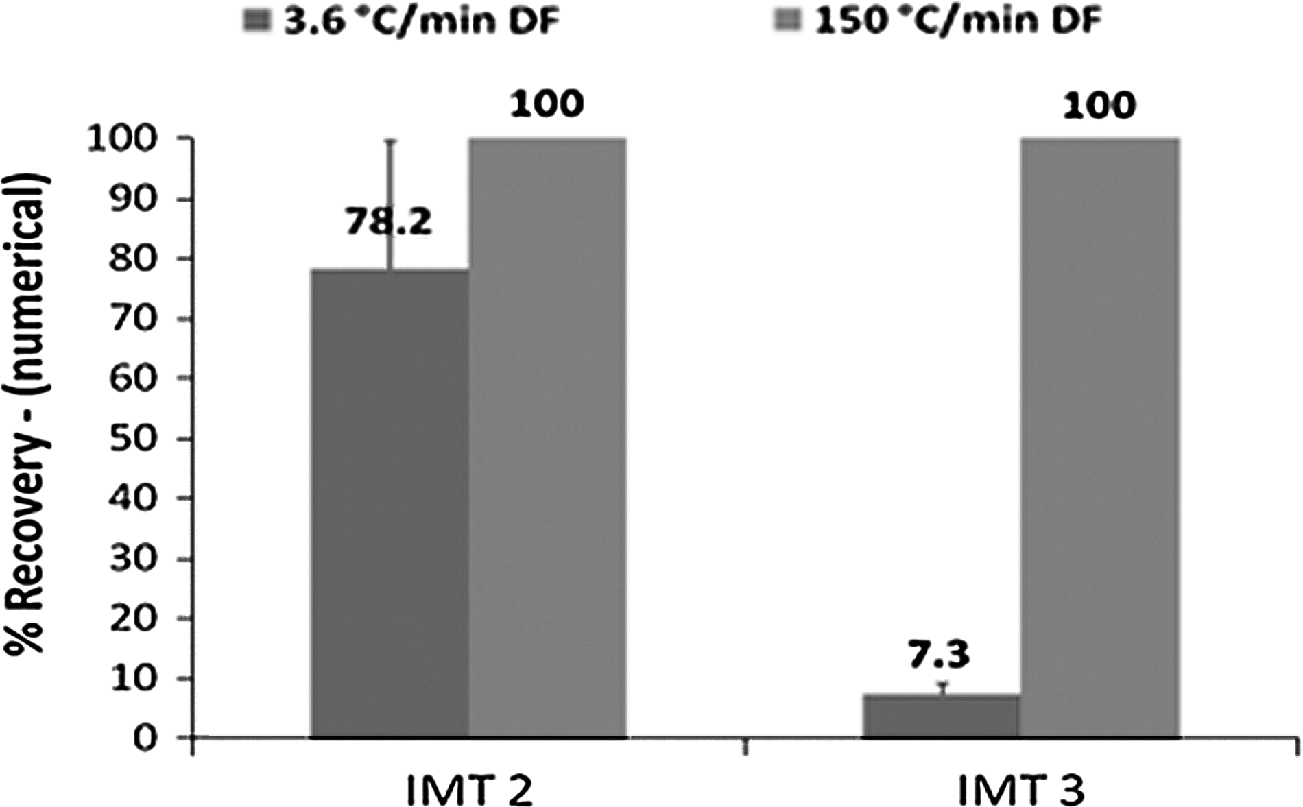
Percentage of cell recovery (numerical) following freeze thawing of low concentration RBC (∼6 million per ml) samples frozen at 150°C/min DF and 3.6°C/min DF in IMT2 or IMT3 freezing solutions. Data are presented as mean±SD (n=12).
Low concentration RBCs samples that were slowly frozen in IMT2 exhibited an increased numerical recovery rate of 78.2%±21.2% in comparison to cells that were frozen in the same fashion with IMT3. Contrary to that, the numerical recovery rate following rapid freezing were the same (100%±0.0%) for both IMT2 and IMT3 samples (Fig. 1). The possibility to freeze slowly is very important as it will produce larger ice crystals which are easier to sublimate during primary drying.
The morphology of RBCs that were frozen in IMT 2 was abnormal (spherocytes, fragmented cells) in both 150°C/min DF and 3.6°C/min DF frozen thawed samples. On the other hand, the frozen thawed samples that were frozen in IMT3 exhibited normal morphology in both rapid and slow DF. The abnormal morphology of the samples frozen with IMT2 might be caused by a high EGCG:cell concentration ratio at this low cell concentration since it was not observed at higher cells concentrations.
The relation between initial cell concentration (prior to freezing) and cell recovery following freeze-thawing and lyophilization (IMT3)
In the following experiment, we have directionally frozen at 150°C/min in IMT3 solution samples containing RBC at 34.5% (3.45·109 cells/mL), 19.3% (1.93·109 cells/mL), 4.6% (460·106 cells/mL), 0.6% (60·106 cells/mL), and 0.08% (8·106 cells/mL) Hct. The samples containing RBC at 34.5%, 19.3%, 4.6%, and 0.08% Hct were frozen and thawed, and samples containing RBC at 4.6%, 0.6%, and 0.08% (low concentration RBCs) Hct were freeze dried and rehydrated with the Dextran-40 rehydration solution.
The recovery and survival rates following freeze thawing of samples containing RBCs at 35.5%, 19.3%, 4.6%, and 0.08% Hct were 53%±8.2%, 82.85%±3.46%, 93.13%±3.16%, and 100%, respectively (Fig. 2).
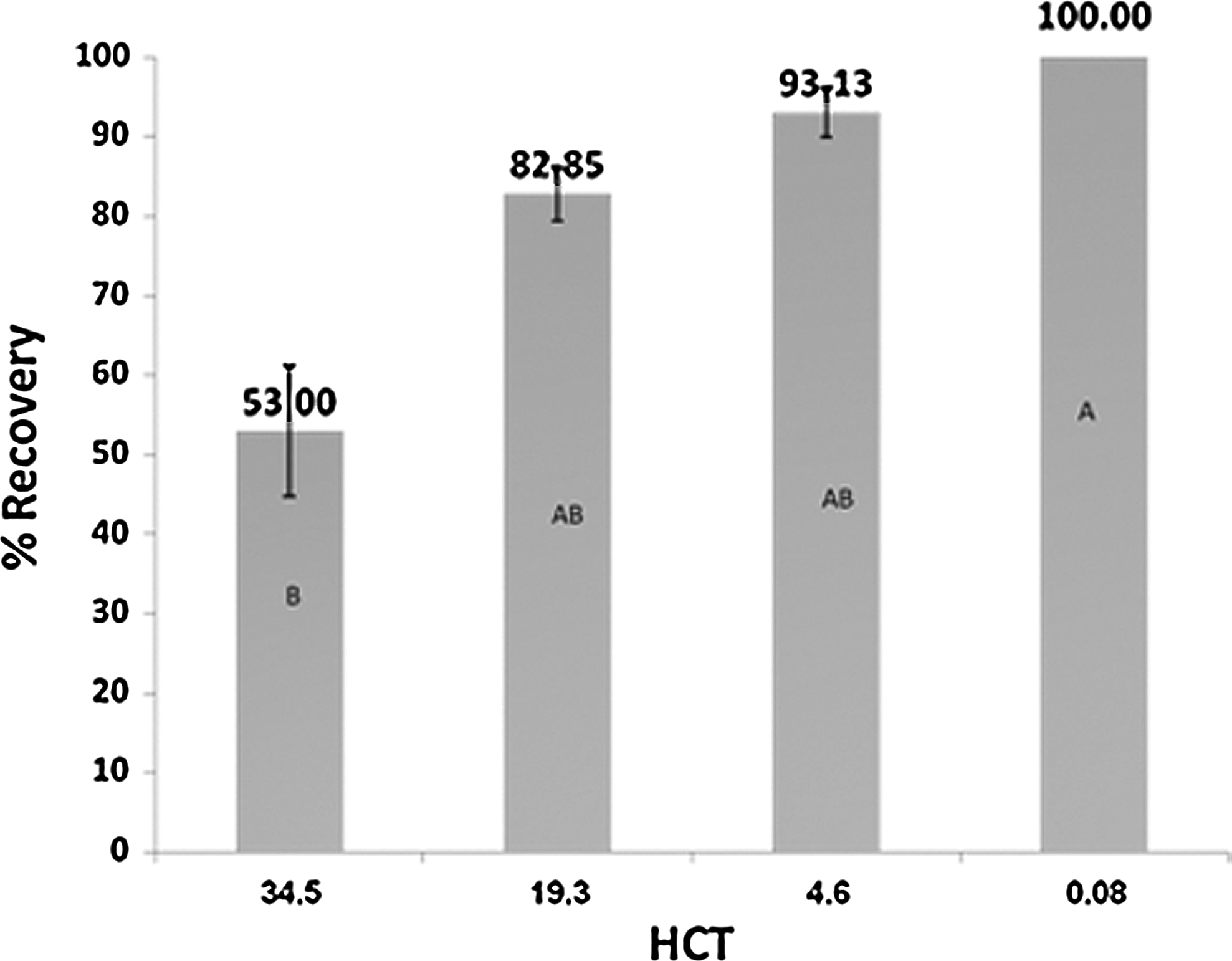
Cell recoveries following freeze thawing of RBCs with IMT3 solution at different hematocrits. Statistical difference is represented by different letters (p<0.05). Data are presented as mean±SD (n=24).
Cell morphology was maintained following thawing. According to the above results, the initial cell concentration highly affects recovery level after freeze thawing; this could be the result of increased mechanical damage which is inflicted on the cells as their number increases. 11 In addition, this trend can also be the result of the decreased ratio of trehalose and HSA (the components of IMT 3) molecules per cell.
The effect of trehalose to RBCs ratio on cell recovery following freeze-thawing
For determining the effect of trehalose to RBC ratio on the post thaw recovery rate RBCs at a Hct of 4% (400·106 cells/mL) and 40% (4·109 cells/mL) were directionally frozen at 150°C/min in IMT3 solution containing 0.1 M, 0.3 M, and 1 M trehalose and 10% (w/v) HSA. For the 40% Hct samples frozen with 0.1 M, 0.3 M, and 1 M trehalose, the trehalose to RBCs ratios were 0.0945, 0.283, and 0.945 mg trehalose to mg RBCs, respectively. For the 4% Hct samples frozen in 0.1 M, 0.3 M, and 1 M trehalose, the trehalose to RBCs ratios were 0.945, 2.83, and 9.45945 mg trehalose to mg RBCs, respectively.
The recovery rates following freeze thawing of samples containing 40% Hct and frozen with 1 M, 0.3 M, and 0.1 M were 71.38%±14.20%, 53.50%±16.73%, and 36.48%±9.98%, respectively (Fig. 3). The recovery rates following freeze thawing of samples containing 4% Hct and frozen with 1 M, 0.3 M, and 0.1 M were 78.21%±4.25%, 92.08%±4.32% and 81.39%±4.8%, respectively (Fig. 3).

Cell recovery percentages of samples containing RBCs at Hct of 40% (4·109 cells/mL) and 4% (400·106 cells/mL) following freeze thawing. The trehalose to lipids ratio is denoted in the yellow quadrates. The samples were directionally frozen at 150°C/min in IMT3 solution with 0.1 M, 0.3 M, and 1 M as indicated. All samples that were frozen at a Hct of 4% were significantly better then samples frozen at a Hct of 40% except the sample that contained 1 M trehalose. Different letters represent statistical difference (p<0.05). Yellow bars indicate the trehalose to RBCs calculated ratios.
The trehalose to RBCs ratio in the 4% Hct sample frozen with 0.1 M trehalose in IMT3 and 40% Hct sample frozen with 1 M trehalose in IMT3 is 0.945945 mg trehalose to mg RBCs, the recovery rates were 71.3±14.2% and 81.4±4.8% for the 40% with 1 M trehalose and 4% Hct with 0.1 M trehalose samples, respectively. These results and the fact that there is an increased recovery rate at trehalose to RBCs ratio of 2.83 mg trehalose per mg RBCs for 4% Hct samples, support the notion that there is an ideal trehalose to RBCS ratio that must be met to yield maximum recovery following freeze thawing and that higher or lower trehalose to lipid ratio will result in decreased recovery rates following freeze thawing. These results indicate that the increased damage that occurs following freeze-thawing in samples containing increased cell concentration in comparison to samples that contain decreased cell concentration is probably due to a combination of the decreased protecting agent (e.g., trehalose) per RBCs ratio and increased mechanical stress due to cell concentration.
Deformability and Hb saturation of RBCs directionally frozen at 150°C/min in IMT3
We also evaluated the cells' deformability in the RheoScan-D ektacytometer (Reho Meditech, Seoul, Korea) (as described in Materials and Methods). The results are shown in Figure 4, and indeed we can see a slight shift of the graph, indicating the cells frozen with IMT3 solution are slightly more rigid than fresh RBCs but they are still within normal range for deformability of RBCs. The overall shape of the Hb saturation curve for fresh pRBCs, pre-freezing, and frozen thawed RBCs samples was similar; however, there was a slight shift to the left (Fig. 5). Oxygen affinity is characterized by the p50. This parameter corresponds to the pO2 at which the Hb is half saturated. The p50 of Hb saturation of the frozen thawed cells was slightly reduced (17.69±1.1) in comparison to the p50 value of the fresh (20.37±0.5) and the pre-frozen (20.62±1.09) samples. This could be the result of decreased 2,3-DPG levels, and the leftward shift indicates that the Hb under study has mildly increased affinity for oxygen so that Hb binds oxygen more easily, but unloads it more reluctantly. This effect is probably reversible and upon 2,3-DPG synthesis, the p50 of Hb saturation of frozen thawed sample will slightly increase and resemble the fresh pRBC value. 23
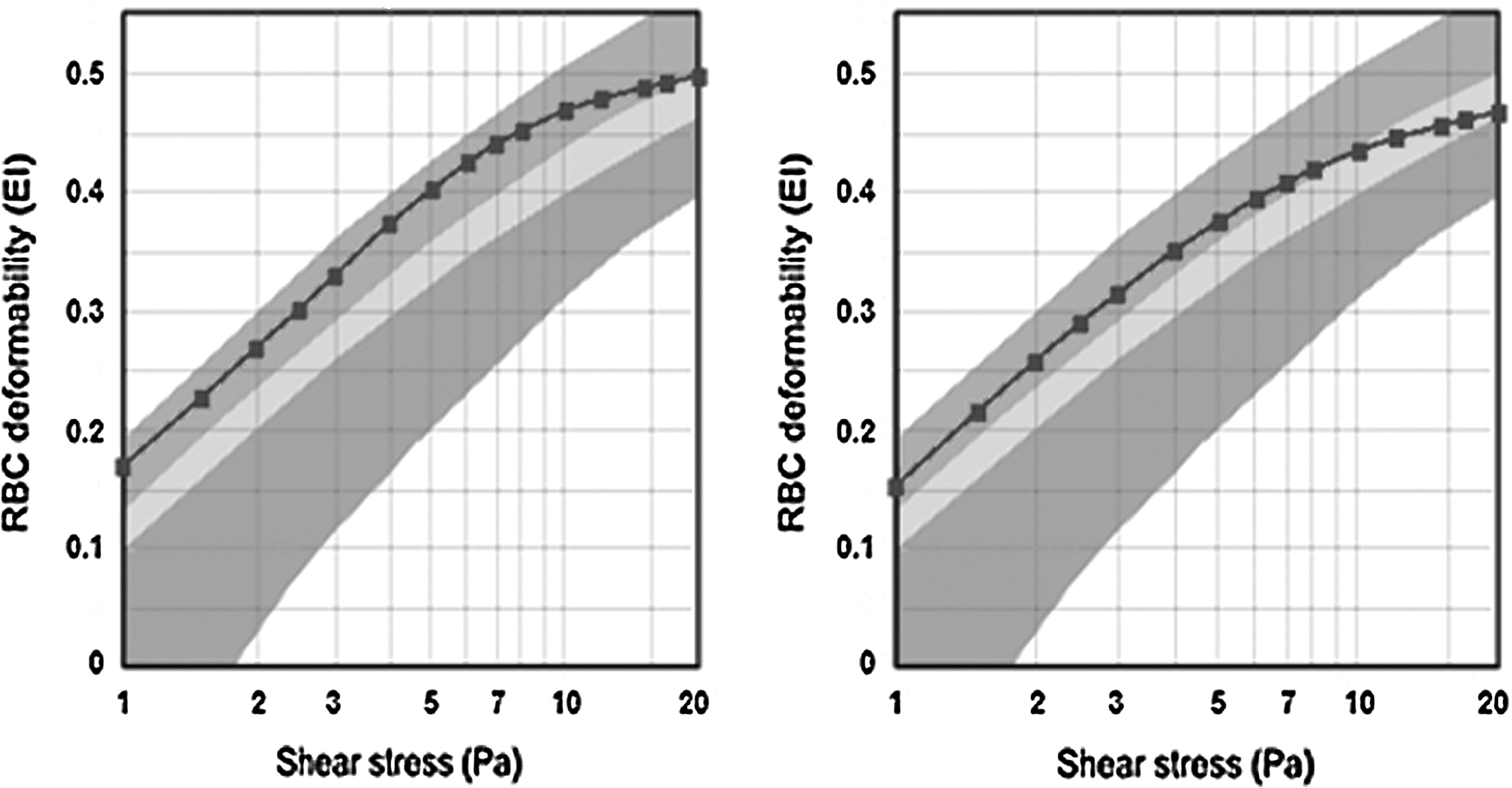
Deformability results of fresh pRBCs and RBCs after freeze thawing with IMT3 solution.
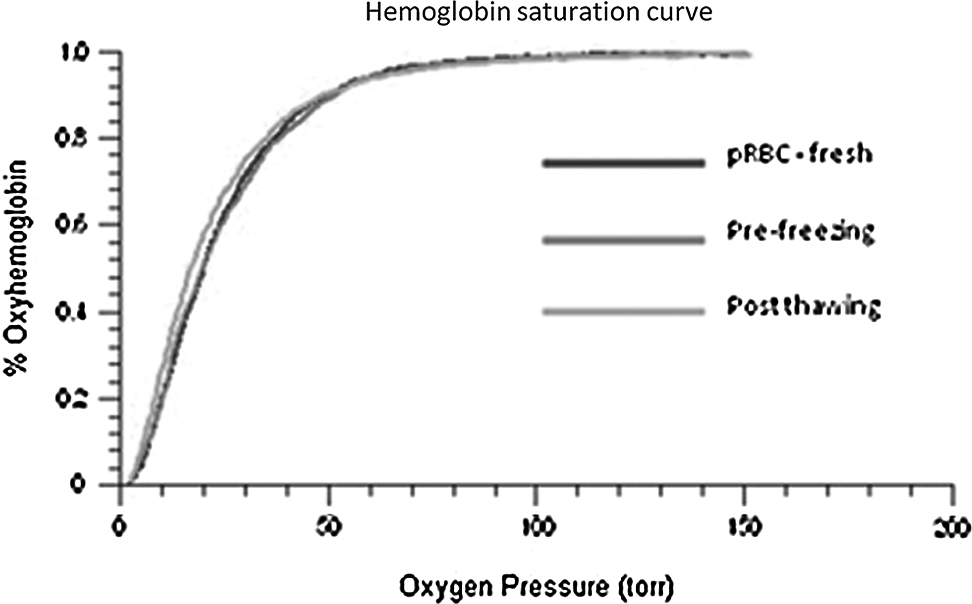
Hb saturation vs. oxygen pressure. The assay was performed with frozen-thawed 4% Hct RBC samples, fresh pRBC, and RBC at 4% Hct with IMT3 freezing solution (pre-freeze).
Lyophilization using RF technology
Our latest technology breakthrough is using RF for drying biologics. RF technology has the ability to homogenize and speedup the drying process and performs this while still keeping the frozen samples at low temperatures.
The utilization of RF technology to improve and speed up the RBC lyophilization process is divided to several iterative tasks:
1. RF simulation and algorithm development: The simulation involves the shape of the RF cavity, the number of RF antennas and position of the RF antennas relative to the sample/samples being lyophilized. 2. RF module design and mechanical design: The RF module design and mechanical design are the implementation of one embodiment of the simulation results. 3. RF lyophilization system: The RF lyophilization system is comprised of two sub systems, the first is a commercial bench top lyophilizer which is in vacuum and mass transfer communication with the second sub system which is an RF cavity with independent power and control units (Fig. 6).
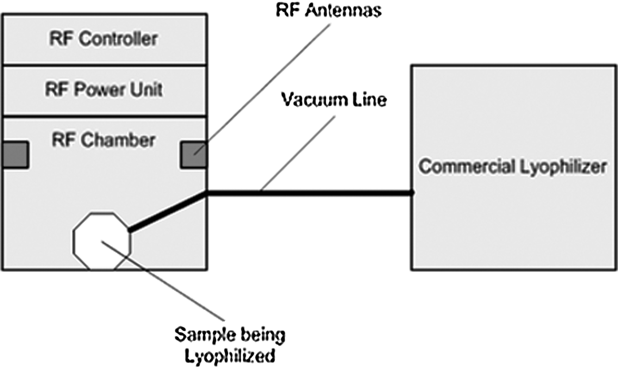
The RF lyophilizing system scheme (Chuno®).
The first iteration of the simulations and the design were completed on April 2011, and we started the preliminary experiments in July 2011.
We lyophilized the IMT3 solution with the following process parameters:
Initial Weight: 50 g (in a single jar) Vacuum (SP): No set point (with no RF power, the pressure is 12mTorr) RF Power: 10 Watt Time: 7 H
For control we used the same quantity of IMT3 solution, attached directly to the lyophilizer through the external tube port.
The results of this experiment were very impressive; the pressure in the lyophilizer during the process was stable (40 m Torr ∼ 50 m Torr), which indicates that the lyophilization process was rather stable. After 7 hours in the RF system, the sample lost 83.0% weight, while the control sample lost 43% weight (Fig. 7), the sample in the RF system does not showed any boiling damages but there were some sort of light caramelization of the cryoprotectant solution.
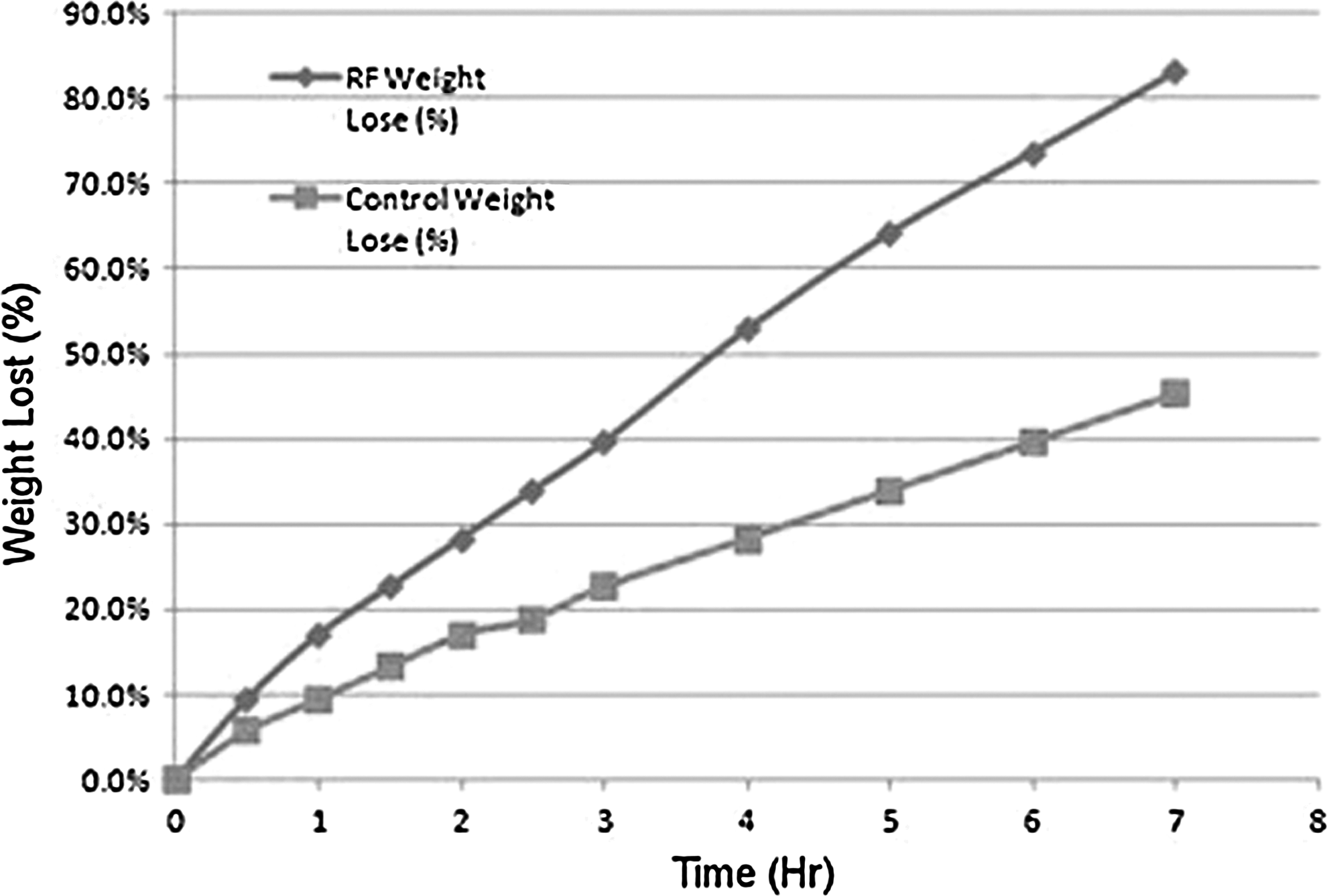
50 mL sample weight loss percentages during drying for 7 hours in the RF lyophilization system vs. control (50 mL sample dried without being exposed to the RF system, only to the lyophilizer).
Our next set of experiments involved RBCs: pRBCs were diluted in a ratio of 1:20 in IMT3 solution. A total of seven test tubes were frozen using the MTG-516 at a cooling rate of 150°C (4°C to −120°C to −130°C at a velocity of 1 mm/sec). Samples were stored in LN and then they were placed inside an open a glass jar that was placed on the shelf of the RF lyophilizing system. The samples were dried for 45 minutes and the RF power was set to 63.1 Watt. After 45 min, samples were taken out and thawed in a water bath heated to 37°C. Each sample was evaluated for free Hb and the remaining volume of the sample was assessed for calculating the percentage of the water loss. Free Hb prior freezing was 0.
From these initial results, we can see that there is a correlation between the percentage of water loss and the post thaw recoveries of the cells; as the water loss increases, the recovery decreases. Nevertheless the results showing that at a water loss of up to 65% the recovery rate was 74.5% (Fig. 8).
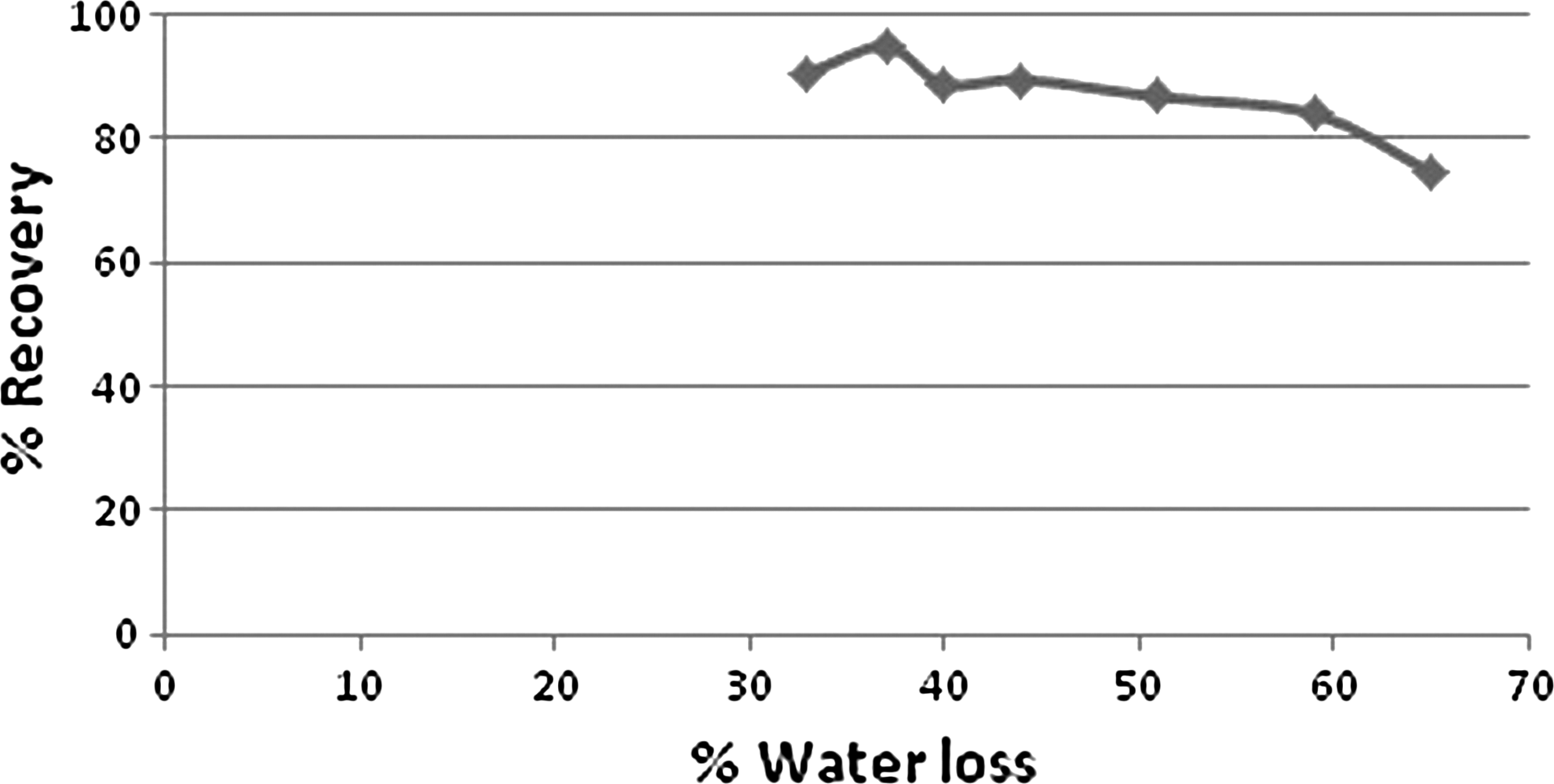
Cells recovery vs. water loss percentage of RBCs samples frozen with IMT3 at a Hct of 4% and dried for 45 min at the RF lyophilization system, followed by thawing in a water bath.
Discussion
Freeze-drying of RBCs has received a lot of attention over the years, and several approaches for lyophilization have been reported,24–30 none of which have been particularly successful. One of the main arguments is that for success in lyophilization, one needs to stabilize the cell membrane and the best method to do this is by using the disaccharide trehalose and introducing it into the cell membrane in order to stabilize both the membrane's sides. 30 We have demonstrated a link between initial cell concentration prior to freezing and cell recovery following freeze thawing. The increased recovery is probably due to reduced mechanical damage 11 in low concentration RBC samples and increased protecting agent (e.g., trehalose) per cell ratio. 31
The optimal solution for freeze drying and rehydration as well as freeze thawing of low concentration RBC sample was found to be IMT3 (trehalose and HSA). The recovery rate of low concentration RBC samples frozen in IMT3 at 1 mm/second and 150°C/min DF following freeze thawing and freeze drying and rehydration with Dextran-40 rehydration solution were 100% and 75%, respectively. Most importantly, cells that were freeze dried in IMT3 were stable and fully survived an 8-fold dilution following rehydration when a low cells concentration was used. Indeed, the optimal trehalose to RBCs ratio which was found to be 2.83 mg trehalose per 1 mg RBCs. This trehalose to RBCs ratio is the ratio that is found when using IMT3 solution at a 4% Hct, as we did in most of our experiments.
However, very poor recovery rates were seen in low RBC samples frozen in IMT3 at 150°C/min DF that were freeze dried and rehydrated with DDW in contrast to the stable 75% recovery that was obtained following freeze drying and rehydration with 20% Dextran-40.
These results support R.P. Goodrich and S.O. Sowemimo-Coker rehydration paradox hypothesis. 24 According to that hypothesis, the loss of membrane surface area that leads to cell lysis of lyophilized cells occurs during rehydration. When the samples are lyophilized, the water is removed, causing the concentration of components inside and outside the cells to increase toward infinity. When the solid is rehydrated, there is a transient gradient for water to re-enter the cells. The size of this gradient with the first drop of water is enormous and decreases as the solid is dissolved. Since the outside of the cell is rehydrated first, the gradient will always go from outside to inside for the water flow. This behavior could have profound consequences for the cells. If the water enters too rapidly, the cells could swell and lyse before they have the opportunity to regulate their volume. This lysis event may be accompanied by a resealing event with a loss of membrane vesicles in the process. The resealed cell would then possess an altered surface area to volume ratio relative to the starting cell.
Apparently the rehydration with the highly viscous 20% Dextran-40 rehydration solution, enables slow water penetration to the cell that does not inflict damage to the cell membrane. These cells survive three consecutive 1:1 v/v dilution cycles with 5 min incubation period in-between each cycle, resulting in an 8-fold dilution which results in only 2.5% Dextran-40 in the final solution; but this occurred only when cells concentration was very low (6·106 cells/mL).
In addition, cell morphology of low concentration RBC samples frozen with IMT3 was good both following freeze thawing and freeze drying and rehydration in contrast to the abnormal morphology exhibited by low concentration RBC samples frozen in IMT2. The increased EGCG per cell ratio in the low concentration RBC samples frozen in IMT2 is probably the reason for the abnormal morphology of the thawed and rehydrated cells, since the morphology of RBC frozen in IMT 2 at 4% Hct (approximately 400·106 cells per ml) following freeze thawing and freeze drying, and rehydration was better and resembled normal morphology. Additionally Chen et al. 32 have recently demonstrated that EGCG presented a prooxidant role on erythrocyte ghost membranes. These results indicated that EGCG was able to bind covalently to sulfhydryl groups of membrane proteins, leading to the formation of protein aggregates with intermolecular cross-linking. Therefore, we also freeze dried low concentration RBC samples in modified IMT 2 which contained 1/100 and 1/50 of EGCG concentration in conventional IMT2, but following rehydration only poor recovery rates were obtained (data not shown).
We also assessed the deformability and Hb saturation of RBC frozen in IMT3 and found only negligible increase in the median osmotic fragility (the NaCl concentration at which 50% of RBC were lysed) of frozen thawed samples in comparison to untreated controls, and mild decrease in the P50 of Hb saturation of frozen thawed samples.
The lyoprotective quality of high molecular weight polymers such as Dextran-40, hydroxyethyl starch (HES), and polyvinylpyrrolidone (PVP) is well documented in the literature, and was attributed to both the increase in the glass transition temperature in the presence of polymers and the water replacement ability they possess.24,33 Goodrich et al. 24 dried RBC preparations in pure carbohydrate or pure polymer preparations, and demonstrated that, although the glass transition temperature of these preparations were never exceeded, the resultant cells were unstable, indicating that both agents are needed for an improved outcome.
In our aim to develop a freeze dried RBCs product, we saw that the damage to the cells begins as soon as the desiccation process begins and it proceeds as the desiccation proceeds. One of the hypotheses concerning why the RBCs are so sensitive to the drying process can be related not only to the mechanical damage caused to the cells during freezing and drying (which we saw to greatly affect the survival of the cells) but also to damage caused to the RBCs structure during lyophilization and rehydration. We have seen that rehydration with 20% Dextran protects the cells from immediate lysis, and it has been suggested that this has to do with slowing the rehydration process and preventing the cells from being over rehydrated and eventually explode, according to the R.P. Goodrich and S.O. Sowemimo-Coker rehydration paradox hypothesis. 24
To shed more light on this subject and further explore the damage that occurs during or following lyophilization, we performed an experiment with increasing cell concentrations directionally frozen in IMT3 at 150°C/min.
In addition, we know that fast cooling rates are best for freeze thawing of RBCs and so far have also yielded best results after freeze drying. But in general, they are less optimal for lyophilization due to the fact that small ice crystals results in a longer and less homogenous drying process. Furthermore, it might be that the RBCs do not survive the slow freezing process because of excessive damage caused to their structure by the exo-osmosis of water during the freezing process. It is well known that there is a reverse correlation between the size of the ice crystals and the interface velocity or the cooling rate, and also the chances for intracellular crystallization to occur increase as the cooling rate is faster.16,17,34,35 This is attributed to less time for exo-osmosis to occur and an increased chance for water to still be inside the cells when freezing, whereas at slow cooling rates water has sufficient time to leave the cells. But there might be damage caused by the increasingly growing osmolarity of the solution as more and more water becomes ice and the remaining solutes concentration increase; the cells shrink as this occurs and possible damage to their structure can happen at high osmolarities. 36
RBCs are small cells (6–8 μm diameter) with a defined structure (biconcave) that is linked to their in vivo function as oxygen suppliers. 37 It might be that their water content is low and that in a fast cooling rate (120°C/min–150°C/min) sufficient for water exo-osmosis, combined with the high cell concentration, results in close to 100% survival. Further, it might be that at slow cooling rates they undergo excessive structural damage due to mechanical damage. 34
To summarize the results of the RF freeze drying, we can see that the RF system can speed the desiccation process by a factor of 2 and even more in some cases. However, we still need to improve uniformity of the energy transmission and better control RF power levels. Additional simulations and algorithm simulations are needed to find optimal positions of the samples inside the device and to improve uniformity of the energy absorption and avoid hot spots.
Nevertheless, from these initial results, the RF drying system looks very promising, and it seems it can reduce the damage caused to the cells at the early stages of drying.
Footnotes
Acknowledgments
We would like to thank Prof. Korenstein and Dr. Ben-Dov from the Tel-Aviv University for performing the rheology analysis. This work was supported by the US Army award number W81XWH-10-2-0126.
Author Disclosure Statement
No competing financial interests exist.