
Abstract
Human biospecimens represent invaluable resources to advance molecular medicine, epidemiology, and biomarker discovery/validation, among other biomedical research. Biobanks typically cryopreserve biospecimens to safeguard their biochemical composition. However, exposing specimens repeatedly to freeze/thaw cycles can degrade their integrity in unforeseen ways. Those biobanks storing liquid samples, thus, regularly make a fundamental compromise at collection time between freezing samples in many small volumes (e.g., 0.5 mL or smaller) or in fewer, larger volumes (e.g., 1.8 mL). The former eliminates the need to expose samples to repeated freeze/thaw cycling, although increasing up-front labor costs, consumables used, and cold storage space requirements. The latter decreases up-front labor costs, consumables, and cold storage requirements, yet exposes samples repeatedly to damaging freeze/thaw cycles when smaller aliquots are needed for analysis. The Rhode Island BioBank at Brown University (RIBB) thoroughly evaluated the performance of an original technology that minimizes a sample's exposure to freeze/thaw cycling by enabling the automated extraction of frozen aliquots from one single frozen parent sample without thawing it. A technology that eliminates unnecessary sample exposures to freeze/thaw cycles could help protect sample integrity, extend its useful life, and effectively rectify and eliminate the aforementioned need to compromise. This report presents the results of the evaluation, and conclusively demonstrates the technology's ability to extract multiple uniform frozen aliquots from a single cryotube of never-thawed frozen human plasma, which faithfully represent the parent sample when analyzed for typical biochemical analytes, showing a coefficient of variability lower than 5.5%.
Introduction
Biospecimens cryopreserved to prolong cell life during short- and long-term storage require thawing before aliquotting. However, repeated freeze/thaw cycles may degrade critical biological molecules (e.g., RNA, proteins) and damage antibodies of interest. Once a sample is compromised outside of its frozen state, it risks viability, increases chances of deterioration, and may compromise and obscure analyses' results.
2
For example, studies have shown that RNA quality degrades with successive freeze/thaw cycles, losing approximately 30% integrity after five freeze/thaw cycles, and more than 50% after nine.
3
An experiment designed to simulate long-term freezing studied how several cancer biomarkers changed in serum samples that were repeatedly frozen and thawed concluded that repeated freeze/thaw cycles affected their concentrations significantly and recommended against repeated thawing of sera.
4
An investigation on how several features—such as collection tubes, length of storage, and number of freeze/thaw cycles—might impact cytokine profiles concluded that “samples for cytokine measurements should not have been subjected to repeated freeze/thaw cycles” in view of how levels rise for some cytokines (IL-4 and TNFα) and drop for others (IL-13, IL-15, IL-17, IFNγ, and CXCL8) after one or more freeze/thawing cycles.
5
A study on the impact of duration of storage and freeze/thaw cycles on plasma samples used in mass spectrometry-based biomarker discovery concluded that exposing the samples repeatedly to freeze/thaw cycles, and particularly after two thaws, resulted in a trend towards increasing changes in peak intensity. They concluded that limiting freeze/thaw cycles is critical to maintaining plasma proteome integrity in terms of maintenance of peak number and intensity.
6
Therefore, it is extremely important that the samples be handled consistently during aliquotting, freezing, and storage. Best Practice from the International Society of Biological and Environmental Repositories (ISBER), therefore, recommends minimizing sampling frequency to avoid/reduce potential molecular damage from freeze/thaw cycling.
2
Conversely, the ability to sample a specimen frequently without exposing it to repeated freeze/thaw cycles would be a welcome development to many scientists concerned about the deleterious effects of several freeze/thaw cycles on their critical samples. Resource availability (e.g., staffing, consumables, institutional real estate) forces biobanks into difficult choices on biospecimen processing and storage to make them most accessible for future novel tests. Each choice, outlined below, confers scientific, quality, cost, and space advantages and disadvantages:
1) After collection, divide samples during processing into large volume aliquots (e.g., 1.8 mL), freeze, and later, thaw them when aliquots are requested for analysis. Material remaining in the parent samples is refrozen until future aliquots are needed. 2) After collection, divide samples into small volume aliquots and freeze. Distribute a single frozen tube when aliquots are requested for analysis. 3) Adopting a hybrid approach, freeze samples into large volume aliquots, thaw them when the first small aliquot is requested for analysis, transfer any remaining sample into multiple small volume aliquots and refreeze.
The first approach is the least expensive one, yet potentially the most damaging to the samples, particularly beyond the first freeze/thaw cycle. Biospecimens in frequently-accessed collections could potentially undergo multiple freeze/thaw cycles before leaving the biobank, harming the specimen in terms impossible to quantify. Furthermore, different samples may undergo dissimilar numbers of freeze/thaw cycles, making “apples to apples” comparisons unfeasible. Approach two is appealing because samples are never thawed at the biobank. However, preparing and storing multiple small aliquots is expensive: up-front labor costs to process samples are substantial, considerable costly cold storage space is needed, and costly laboratory supplies are consumed. The hybrid approach ensures a single freeze/thaw cycle per aliquot at the biobank. In studies in which only a portion of specimens collected are eventually sent out for analysis, the hybrid approach is a particularly attractive choice; the costs to create multiple aliquots are the same as in approach two, however they are postponed until the first small aliquot is requested and limited to only the samples sent out for analysis.
This article will present key performance results from the evaluation of a novel Frozen Sample Aliquotter technology designed to help biobanks eliminate the inherent compromises between quality and costs in these approaches, allowing them to eliminate a sample's repeated exposure to freeze/thaw cycles at the biobank and allay concerns about sample quality and resulting data integrity. The Frozen Sample technology was conceived by Dale Larson while at the Harvard Medical School. The technology's initial development at the Harvard Medical School was funded by grants from the Dana Farber Harvard Cancer Center and the National Cancer Institute (R21CA114167).
The technology consists, in essence, of a specialized rotary drilling system in which a coring probe extracts multiple frozen cores from one frozen sample, and deposits the still-frozen cores into a separate tube for downstream analysis. The extracted core(s) and the parent remain frozen throughout the process. After frozen cores are extracted, the remainder of the parent sample may be returned to the freezer, still frozen, to wait until a new aliquot extraction is requested.
This research was designed to answer three questions:
1) Is a frozen core of plasma extracted from a frozen sample intrinsically the same as the parent sample? 2) Are both these samples intrinsically the same as plasma prepared by typical freeze/thawing methods? 3) Is there any degradation to the remainder of the initial sample after a frozen aliquot is removed?
At the time of this research, prior testing of the technology's ability to extract volumetrically-uniform cores consistently from frozen plasma samples at temperatures of −70°C and below had been presented at the ISBER 2008 Annual Meeting (Table 1).
Volumetric uniformity was tested using a gravimetric method. Frozen cores were extracted from bovine serum (purchased from Lampire Biologicals; www.lampire.com) and the difference between the empty mass of a destination vial and the mass with the frozen aliquot was recorded using a Mettler balance. The coring probe used for this test was designed to have an internal volume of approximately 0.120 mL (the coring probe used on the testing at RIBB was designed to have an internal volume of approximately 0.80 mL).
Materials and Methods
The first-generation experimental platform of the Frozen Sample Aliquotter technology was evaluated for any deleterious effect of the technology on the samples by assessing the degree of agreement among assay values of three sets of human plasma:
1) Frozen plasma cores extracted from frozen samples using the Frozen Sample Aliquotter. 2) Frozen plasma remaining in the parent samples after extracting multiple frozen cores. 3) Conventionally-pipetted plasma aliquots (controls).
All three sets of aliquots experienced exactly one freeze/thaw cycle before being analyzed for four markers: total cholesterol, triglycerides, glucose, and immunoglobulin G (IgG) at the Clinical and Epidemiologic Research Laboratory (CERLab), Department of Laboratory Medicine, Children's Hospital Boston, MA. These assays are approved by the Food and Drug Administration for clinical use, and the CERLab is certified by the Centers for Disease Control and Prevention/National Heart, Lung, and Blood Institute Lipid Standardization Program. The analytes chosen for this project were recommended by the CERLab for two reasons: they are representative of a broad range of molecule types and the assays perform very reproducibly. The CERLab's typical day-to-day CVs are 1.7% for cholesterol, 1.8% for triglycerides, 1.7% for glucose, and 3.0% for IgG. Assays with very low variability were chosen in order to be able to detect possible variation in the assay results due to the action of coring the plasma. The purpose of this study was not to test volatile assays that would likely be affected by freeze to thaw cycling.
The Frozen Sample Aliquotter
The first-generation experimental platform was designed to test basic coring functionality, cryotube handling and manipulation, and temperature control for the Frozen Sample Aliquotter. Design parameters for the experimental platform were prioritized to demonstrate the ability to (i) extract multiple homogenous and volumetrically-uniform frozen cores from a frozen parent sample hands-free; (ii) deposit the frozen cores automatically into pre-selected destination vials; (iii) clean the coring probe between samples to prevent contamination by carryover; and (iv) maintain samples at a temperature of −40°C.
This experimental platform's automation included systems to core samples and to dispense frozen cores, integrated cleaning and thermal control systems, and user-defined worklists. In this platform, manual steps were required to place the vials into coring and deposition positions, de-cap and re-cap them before and after coring, and return them to the corresponding cold trays.
Operating principles
The heart of the Frozen Sample Aliquotter is a medical-grade stainless steel coring probe designed to extract volumetrically-uniform frozen cores from, in this case, a plasma sample. The coring probe drills the full depth of the frozen sample entirely hands-free, controlled by a user-defined worklist. Upon withdrawal from the sample, the coring probe automatically moves to the appropriate destination vial, and deposits the frozen core into it. This instrument is not designed to be a volumetrically-controlled liquid handler; the volume of a core is determined by the diameter of the coring probe, the type of tube used, the fill level of the material in the tube, and the depth to which the coring probe descends. In this study, a 1.8 mL cryotube filled with plasma to the recommended level, and the coring depth determined the volume of a frozen core to be approximately 80 μL each.
Sample management
The CERLab required a minimum of 0.100 mL to conduct the planned assays. Therefore in order to provide sufficient plasma, two cores totaling approximately 0.160 mL (2X approximately 0.80 mL) were sent in each vial for analysis. The study was configured to extract six frozen cores individually from each parent sample, and to dispense two frozen cores at a time into three separate destination vials.
The sample coring process began by loading frozen (‘parent’) plasma vials and empty (‘destination’) vials into cooled aluminum trays; the destination vials were in place to receive the extracted frozen cores. The trays sat on platforms (one designated as the ‘parent’ side; the other, as the ‘destination’), each of which was able to hold multiple trays. Within each tray, the vials securely rested in cylindrical sleeves. Figure 1 presents for illustration a high-level view of a ‘parent’ tray, coring station, and coring probe in the experimental platform.

Sample tray illustration for the experimental platform. The sample coring process began by loading frozen (‘parent’) plasma vials and empty (‘destination’) vials into cooled aluminum trays. In the experimental platform, the trays sat on platforms (one designated as the ‘parent’ side; the other, as the ‘destination’), each of which was able to hold multiple trays. The vials securely rested in cylindrical sleeves within each tray. Figure 1 presents a high-level view of a ‘parent’ tray in the experimental platform, which shows the ‘parent’ vials resting in the sleeves, the coring station, and coring probe.
For this evaluation, the sample temperature was set at −40°C. To maintain this temperature, a commercially-available immersion cooler was coupled to a cooling block that sat in very close proximity to the platforms. Coolant and dry gas (argon) flowed through the cooling block. The cooling block was underneath and in close proximity to the trays, thus the cryotubes were also conductively cooled by the trays and sleeves. The argon also provided a cold blanket above the vials and a low-humidity environment to avoid condensation on/in open vials.
In between the coring of individual parent samples, the coring probe underwent a programmable cleaning cycle. The cleaning subsystem consisted of a port into which the coring probe was automatically inserted and sealed while cleaning reagents were pumped within and around it to wash it and eliminate carryover between samples. Compressed air was used to dry the probe.
Experimental approach
A three-arm study was performed to address the degree to which (i) frozen plasma cores extracted with this technology (Cores) and (ii) the remaining (Remainders) plasma in the parent vial after six frozen cores were extracted are representative of (iii) plasma prepared by the typical method of thawing and aliquotting (Controls).
Frozen units of EDTA plasma from 50 male and female donors were purchased from Valley Biomedical, Winchester, VA. Units were approximately 225 mL each, and were stored at −20°C prior to shipping to RIBB.
For this study, twenty of the fifty units from both fasting and nonfasting diverse individuals (Table 2) were chosen for testing and were expected to provide a wide range of concentrations of analytes such as cholesterol and triglycerides. These units were from 10 males and 10 females between 20 and 51 years of age (average age of 37.6). Only 15 of the 20 donors reported their weight and height, allowing their BMI to be calculated (ranging between 17.6 and 49, with average of 31.5). Each of the 20 units of plasma was thawed, mixed gently in a glass dish set in ice on a stir plate, and aliquotted into multiple homogeneous aliquots. Forty parent vials containing 1.8 mL were aliquotted into Nunc 1.8 mL cryotubes (Mfcr. No. 368632), and ten vials (Controls), containing 200 μL each, were aliquotted from each unit of plasma. Each vial was uniquely labeled, frozen in the vapor phase of liquid nitrogen at −170°C, and tracked using Freezerworks Sample Management System (Dataworks Development, Inc., Mountlake Terrace, WA). Control vials were inadvertently not prepared for the first five units.
Parent vials from 20 different donors were used for coring using the Frozen Sample Aliquotter. Six frozen cores (approximately 80 μL each) were taken from the perimeter of each of the frozen parent samples. Two frozen cores each were combined into one destination vial—resulting in three destination vials per parent sample (approximately 160 μL each). The following steps were followed to extract frozen cores:
1) Bring the system to −40°C; 2) De-cap the empty destination vial manually and place it in the destination station; 3) De-cap the parent vial manually and place it in the coring station; 4) Run the software worklist controlling the automated routine; 5) Remove destination and parent vials from their processing stations and re-cap them; 6) Run the probe cleaning cycle; 7) Repeat steps two through six until all parent tubes were processed; 8) Place the processed parent and destination vials back in the vapor phase of liquid nitrogen.
After all frozen parent samples were processed, a Reproducibility study and a Diversity study were performed. Both the Reproducibility Study and the Diversity Study were designed to test whether cores taken from a vial (Cores), the sample remaining in the vial (Remainder), and an aliquot that did not undergo the coring process (Controls) were representative of each other. However, the Reproducibility Study used many replicates of a small number of plasma units and was specifically designed to test whether assay results of Cores and Remainders prepared from a single donor were consistently reproducible. The Diversity Study used only two replicates of plasma from people who presumably had different levels of analytes in their blood.
A batch of vials that included the frozen core vials, remainder vials, and control vials and Quality Control (QC) vials were shipped on dry ice to CERLab and measured for total cholesterol, triglycerides, glucose, and IgG. (These assays are approved by the U.S. Food and Drug Administration for clinical use.) All test vials underwent a single thaw cycle at the time of testing in the CERLab. The QC samples were included to test the variation of the assays conducted in the CERLab. They were conventionally-prepared plasma aliquots from two different plasma units; eleven replicates from QC1 and 10 replicates from QC2 were included for analysis.
Reproducibility Study
The Reproducibility Study used EDTA plasma from three donors. Ninety-six tubes were sent to CERLab for analysis in this arm of the study (Fig. 2):
Reproducibility Study procedure. Ten frozen plasma samples from each of three donors were cored using the Frozen Sample Aliquotter (30 tubes total). Six cores were extracted from each frozen parent sample. The frozen cores taken from each sample were deposited in three destination tubes (two frozen cores were combined in each destination tube). In total, 96 tubes were sent to CERLab to be assayed, including 60 tubes with frozen cores (2 tubes x 10 parent samples x 3 donors), six controls (2 controls x 3 donors), and 30 parent tubes with remaining parent sample after coring (10 tubes x 3 donors). • Three donors each having ten parent vials processed, and frozen cores placed into two destination vials (3×10×2=60 vials) • Ten parent vials with remaining plasma after processing, from each donor (3×10=30 vials) • Two conventionally-prepared control vials for each donor (2×3=6 vials)
Diversity Study
The Diversity Study used EDTA plasma from seventeen donors. Seventy-five tubes were sent to CERLab for analysis in this arm of the study (Fig. 3):
Diversity Study procedure. One frozen plasma sample from each of 17 donors was cored using the Frozen Sample Aliquotter (17 tubes total). Six cores were extracted from each frozen parent sample. The frozen cores taken from each sample were deposited in three destination tubes (2 frozen cores were combined in each destination tube). In total, 75 tubes were sent to CERLab to be assayed in the Diversity Study, including 34 tubes with frozen cores (2 tubes x 1 parent samples x 17 donors), 24 controls (2 controls x 12 donors), and 17 parent tubes with remaining parent sample after coring. • Seventeen donors each having one parent vial processed and frozen cores placed into two destination vials (17×2=34 vials) • Plasma remaining in each of the processed parent vials from each of the donors (1×17=17 vials) • Two conventionally-prepared control vials for twelve (12) of the 17 donors (2×12=24 tests). As noted above, control vials were not prepared for the first five units.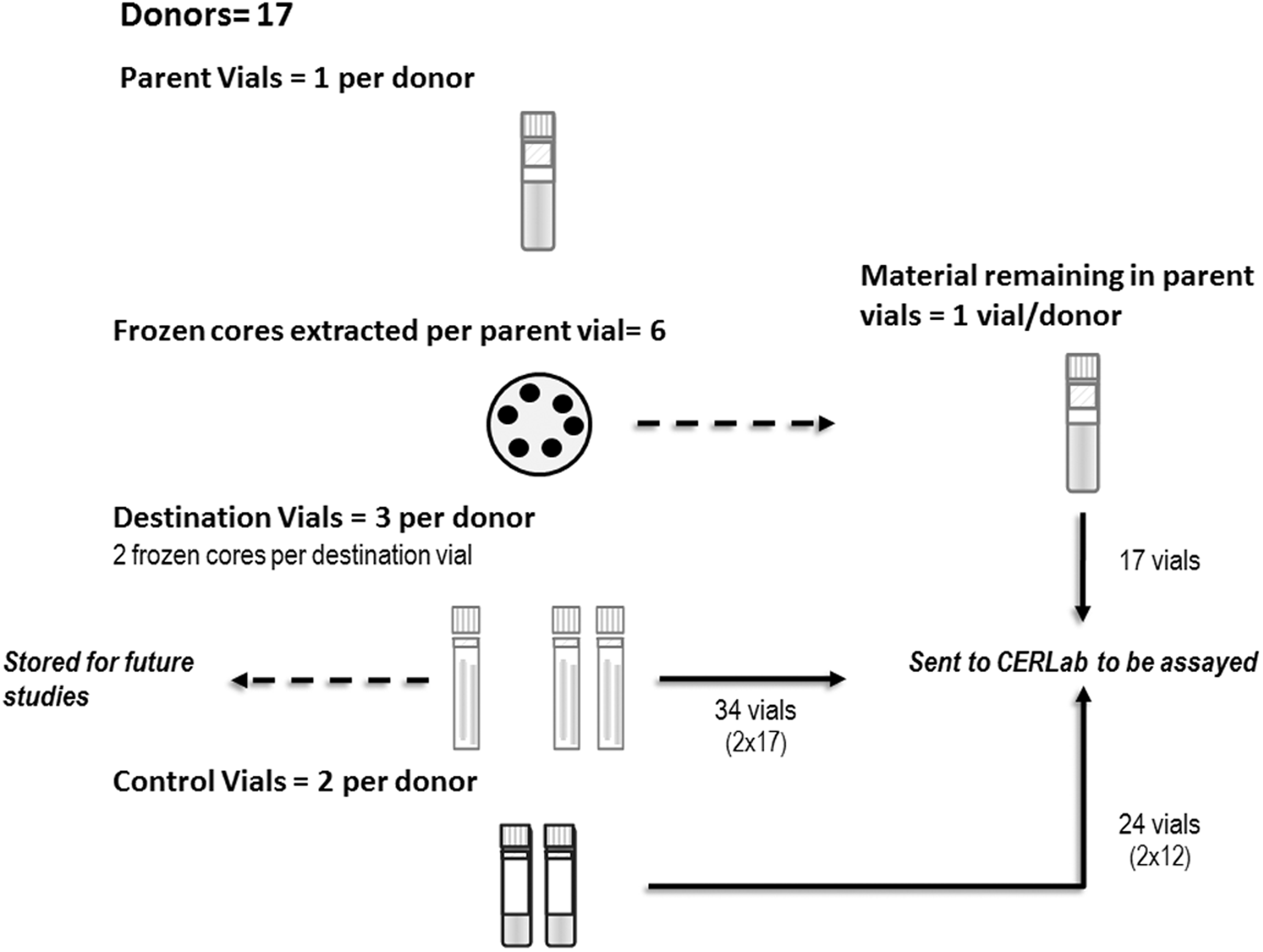
Results
Reproducibility and diversity studies
The assay results for the blinded Quality Control samples are presented in Table 3. The average CV for the analytes was 3.4%, ranging from 1.9% to 6.8% (cholesterol 2.0%, triglycerides 6.8%, glucose 1.9%, and immunoglobulin-G 2.8%).
Avg, average; Chol, total cholesterol; CV, coefficient of variation; Glu, glucose; IgG, immunoglobulin G; SD, standard deviation; TG, triglycerides.
The assay results for donors 1–17 are shown in Table 4 for the Diversity Study. The assay results for donors 18–20 are shown in Table 5 for the Reproducibility Study. In both studies, the results for Cores from the same donor were averaged, as were results for the results for the Remainders and the Control vials. Standard deviation (SD) and Coefficient of Variation (CV) were calculated for the three donors in the Reproducibility Study (donors 18, 19, 20). In Table 6 (Ratios), the values were normalized, by dividing the values for both the frozen Cores and the Remainder sample by the values for the Controls. Across all donors, the average concentration of the analytes was 3% higher in the Cores and 0.6% lower in the Remainders, as compared to the Control samples (Table 6).
Assay results and statistics for all Frozen Cores, Remainder plasma and Control vials of plasma in the Diversity Study. All plasma tested for Cholesterol, Triglycerides, Glucose, and Immunoglobulin G. Note that control aliquots were inadvertently not prepared for donors 1–5 (*), and were therefore not included in this table.
Avg, average; Chol, total cholesterol; Cores, frozen material extracted from parent vial by Frozen Aliquotter instrument; CV, coefficient of variation (%); Glu, glucose; IgG, immunoglobulin G; Remainder, remaining frozen plasma left in parent vial after cores were extracted; SD, standard deviation; TG, triglycerides.
Assay results and statistics for all Frozen Cores, Remainder plasma, and Control vials of plasma in the Reproducibility Study. All plasma tested for Cholesterol, Triglycerides, Glucose, and Immunoglobulin G.
Avg, average; Chol, total cholesterol; Control, 200 μL of plasma conventionally prepared; Cores, frozen material extracted from parent vial by Frozen Aliquotter instrument; CV, coefficient of variation (%); Glu, glucose; IgG, immunoglobulin G; N, number of values used in calculations; Remainder, remaining frozen plasma left in parent vial after cores were extracted; SD, standard deviation; TG, triglycerides.
Cholesterol, triglycerides, glucose, and immunoglobulin G for donors 6–20. Note that control aliquots were inadvertently not prepared for donors 1–5, and were therefore not included in this table.
Control, 200 μL plasma conventionally prepared; Cores, frozen material extracted from parent vial by Frozen Aliquotter instrument; Remainder, frozen plasma remaining in parent vial after cores were extracted.
The results for the Reproducibility Study in which ten vials of plasma each from donors 18, 19, and 20 were cored and then analyzed along with the remaining plasma and controls are presented in Table 5. The Coefficients of Variation (CV) in the Reproducibility study were 5.4%, 3.3%, and 1.7%.
Discussion
The assay results in both the Reproducibility and Diversity Studies showed consistent agreement between frozen cores extracted with the Frozen Sample Aliquotter as well as the Remainder parent sample when compared to Controls. The average ratio between the Cores compared to Controls (+3%) and the Remainders compared to Controls (-0.6%) is negligible when considering that the average CV for these assays was 3.4%. This indicates that the quality and characteristics of the parent sample were preserved and not detrimentally affected by the coring process. Since the material remaining in the parent sample after frozen coring is in fact the majority of the sample, this is a critical attribute of the technology.
Thus, the Frozen Sample Aliquotter technology:
• Delivers a means to improve operations to biobanks that freeze large-volume samples at collection time. ○ A single large volume (e.g., 1.8 mL) of plasma can be accessed multiple times for testing without subjecting it to repeated, damaging freeze/thaw cycles. • Provides a solution to greatly reduce consumables, labor, and costly freezer storage space for biobanks that freeze their samples in small volumes or have adopted the aforementioned “hybrid” approach. ○ The technology allows biobanks to store biospecimens in higher volume samples and to make aliquots for analysis at the time they are requested for testing rather than at the time of initial collection or first thaw.
This technology may not benefit biobanks that store specimens in very small volumes (i.e., ‘lab-ready’ volumes). However, it is of great benefit to the hundreds of biobanks that have legacy collections of plasma and serum stored in large volumes (i.e., 1.8 mL cryovials). The evaluation demonstrated that the Frozen Sample Aliquotter technology can successfully extract multiple consistently homogeneous frozen cores from a single 1.8 mL vial of frozen plasma without thawing the parent sample.
Footnotes
Acknowledgments
RIBB received the test platform used in this study from CryoXtract Instruments, LLC. The test platform was developed in collaboration with the Charles Stark Draper Laboratory, Inc., where Mr. Larson currently is Director of Biomedical Engineering. Steve Bellio and Linda Maloney comprised the team working on the test platform under Mr. Larson's supervision at The Charles Stark Draper Laboratory.
Author Disclosure Statement
No competing financial interests exist.
The Frozen Sample technology was conceived by Dale Larson while at the Harvard Medical School. The technology's initial development at the Harvard Medical School was funded by grants from the Dana Farber Harvard Cancer Center and the National Cancer Institute (R21CA114167).