
Abstract
Formalin fixation is known to inactivate most viruses in a vaccine production context, but nothing is published about virus activity in tissues treated with alternative, non-crosslinking fixatives. We used a model assay based on cell culture to test formalin and PAXgene Tissue fixative for their virus-inactivating abilities. MDCK, A549, and MRC-5 cells were infected with Influenza A virus, Adenovirus, and Cytomegalovirus, respectively. When 75% of the cells showed a cytopathic effect (CPE), the cells were harvested and incubated for 15 min, or 1, 3, 6, or 24 hours, with PBS (positive control), 4% formalin, or PAXgene Tissue Fix. The cells were disrupted and the released virus was used to infect fresh MDCK, A549, and MRC-5 cells cultured on cover slips in 24-well plates. The viral cultures were monitored for CPE and by immunocytochemistry (ICC) to record viral replication and infectivity. Inactivation of Adenovirus by formalin occurred after 3 h, while Influenza A virus as well as Cytomegalovirus were inactivated by formalin after 15 min. All three virus strains were inactivated by PAXgene Tissue fixative after 15 min. We conclude that PAXgene Tissue fixative is at least as effective as formalin in inactivating infectivity of Influenza A virus, Adenovirus, and Cytomegalovirus.
Introduction
Not much is known about the stability of viruses in tissue or cytological specimens, but Influenza A virus has been found to be infective on smooth surfaces and bank notes for several hours. The duration of infectivity is prolonged up to 24 h when the virus is embedded in protein, such as dried mucus or culture medium. 3 This indicates that virus infectivity is highest when the virus is stored in the host environment. As long as tissue is not chemically fixed, virus infection can occur by exposure to aerosols (e.g., opening a tissue container) or by percutaneous exposure (e.g., scalpel stick accidents during grossing). In the field of vaccine preparation, formalin is widely used to inactivate viruses, although accidents caused by incomplete inactivation have been previously reported. 4 As soon as tissue is thoroughly fixed in formalin, most viruses are fully inactivated. 5 However, tissue fixation takes several hours, depending on the size of the specimen. The average penetration rate of formalin into tissue is 2 mm/h; large organs or specimens can take up to 24 h or longer to be completely fixed. Some viruses are very resilient and are only inactivated after long exposure to glutaraldehyde, 6 indicating that tissue fixation in formalin may not be sufficient to assure a safe working environment for pathologists.
PAXgene Tissue fixation reagent (PAXgene) is a novel, commercially available fixative (provided as ready-to-use solution) that is based on an alcohol/acid mix which results in tissue morphology and antigenicity comparable to that of formalin fixation. The major advance is that macromolecules are preserved in a more native state compared to macromolecules derived from formalin-fixed tissue. In proteomic and genomic assays, proteins and nucleic acids derived from PAXgene-fixed, paraffin-embedded (PFPE) tissues react as those derived from fast frozen tissues.7–10 Recently, Cadoret et al. 11 showed that DNA isolated from PAXgene-fixed tissue of Atlantic salmon gills could be used to detect Neoparamoeba perurans DNA by polymerase chain reaction (PCR). Also, conventional histological staining and immunohistochemistry (IHC) could be performed on PAXgene-fixed tissue to visualize the amoeboid parasite in salmon gills.
Although alcohol can be used to decontaminate virus-contaminated surfaces, 12 it is not known if inactivation by alcohol-based tissue fixatives occurs. Since PAXgene fixation leaves macromolecules in their native form,7–10 it could be that virus could become re-activated upon rehydration. Formalin can be used to inactivate viruses for vaccine production, 4 and recently Kading et al. described inactivation of Rift Valley fever virus during paraformaldehyde fixation of mosquito specimens. 13 To provide a standardized method to monitor virus inactivation, we chose to use this virus cell culture approach to find out whether the novel fixative would inactivate viruses and if the viruses would remain inactive upon rehydration.
We used Influenza A virus (RNA virus, Orthomyxovirus, Influenzavirus A) and Adenovirus (DNA virus, Adenoviridae, Human adenovirus type 2) as model viruses in the cell culture virus inactivation assays because these viruses can be transmitted in aerosols that may form during tissue handling,14,15 and they are known to remain active for hours to days outside the host environment. 3 Human cytomegalovirus (DNA virus, Herpesvirales, Herpesviridae, Cytomegalovirus) was chosen because of its overall prevalence in nearly 60% of the population in the U.S. 16 It has the ability to infect a variety of human cell types such as fibroblasts, smooth muscle cells, macrophages, and cells of the bone marrow. 17 In addition, its ability to establish a lifelong latency after primary exposure in specific types of the myeloid lineage16,18 makes it relevant for investigating safety matters in tissue processing.
Materials and Methods
Primary infection
A monolayer of MDCK cells (Madin-Darby Canine Kidney Cells, ATCC, CCL-34) in a 75 cm2 culture flask was incubated with 0.01 multiplicity of infection (MOI) Influenza A virus (H3N2, reference strain A/Perth/16/09) containing EMEM medium (Eagle's Minimal Essential Medium, Lonza, Breda, The Netherlands) supplemented with 10% fetal calf serum (FCS), 100 IU/mL penicillin, 100 μg/mL streptomycin, 2 mM glutamine, 1.5 mg/mL sodium bicarbonate (Cambrex), 10 mM HEPES (N-2-hydroxyethylpiperazine-N'-2-ethanesulfonic acid, Lonza) and nonessential amino acids (MP Biomedicals Europe, Illkirch, France) for 1 h at 37°C. The medium was removed, the cells were washed with PBS (phosphate buffered salt solution, generic) and new medium was added. After 2 days of incubation 75% CPE (cytopathic effect) was reached.
A monolayer of A549 cells (human lung carcinoma cells, LGC Promochem, United Kingdom, ATCC-CCL-185) in a filter top 162 cm2 flask was incubated with 0.01 MOI Adenovirus type 2 (human clinical isolate from Erasmus MC diagnostic virology−80°C archive) containing medium (Ham's F12 Medium, Lonza) for 1 h at 37°C, after which 40 mL of medium was added. After 4 days of culture 75% CPE was reached.
MRC-5 cells (human lung fibroblast cells, LGC Promochem, Germany, ATCC #CCL-171) were cultivated in 9×175 cm2 cell culture flasks with EMEM+GlutaMax (Gibco, Life Technologies, UK) supplemented with 10% FCS (Gibco) and 1% Penstrep (Gibco) at 37°C and 5% CO2 until 60%–70% confluency. Infection was performed in 6 flasks with 2 mL suspensions of human cytomegalovirus AD 169 (HPA #622) (900 pfu/mL) per flask (0.003 MOI). Multiple negative controls without virus infection were performed for every cell line. Cells were continuously cultured undergoing one passage because of high density of cells until 75% CPE was observed (10–14 days) (Fig. 1,I).
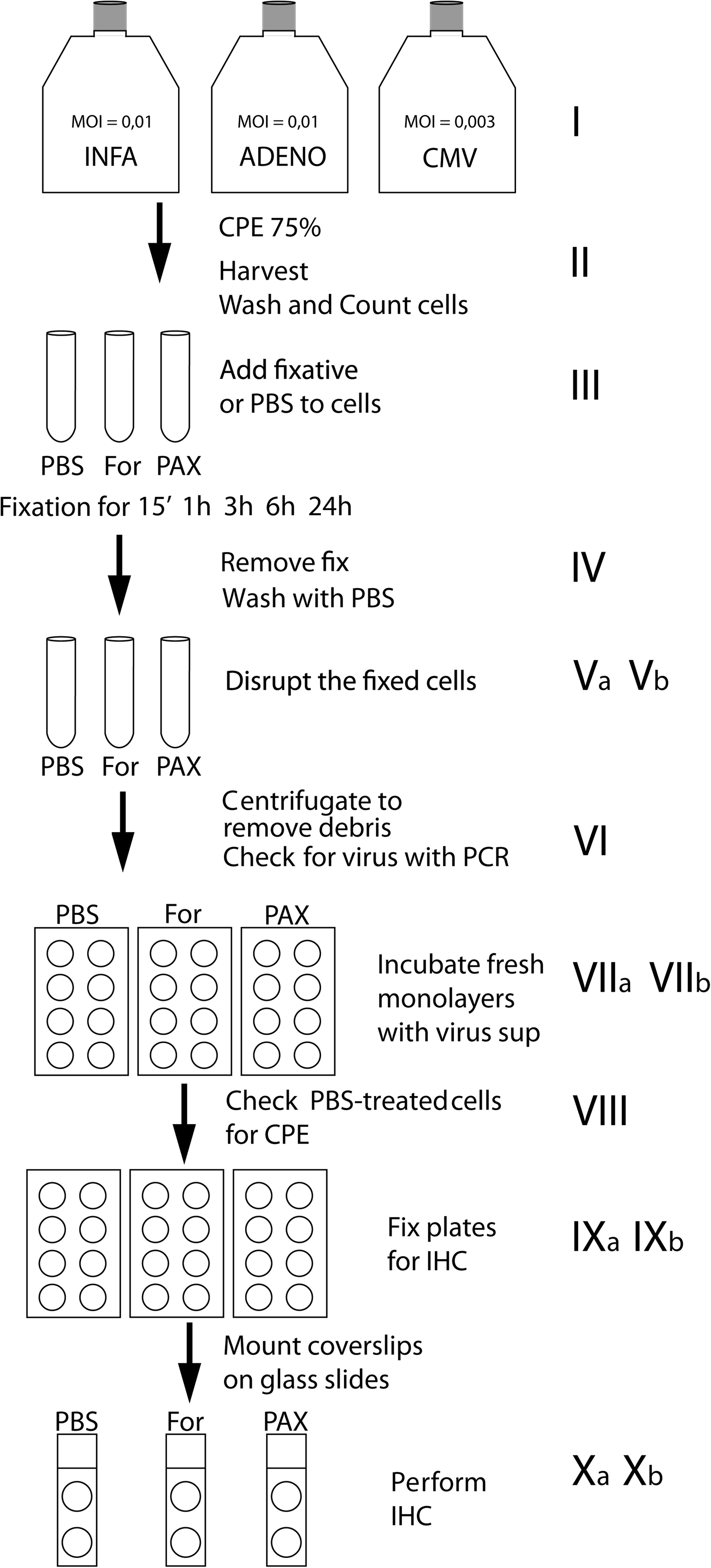
Experimental workflow.
Cell harvest, fixation, and disruption
After CPE was observed, cells were harvested using 0.05% Trypsin-EDTA (Gibco). Cells were collected, centrifuged for 5 min at 200×g, washed in PBS and counted. Duplicate aliquots of 1·105 cells/mL were made, centrifuged at 200 g for 5 min in order to discard excess medium (Fig. 1,II), and fixed at room temperature with 10 mL of formalin (4% buffered formalin, Klinipath, The Netherlands), PAXgene (Paxgene Tissue System, PreAnalytix GmbH, Hombrechtikon, CH), or incubated with PBS (positive control) for 15 min, 1, 3, 6, and 24 h (Fig. 1,III). After fixation or incubation with PBS, cells were centrifuged (200×g for 5 min) to remove the fixative/PBS (Fig. 1,IV). For influenza A and adenovirus (Fig. 1,Va), the pelleted cells were resuspended in 3 mL medium and subsequently disrupted by vortexing in glass bead tubes (UTM kit 3 mL, Copan, ITK Diagnostics BV, The Netherlands) to release intracellular virus. For CMV (Fig. 1,Vb), cells were disrupted in a Gentle Macs Dissociator (Miltenyi, Germany), a slightly different procedure. The suspension obtained was centrifuged (500×g for 5 min) to remove cell debris (Fig. 1,VI).
Virus quantification and infection of recipient cells
After fixation and disruption of the virus positive cells, PCR was performed to prove presence of virus in the supernatant. Since the same amount of supernatant (derived from the same number of cells) was used for PCR, cycle threshold (Ct) values could be used to show that in all cases the same amount of virus was introduced into the recipient cell culture (Fig. 1, VI).
Influenza A-specific PCR performed on 200 μL of the supernatant confirmed the presence of this virus, with average Ct values of 15.46. Adenovirus-specific PCR 20 performed on 200 μL of the supernatant confirmed the presence of this virus, with average Ct values of 12.96. CMV DNA-specific PCR performed on 200 μL supernatant using QIAamp MinElute Virus Spin Kit for isolation of viral nucleic acids and artus CMV RG PCR Kit (both from Qiagen, Hilden, Germany), according to the protocol provided with the kit, confirmed the presence of CMV in all samples (average Ct 22.88), except the negative controls.
The Influenza A virus supernatant was transferred to fresh MDCK cells (1 mL/well) on cover slips in 24-well plates (in duplicate). Negative control wells were cultured in the presence of culture medium only. The adenovirus supernatant was added to recipient A549 cells on cover slips in 24-well plates (1 mL/well, duplicate wells). Medium without virus was added to the negative control wells (Fig. 1,VIIa). 1.5 mL of each CMV supernatant was used for reinfection of a MRC-5 monolayer with 60%–70% confluency in a 25 cm2 flask (Fig. 1,VIIb). Cells were incubated until 50% CPE was observed in the positive control wells (Fig. 1,VIII).
Detection of virus activity with immunocytochemistry
Medium was removed from the wells, the wells were washed with PBS, and the cover slips were fixed in 80% acetone (influenza A and adenovirus, Fig. 1,IXa) or methanol/acetic acid (CMV, Fig. 1,IXb) for ICC. The influenza A ICC procedure was adapted from the one step fluorescence kit, without further need of optimization. After air drying overnight, the cover slips were incubated for 30 min at room temperature with monoclonal mouse anti-influenza A virus-FITC antibody (Imagen™ Influenza Virus A, REF K610511-2,Oxoid), diluted 1:3 in normal antibody diluent (NAD, ScyTek Laboratories, Logan, Utah).
The adenovirus ICC (peroxidase-DAB) procedure was also adapted from the one-step fluorescence kit. Background staining, predominantly found in dividing cells, was eliminated by diluting the primary antibody until negative control cells were free of any background. Adenovirus ICC was performed with a 1:160 diluted monoclonal mouse anti-adenovirus-FITC antibody (Imagen™ Adenovirus, REF K610011-2. Oxoid). Slides were repeatedly washed with PBS/0.05%Tween, and the primary antibody was detected by peroxidase-labeled EnVision (Chemvision, Dako, Denmark). The peroxidase label was visualized with DAB (Chemvision) and the cells were counterstained with haematoxylin (Fig. 1,Xa).
For CMV, contrary to the other assays, the cover slips were not air dried overnight but washed in PBS prior to the ICC procedure. 1:50 diluted monoclonal mouse anti-CMV (M085401, Dako) was incubated for 60 min. After repeated washing, the second antibody EnVision Ready-To-Use (Dako) was incubated for 30 min, washed again, and staining was performed with AEC Substrate System (Ready-To-Use) (Dako, 3-amino-9-ethylcarbazole that forms a red end-product at the site of the target antigen). The reaction was stopped with water, and slides were counterstained with hematoxylin for 1 min. Slides were covered with Aquatex (Merck, Germany) (Fig. 1,Xb).
Results
Figure 2 shows formalin- and PAXgene-inactivated influenza A virus within 15 min of fixation. No viral activity was found after 1, 3, 6, and 24 h of fixation (data not shown). The positive control showed a strong signal, while the negative control showed no virus-specific signal, nor any background staining. The monolayer of MDCK cells to which formalin-fixed virus was applied was no longer intact and most cells looked damaged and deformed. The deformed cells were virus negative. However, while viable cells were also detected, there was no specific ICC signal indicating absence of active virus.
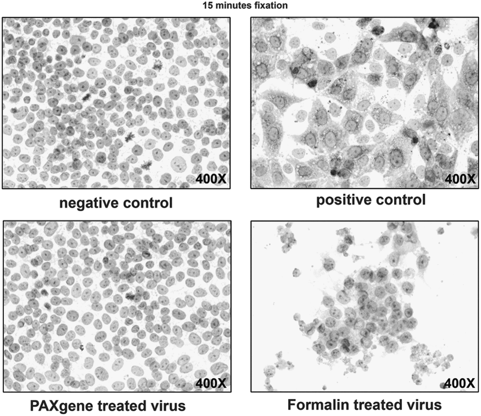
Inactivation of Influenza A, The upper panel shows the negative and positive control MDCK cells stained for Influenza A virus. The lower panel shows staining of monolayers inoculated with virus that was fixed for 15 min with PAXgene and formalin. Original magnification 400X.
When A549 cells infected with Adenovirus were fixed with formalin, total inactivation was observed only at 6 and 24 h of fixation. PAXgene inactivated Adenovirus within 15 min (Fig. 3). Human CMV was inactivated by both formalin and PAXgene after 15 min incubation (Fig. 4). The MRC-5 monolayer showed a mild degree of cell damage after formalin-treated cell supernatant was applied. The amount of damaged cells seems to be dependent on the duration of formalin incubation. PBS control samples showed strong signals with immunohistological staining. No signals of active CMV by immunohistological staining were seen after PAXgene and formalin fixation from 15 min onwards (data not shown).
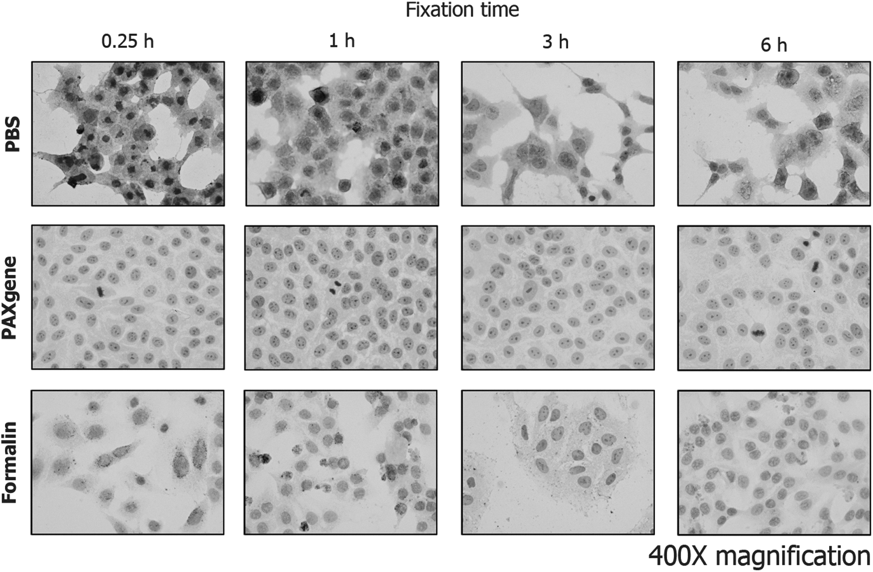
Inactivation of Adenovirus. The upper row shows the positive control A549 cells stained for Adenovirus. The middle row shows staining of cells inoculated with PAXgene-fixed virus, and the bottom row shows staining of cells inoculated with formalin-fixed virus. Cells inoculated with virus that was fixed with formalin for 3 h show weak staining. After 6 h of formalin fixation, no staining is observed. Original magnification 400X.

Inactivation of Cytomegalovirus. The upper panel shows the negative and positive control MRC-5 cells stained for Cytomegalovirus. The lower panel shows staining of monolayers inoculated with virus that were fixed for 15 min with PAXgene and formalin. Original magnification 400X.
Discussion and Conclusion
The aim of this study was to test the virus inactivating properties of formalin and PAXgene in a virus culture assay. Since we found no literature with evidence or insight regarding inactivation of virus in tissue samples at the time these experiments were planned, we designed a cell culture model to investigate whether formalin and PAXgene would inactivate several viruses. This cell culture model does not resemble solid tissue fixation precisely, but it does allow us to gain insight into how different virus strains react to tissue fixatives while actively infecting a cell layer. Any extrapolation of this cell culture data to solid tissue fixation is based on known dynamics of solid tissue fixation and commonalities between cell fixation and solid tissue fixation. All model viruses remained infective for at least 24 h in a PBS solution at room temperature. Therefore, we may conclude that virus also remains infective for at least 24 h in unfixed tissue specimens. The penetration rate of formalin is around 2 mm per hour. Large clinical tissue specimens are almost always cut open to enhance fixation, but some parts of the specimen could remain too thick to allow thorough fixation overnight. Our observation that the time needed to inactivate viruses differs between type of virus and type of fixative, argues for vigilance when working with large solid organs, even after overnight gross fixation.
We have chosen ICC instead of PCR as the virus replication read out system. With PCR, differentiation between infective and inactivated virus is not possible. PCR was only used to show that virus was present in the medium used for re-infection of the recipient cells. As the PCR kits are not validated for use with cell culture material, calculating copy numbers could be misleading. Since we have taken equal amounts of material for PCR, the Ct values do serve as a measure for the amount of virus put into the recipient culture and confirm that equal amounts of virus were actually present in the supernatants.
To avoid false positive ICC results (i.e., detection of fixed virus-positive donor cells), the fixed cells and control cells were disrupted before the inoculation of fresh recipient cells. The culture was ended when the positive control wells showed 50%–75% CPE. Prolonged culture would have resulted in complete lysis of the positive control cells, which would eliminate positive ICC because of lack of cells. The damaged cell layers observed after treatment with formalin-fixed virus could be explained by a cytotoxic effect of residual formalin cross links in the virus homogenate. The number of affected cells seems to increase in correlation with formalin fixation/inactivation time, indicating that an increase of cross links or any chemical alteration in the cells may lead to loss of cells in the culture. Nevertheless, we did find viable, healthy looking cells in these wells; both these and the affected cells were virus negative. It is safe to conclude that, although a cytotoxic effect may have taken place, the virus was unable to infect the remaining cells and therefore was inactivated by formalin fixation.
Inactivation of Adenovirus by formalin seems to be a slower process than inactivation by PAXgene. This may be due to the fundamental difference between the two methods of fixation and the different virus structures. In the case of influenza A and CMV, the virus envelope provides more molecules for formalin cross links. The non-enveloped adenovirus is more resilient to formalin inactivation as fewer surface molecules are available for cross linking. 21 Formalin forms cross links between proteins and nucleic acids, whereas alcohol-acetic acid-based fixatives like PAXgene precipitate macromolecules. Since the standard formalin solution contains only 4% formaldehyde, the number of molecules in the aqueous solution may be insufficient to form cross-links in small virus particles in a short period of time. The high alcohol concentration (not disclosed but at least 50% to 70%) in PAXgene may deliver an abundance of molecules to precipitate the virus proteins. More research in the field of virus-induced pathology is needed to explore the benefits of PAXgene for this specific subject. However, previously published articles show that histological techniques as well as proteomic and genomic techniques work with PAXgene-fixed tissues.7–10 The possibility of detecting parasite DNA in DNA extracted from PFPE salmon gills 11 suggests that in future experiments, virus RNA, DNA, and proteins may be easily detected in PFPE tissues.
In conclusion, our experiments show that the viruses investigated, Influenza A, Adenovirus, and Cytomegalovirus, can be inactivated by PAXgene tissue fixative and formalin. When virus was rehydrated after PAXgene fixation (i.e., resuspension in medium), re-activation did not occur. Therefore, PAXgene is, at least regarding inactivation of these three viruses, a safe replacement of formalin in clinical pathology laboratories.
Footnotes
Acknowledgments
The research leading to these results received funding from the European Union Seventh Framework Programme [FP7/2007–2013] under grant agreement n° 222916. The hCMV part was supported by the Christian Doppler Research Association, Federal Ministry of Economy, Family and Youth, National Foundation for Research, Technology and Development, Sensengasse 1, 1090 Vienna, Austria. We thank all SPIDIA partners, the Rotterdam Diagnostic Virology laboratory, and the Graz Virology laboratory for their support. Special thanks to Frank van der Panne for his excellent illustrations.
Author Disclosure Statement
No competing financial interests exist.