
Abstract
Biobanks of fresh, unfixed human normal and malignant tissues represent a valuable source for gene expression analysis in translational cancer research and molecular pathology. However, the success of molecular and cellular analysis in both clinical and translational research is strongly dependent on the collection, handling, storage, and quality control of fresh human tissue samples. The aim of this study was to evaluate an innovative vacuum-based refrigerated system, as a logistically feasible technology to increase the collection of tissue specimens, preserving the integrity of cellular and molecular components. We tested randomly-selected tissues stored under vacuum at 4°C by using endpoints important for research and diagnosis, including tissue morphology, epitope stability, and RNA integrity. Gene expression was evaluated by qualitative and quantitative RT analysis of selected housekeeping and tissue-specific genes. Tissue morphology and overall protein stability were generally well preserved, being compromised only in gallbladder tissue. By contrast, phosphoprotein and RNA analysis demonstrated a time-dependent degree of degradation, with progressive loss of stability from 24 to 72 hours. However, this reduction in RNA quality did not represent a limitation for successful expression analysis of selected genes. Indeed, a comparative qualitative and quantitative RT-PCR analysis showed that RNA extracted from tissues stored under vacuum is suitable for gene expression profiling, but requires highly sensitive technologies, such as quantitative RT-PCR. These data suggest that the refrigerated vacuum-based system represents a suitable and feasible technology for routine transport of fresh specimens from surgery to biobanks, thus increasing the opportunity to collect biospecimens.
Introduction
R
The purpose of tissue banks is to enhance the quality and the speed of both basic and translational research, 2 providing unique resources to study molecular changes in the in situ environment of cancer. Thus, storage of tissues with intact morphology, protein, DNA, and RNA for research or diagnosis is the main goal of human tissue biobanks. 6 Biospecimen quality is vital for tissue biobanks, being dependent on standardized and established handling processes.7–9 Samples are generally obtained immediately after excision and prior to fixation, to ensure optimal preservation of protein and nucleic acids. 2 Even though formalin-fixed, paraffin-embedded tissues are adequate for some morphological procedures, the analysis of frozen tissue samples is required for most molecular diagnostic and research applications, which need intact genomic DNA, RNA, or proteins.5,10,11 A major objective of molecular and proteomic profiling is to represent the in vivo state rather than a modified state induced by preanalytical variables. 12 Indeed, after resection, tissue is still alive, and this favors detection of dramatic changes in gene expression and protein profiles, especially phosphoproteins, due to ischemia, absence of vascular perfusion, hypoxia, or temperature changes.13,14 Thus, at present, cryopreservation is considered the gold standard for preserving tissue for both genomic and proteomic molecular research.
Although the importance of the quality of banked tissues has been recognized, to date little information exists on the quality of collected samples and innovative strategies for improving their preservation. 8 Therefore, despite the advances in biobanking technologies, important limitations remain. One of the major problems is the need to increase the quantity of biospecimens. 15 Generally, biobanks are located in hospitals, and the number of samples that it is possible to collect is restricted by the number of cases and other factors such as the size of cancer tumor. Therefore, biobanks need to collaborate and join their collections, by establishing central biobanks for collecting tissue specimens from multiple hospitals. In such a perspective, standardized technologies for the transport and preservation of tissue samples are needed. Recently, vacuum-based refrigerated systems have been proposed as an alternative to formalin fixation and as a logistically feasible method to preserve tissues in the interval between surgery and sample processing and biobanking.16,17 This provides a solution to facilitate the transport from collaborating hospital to the central tissue biobank and would increase the number of samples available for translational research studies. 18
The IRCCS-CROB Basilicata Biobank collects fresh frozen tissues of various human organs, including normal and tumor tissues, directly after routine surgery. In order to increase the number of cryopreserved samples for research studies, we tested a vacuum-based refrigerated system to transfer specimens from the surgical theater of surrounding hospitals to the Pathology Unit of the Central Institution. In such a context, the purpose of the present study was to verify the preservation of refrigerated tissues maintained under vacuum for different time points. The study was carried out using endpoints important for research and diagnosis, including the evaluation of morphology and epitope stability, RNA integrity, and gene and protein expression.
Materials and Methods
Collection of specimens and experimental design
Thirty six samples of colon, gallbladder, thyroid, uterus, ovary, and other organs were collected and stored by using the high vacuum biospecimen transfer system (TissueSAFE®, Milestone). Large surgical specimens were collected immediately after surgical removal at the Hospital of Melfi, opened, cleaned with appropriate sponges (they were not washed to avoid the formation of ice crystals during cryopreservation), and stored in vacuum-packed plastic bags with an identification label, and kept under vacuum at 4°C for 24, 48, or 72 h. Specimens were transferred to the Pathology Unit of the IRCCS-CROB and processed by a pathologist to obtain tissue fractions for the biobank, prior to fixation and further processing of the remaining tissue for standard diagnostic procedures.
Among these specimens, 27 samples were randomly selected to evaluate the preservation of tissue morphology, protein stability, and RNA quality. These specimens were grouped in three representative pools based on the length of vacuum storage at 4°C (24, 48, and 72 h). In addition, a pool of control samples snap-frozen immediately after the surgical removal at the IRCCS-CROB Basilicata Biobank without vacuum preservation (time point 0 h) was selected: 4 colon, 3 uterus, 2 ovary, and 2 thyroid tissue samples. Table 1 lists the histopathological characteristics of the entire tissue collection and the samples randomly selected for this study. Handling of human DRG tissue was carried out according to legal provisions and rules of IRCCS CROB, and written informed consent was obtained from all the subjects before sampling the tissues.
Histopathological characteristics of tissue samples stored under the refrigerated, vacuum-based system for 24, 48, or 72 h and tissue samples frozen immediately after surgery. The samples selected for further quality evaluation are highlighted in
Tissue morphology and immunohistochemistry
Five-micron sections were stained for standard hematoxylin/eosin (H&E), according to routine procedures and evaluated microscopically for the assessment of tissue morphological preservation. Immunohistochemistry (IHC) detection of different antigens was performed to evaluate the preservation and the integrity of specific epitopes, as reported in Supplementary Methods (supplementary material is available online at www.liebertpub.com).
Protein extraction and immunoblot analysis
Proteins were extracted by homogenization of 50–100 mg tissue specimens with the T10 basic IKA® Ultra-Turrax in cold lysis buffer [20 mM Tris pH 7.5 containing 300 mM sucrose, 60 mM KCl, 15 mM NaCl, 5% glycerol, 2 mM EDTA, 1% (v/v) Triton X-100, supplemented with protease (Halt™ Protease Inhibitor Cocktail, Thermo Scientific), and phosphatase (Halt™ Phosphatase Inhibitor Cocktail, Thermo Scientific) inhibitors] and further sonication for 30 seconds on ice. Immunoblot analysis was performed as previously reported. 19
Qualitative and quantitative RT-PCR analysis
The expression of the following genes was evaluated by qualitative and/or quantitative RT-PCR analysis as reported in Supplementary Methods: Glyceraldehyde-3-phosphate dehydrogenase (GAPDH), β-actin (β-ACT), Abelson (ABL), S100A13, p22 Sorcin, Vacuolar Protein Sortin 41 (VPS), TRAP1, and F1ATPaseB. Primer details are reported in Supplementary Table S1.
Results
Tissue morphology and epitope integrity are preserved under the vacuum-based refrigerated system
Randomly selected tissues stored under the vacuum-based refrigerated system (Table 1) were assessed for tissue morphology with standard H&E staining and epitope integrity with IHC staining for tissue-specific antigens. Figure 1 reports a panel of H&E and IHC images of five specimens representative of our analysis. Indeed, the integrity of cellular morphology and tissue structure was preserved in the majority of experimental groups. Tissue samples vacuum-stored at 4°C for 24 and 48 h exhibited good cellular and tissue structure, with preservation of the histological zonation and uniformity, as well as a defined intensity and distribution of cellular epitope-specific staining, with minimal or absent background (Fig. 1A, a–f and Fig. 1B, l–m). More specifically, the analysis of uterus tissues vacuum-stored for 24 and 48 h revealed the integrity of the simple ciliated epithelium (Fig. 1A, a and d), as well as a high degree of protein stability upon staining with anti-cytokeratin and anti-ERα antibodies (Fig. 1A, b–c and e–f). By contrast, the stability and the integrity of tissue samples vacuum-stored at 4°C for 72 h was tissue source-dependent: the vast majority of tissues showed an acceptable morphological structure and a preserved epitope stability as observed in thyroid tissue (Fig. 1A, g–i, sample n. 22), whereas borderline morphologic integrity, with loss of cell definition, extensive cell lysis, and lack of defined epitope-specific staining was observed in gallbladder tissue vacuum-stored for 72 h (Fig. 1B, n–o).

Hematoxylin/eosin staining and immunohistochemistry quality control.
Phosphoprotein stability is affected by vacuum-based refrigeration
To further investigate the impact of vacuum preservation on protein stability, we analyzed, by immunoblot, the expression of specific proteins selected based on their wide distribution in human tissues (i.e., Sorcin, AKT, ERK1/2, and GAPDH).12,20,21 Furthermore, we evaluated phosphoAKT and phosphoERK1/2 levels in order to analyze the preservation of post-translational modifications (phosphorylation) upon vacuum-based refrigeration. Figure 2A reports the immunoblot analysis of 14 samples representative of our series. A tissue- and time-dependent variability of protein expression was observed in samples stored under vacuum for different time points, Sorcin and GAPDH being the more stable epitopes (Fig. 2A). By contrast, the stability of phosphoAKT and phosphoERK1/2 was more significantly affected by the length of vacuum preservation, as shown by gallbladder and omentum tissues stored under vacuum for 48 or 72 h (Fig. 2A).
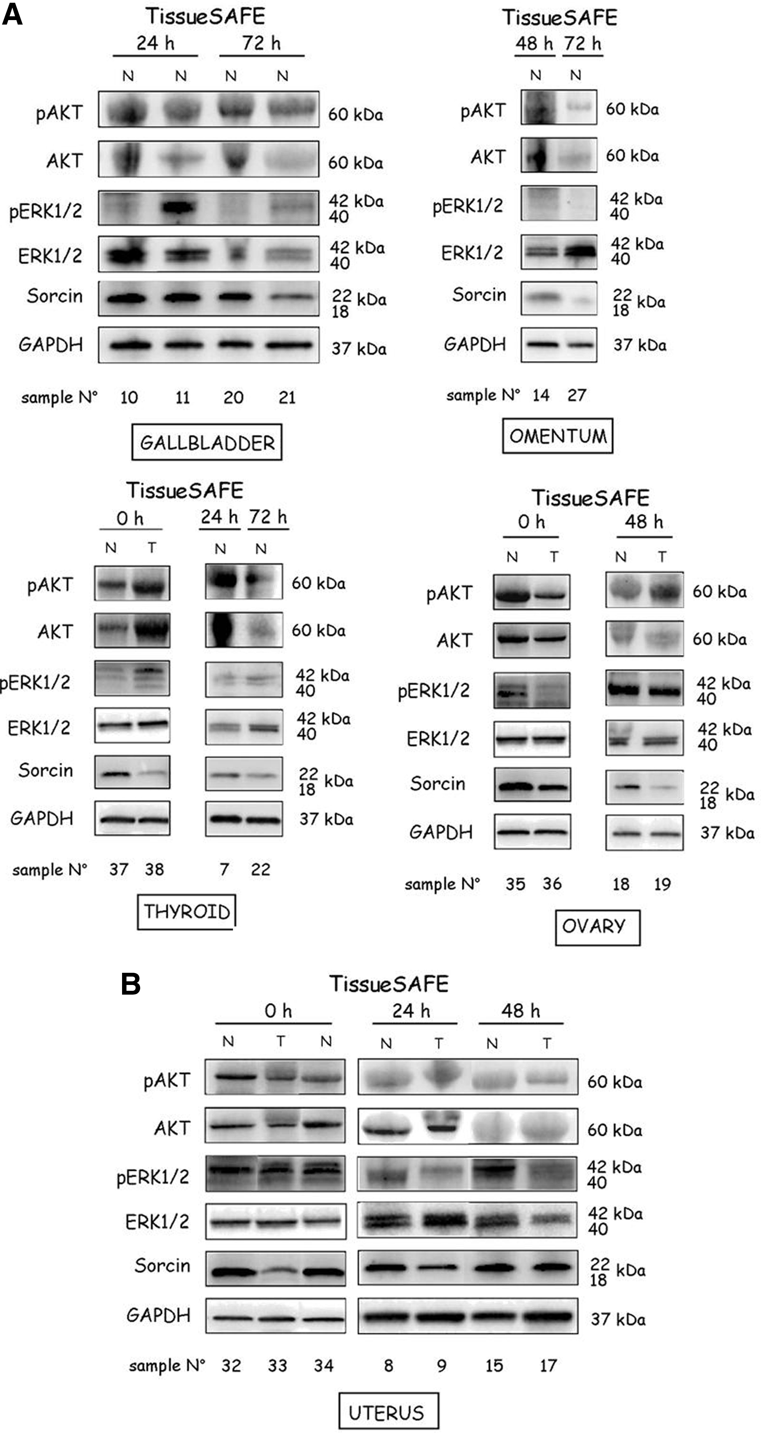
Protein and phosphoprotein stability under vacuum preservation. Total cell lysates from gallbladder, omentum, ovary, thyroid
Since this analysis compared human tissues from different sources, and was affected by tissue-specific gene expression variability, we further analyzed this issue in uterus (Fig. 2B) and colon (Fig. 3) samples stored under vacuum for different time points, in comparison with control tissues frozen in LN2 vapor immediately after surgery (time point 0 h). Indeed, uterus specimens showed well-conserved Sorcin and GAPDH stability (Fig. 2B). Consistently, normal colon mucosas and carcinomas vacuum-stored at 4°C for 24 (Fig. 3A, g and j) and 72 h (Fig. 3A, m and p) exhibited optimal preservation of cellular and tissue structure as compared to snap-frozen samples (Fig. 3A, a and d), as previously observed in uterus samples (Fig. 1A, a–f). Furthermore, the immunostaining for cytokeratin and CDX2 (Fig. 3A, b–c, e–f, h–i, k–l, n—o, and q–r) and the immunoblot analysis of Sorcin and GAPDH (Fig. 3B) exhibited well-conserved epitope stability. By contrast, phosphoAKT and phosphoERK1/2 immunoblot analysis showed a time-dependent loss of protein stability under vacuum preservation either in uterus (Fig. 2B) or in colon (Fig. 3B) samples, suggesting that protein post-translational modifications are likely to be more susceptible to degradation under vacuum storage.

Protein and phosphoprotein stability in colon samples under vacuum preservation.
RNA stability under the vacuum-based refrigerated system is time- and tissue source-dependent
RNA purity was assessed by UV spectrophotometric analysis. As reported in Supplementary Table S2, the quality of the extracted RNAs was comparable and close to the expected A260/A280 for samples vacuum-stored for 24, 48, and 72 h.
The RNA integrity was evaluated by 1% agarose gel electrophoresis based on the visualization of visible intact bands at two different positions (i.e., 4.8 and 1.8 kb), which represent, respectively, the 28S and 18S ribosomal RNA subunits. As reported in Figure 4, the majority of samples demonstrated the presence of two defined bands corresponding to the ribosomal RNA subunits. However, a time-dependent degree of degradation was observed, with progressive loss of RNA stability from 24 to 72 h of vacuum preservation. This is more evident from analysis of RNA stability in uterus and colon samples stored under the vacuum system for different time points, in comparison with control tissues frozen immediately after the surgical removal (Fig. 4). It is also noteworthy that the tissue source of RNA needs to be taken into account, since the quality of RNA extracted from thyroid tissue vacuum-stored for 72 h exhibited a better preservation compared to RNA extracted from other tissues vacuum-stored for shorter period of time (Fig. 4). Consistent with morphologic and protein stability data, the RNA obtained from gallbladders vacuum-stored for 72 h exhibited no evidence of the ribosomal subunits (Fig. 4).
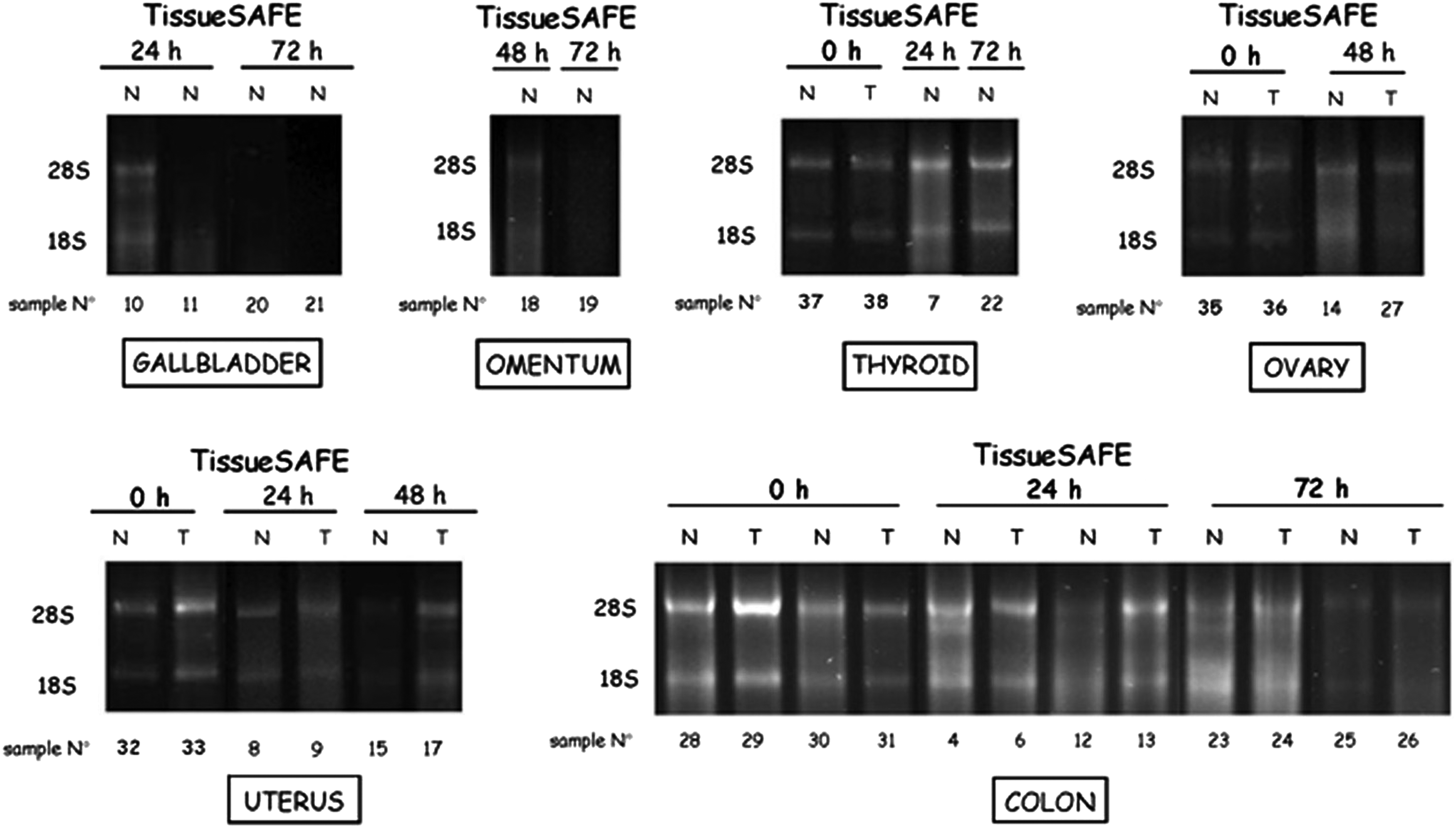
RNA quality assessment by agarose gel electrophoresis. The integrity of RNA was assessed on the basis of the visualization of the 28S and the 18S ribosomal RNA subunits in RNA extracted from gallbladder, omentum, thyroid, ovary, uterus, and colon tissues stored under vacuum for different time points (24–72 h) or control snap-frozen immediately after surgery, without using the vacuum-based preservation system (0 h). The numbers associated with each sample are the same as reported in Table 1.
RNA extracted from vacuum-stored tissues is suitable for gene expression analysis
In order to evaluate whether RNA obtained from tissues stored under the vacuum system is suitable for gene expression analysis, qualitative RT-PCR analysis was performed to study the expression of three housekeeping genes (GAPDH, ß-ACT, and ABL), as well as S100A13, p22 Sorcin, and VPS, three genes with a tissue-dependent expression profile.20–24 As reported in Figure 5, the vast majority of tissues, except the gallbladders vacuum-stored for 72 h, exhibited distinct amplification products specific for GAPDH and ß-ACT upon qualitative RT-PCR analysis. By contrast, the expression of the third housekeeping gene, Abelson, was generally weak and absent in some tissues, whereas the S100A13, p22 Sorcin, and VPS amplification products were not detected in all samples stored under vacuum for different time points. Once more, the gallbladders kept under vacuum for 72 h did not exhibit the S100A13, p22 Sorcin, and VPS amplification products. The RNA from colon tissues exhibited distinct amplification products for all genes (Fig. 5).
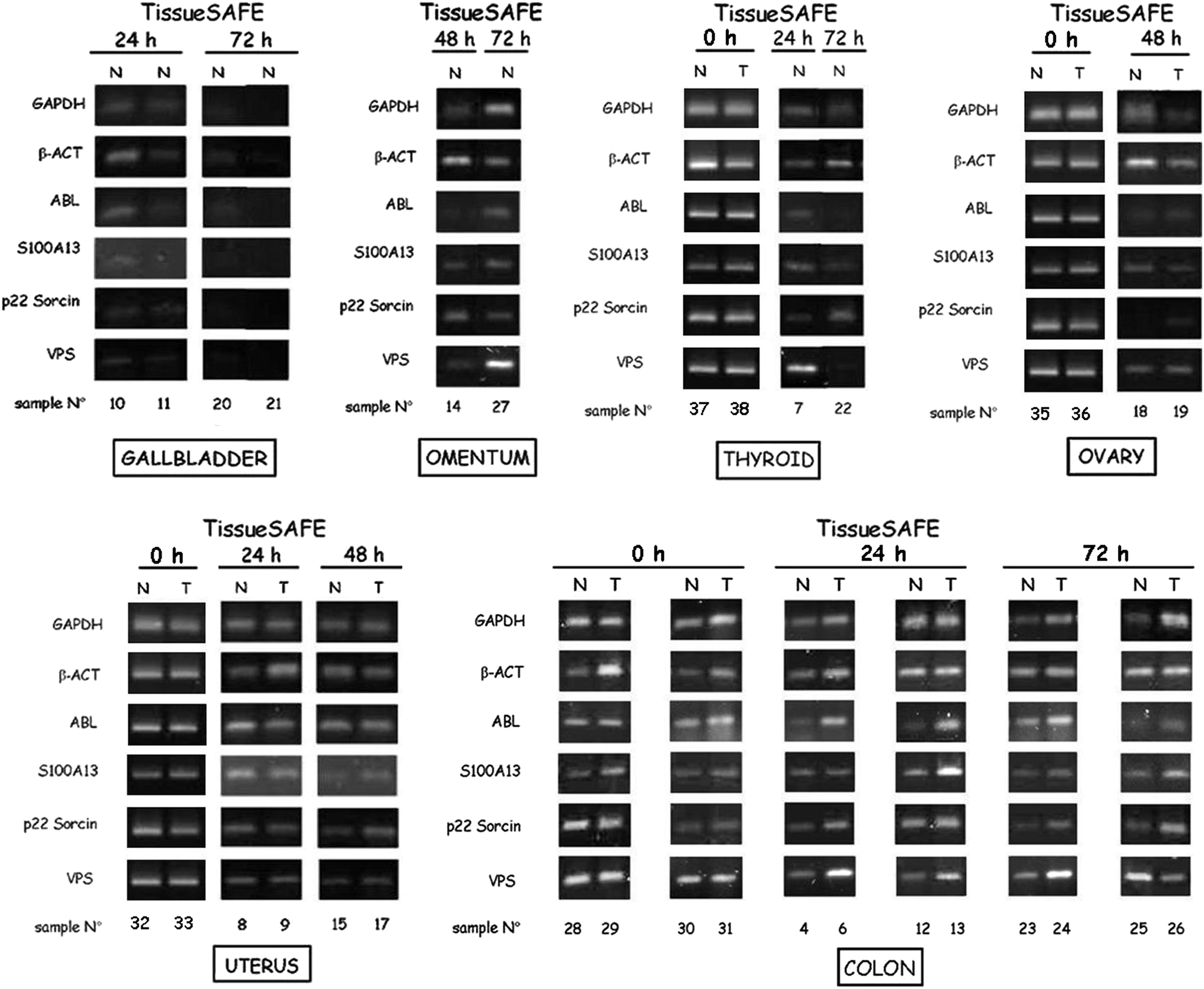
Qualitative RT-PCR analysis. GAPDH, ß-actin (ß-ACT), Abelson (ABL), S100A13, p22 Sorcin, and VPS expression analysis on RNAs extracted from gallbladder, omentum, thyroid, ovary, uterus, and colon tissues stored under vacuum for different time points (24–72 h) or control tissues snap-frozen immediately after surgery, without using the vacuum-based preservation system (0 h). The numbers associated with each sample are the same as reported in Table 1.
These differences in the gene expression profiles may be dependent on either the quality/stability of the RNAs or the tissue variability of gene expression. Thus, based on qualitative RT-PCR data showing apparent lack of expression in some tissue samples, we re-evaluated the expression of S100A13, p22 Sorcin, and VPS genes by quantitative PCR. 25 Furthermore, the expression of two additional target genes (TRAP1 and F1ATPaseB) was studied to further corroborate our analysis. Indeed, by using a more sensitive technology, the expression of S100A13, p22 Sorcin, and VPS was detectable in all tissue specimens that had shown low or negative expression in qualitative analysis, except the gallbladders stored under vacuum for 72h (Table 2). Consistently, TRAP1 and F1ATPaseB expression were similarly detectable in control and vacuum-preserved specimens, except the gallbladder vacuum-stored for 72 h (Table 2).
ΔCts of p22 Sorcin, VPS, S100A13, TRAP1 and F1ATPaseB genes in tissues stored under vacuum for different time points. ΔCts are not reported in samples with undetectable expression of target genes. The numbers associated to each sample are the same reported in Table 1.
The same analysis was performed on RNA extracted from colon (Fig. 6A) and uterus (Fig. 6B) tissues, these representing more homogeneous cohorts of samples suitable for a statistical evaluation of the difference in the ΔCts between tissues vacuum-stored and control tissues snap-frozen immediately after surgery. Indeed, the Student's t-test did not show a statistical difference in the ΔCts of p22 Sorcin, VPS, S100A13, TRAP1, and F1ATPaseB between colon (Fig. 6A) and uterus (Fig. 6B) tissues stored under vacuum and controls cryopreserved after surgery. These results suggest that a sensitive gene expression analysis can overcome the vacuum-induced loss in RNA stability.

Quantitative RT-PCR analysis in colon and uterus tissues.
Discussion
Advances in genomics, proteomics, and biomarker studies have catalyzed transformation in the field of cancer research. A result of this process is the need for well-characterized biospecimens to support the needs of the cancer research community and the development of standard biobanks with the cooperation of surgeons, pathologists and molecular biologists. Storage of frozen tissues with intact morphology, proteins, DNA, and RNA for use in research and diagnostics is the main goal of a biobank. Avoiding RNA and protein degradation is a major challenge in this process. In this context, quality control indicators are needed to monitor tissue and molecular stability. Since RNA is believed to be the most fragile molecule of unfixed tissues, preservation of RNA integrity can be used as a general quality indicator in fresh frozen tissue biobanks.26,27 Indeed, physical trauma and “warm ischemia” during surgery (from blood vessel ligation to surgical excision time), “cold ischemia” (from excision to freezing), preservation treatment of the tissue, type and length of storage, and specimen type are just some of several factors affecting the RNA quality and stability.28,29 Furthermore, it has been suggested that warm ischemia during surgery may result in significant gene expression changes, and results based on the analysis of partially degraded RNA may be not reliable and must be interpreted with extreme caution. 30
The examination of protein biomarkers, particularly phosphoproteins, is another critical step in validation of tissue quality. 12 Phosphoprotein markers of cell signaling have been widely studied in both translational research and clinical settings, making their determination crucial for effective treatment decisions. Response to EGFR inhibitors in lung cancer or glioblastoma and trastuzumab in breast cancer has been proposed to be predicted by measurement of phosphoprotein markers, including phosphoAkt and phosphoERK. 31 In this context, preanalytical factors, such as ischemic time, can have a significant impact on the ability to elucidate signaling pathways in tissues. 32
Based on these premises, the IRCCS-CROB Basilicata Biobank developed a standardized tissue acquisition/processing method aimed at reducing the time (less than 30 min) between tissue excision and freezing in LN2 vapor. However, this standard procedure is not suitable for the processing of tissue samples collected in surrounding hospitals. Thus, in this study, we tested a vacuum-based refrigerated system to preserve tissues during the interval between surgery and biobanking to increase the quantity of biospecimens collected. The study was designed to evaluate the effect of refrigerated vacuum-storage on either protein or RNA stability of randomly selected surgical specimens with respect to different time points and tissue sources. The results suggest that i) the integrity of the tissue structure and the overall epitope stability is generally well preserved, independently from the length of vacuum storage, whereas ii) the RNA and the phosphoprotein stability is affected in a time-dependent manner, and iii) there is a variability in RNA and phosphoprotein stability that is likely dependent on patient heterogeneity and tissue source.
These results need to be interpreted in the general scenario of the different approaches to tissue preservation for biobanks. Because cryopreservation or refrigeration are known to be the gold standards for molecular diagnostics and research, 12 we questioned whether adding vacuum sealing to low temperature may improve tissue preservation. Indeed, vacuum preservation without refrigeration was proven to offer advantages in terms of convenience for staff, tissue preservation, and costs. 17 By contrast, no preserving effect of vacuum sealing was observed with respect to cellular morphology, epitope stability, or RNA integrity, whereas tissue storage at 4°C was shown to offer good preservation, independently of vacuum sealing. 18 More recently, the combination of vacuum sealing and cooling at 4°C was demonstrated to be effective in preserving tissue structure and RNA stability. 16 Bussolati and co-workers suggested that vacuum-sealing preserved tissue, since the absence of air decreased autolytic processes, thus favoring the stability of proteins and RNA. 16 In this context, our study did not address the value of the combination of vacuum and low temperature preservation with respect to refrigeration alone, but it is the first to address the issues of the stability of post-translational modifications (i.e., protein phosphorylation) and the feasibility of gene expression analysis in tissues stored under vacuum at 4°C. Our results suggest that specific proteins may undergo a more rapid degradation under vacuum preservation at 4°C, and thus caution needs to be taken when analyzing vacuum-stored samples for protein post-translational modifications. Furthermore, the analysis of the expression of a variety of housekeeping and tissue-specific genes in specimens stored under vacuum or snap-frozen soon after excision (within 30 min) clearly suggests that RNA obtained from these tissues is suitable for gene expression analysis, but this requires sensitive technologies such as quantitative PCR. This is particularly evident in the cohort of colon and uterus samples, where it was possible to compare gene expression data between vacuum-stored specimens and control samples frozen immediately after surgery. Indeed, no major differences were observed between these tissue samples in terms of expression of target genes, thus supporting the evidence that quantitative RT-PCR is suitable for vacuum-stored tissues and likely overcomes the loss in RNA quality due to vacuum preservation. Finally, our results exhibit a correlation between preservation of cellular morphology and RNA stability, suggesting that the preservation of cellular structure is a prerequisite for molecular stability.
It is important to note that a weakness of our study is the comparison of non-vacuum packed with vacuum-packed specimens from different patients, instead of using aliquots of tissues from the same specimen. While this issue represents a major drawback for the interpretation of our data, it suggests that further studies are required to definitively assess the value of vacuum/low temperature preservation with respect to either snap-freezing or low temperature alone for the routine transport of tissues from collaborating hospital to the central tissue biobank.
The issue of RNA stability is extremely relevant from the perspective of using these samples for clinical diagnostic or translational research purposes, especially for genome-wide expression analysis by microarray technologies. Indeed, approximately 80% of the total RNA is ribosomal, while 15% is transfer RNA, with protein-encoding mRNA representing only a small portion. 15 Therefore, the stability of total RNA significantly affects the expression profiles of specific genes, and once degradation occurs, even if it is marginal, gene expression results have to be interpreted with extreme caution. From these perspectives, our quality assessment data suggest that RNA extracted from tissues stored under vacuum for prolonged time periods (48–72 h) may be partially degraded and likely is not suitable for wide-genome gene expression analysis by microarray technologies. However, in this context, our results are not conclusive and further analyses are needed.
Finally, a potential confounding factor affecting protein and RNA stability is represented by the tissue source and the inter-patient variability. Indeed, gallbladder exhibited a particular vulnerability compared to other tissues (i.e., thyroid gland) under vacuum preservation conditions. While this issue may be more relevant for the preservation of normal tissues with respect to tumor samples, it suggests that tissue-dependent stability needs to be strictly controlled when using such a technology on a wide scale.
In conclusion, this study supports the use of refrigerated vacuum preservation during routine transport of fresh specimens from surgery to biobanks, since the majority of tissues were proven to be stable under these conditions and suitable for standard diagnostic and research morphological evaluation and gene expression analysis by quantitative PCR. However, such a technology requires standardized control procedures respect to the length of storage and the specimen type.
Footnotes
Acknowledgments
Special thanks to the Pathology Unit of Melfi Hospital for providing surgical tissue specimens.
Author Disclosure Statement
The authors declare that they have no financial conflict of interest.
This work was supported by the Associazione Italiana per la Ricerca sul Cancro (AIRC) (Grant IG8780) and by the Italian Ministry of Health (Grant GR-2010-2310057) to ML.
References
Supplementary Material
Please find the following supplemental material available below.
For Open Access articles published under a Creative Commons License, all supplemental material carries the same license as the article it is associated with.
For non-Open Access articles published, all supplemental material carries a non-exclusive license, and permission requests for re-use of supplemental material or any part of supplemental material shall be sent directly to the copyright owner as specified in the copyright notice associated with the article.