
Abstract
The aim of this study was to evaluate the efficiency of different media in the in vitro culture of bovine preantral follicles that were used either fresh or following slow freezing treatment. Frozen and fresh noncultured or cultured ovarian fragments were processed for histological, viability, and cell proliferation analyses. For cryopreservation, a solution containing 1.5 M ethylene glycol was frozen in a programmable biological freezer. After thawing, a portion of the samples was destined for frozen controls. The remainder were cultured in vitro for 5 days in three media: α-MEM, McCoy, or M199. Samples from these culture media were collected on days 1 and 5 for quantification of reactive oxygen species (ROS) and for hormonal assays. In fresh-cultured tissues, the percentage of morphologically normal follicles was significantly higher when cultured in M199 compared to that in the other media. In frozen-cultured tissues, McCoy medium was significantly superior to the other media, and was the only treatment that helped in maintaining the viability similar to fresh and frozen controls. Upon quantification of the nucleolus organizer region, we observed greater proliferation of granulosa cells in the frozen-cultured tissues with McCoy medium, and lesser proliferation in fresh-cultured tissues only with α-MEM. In frozen-cultured tissues, ROS levels were highest at day 1 and progressively reduced during culture, independent of the media used. In conclusion, under the conditions used in this study, the M199 and McCoy media are recommended for the culture of follicles derived from fresh and frozen ovarian tissues, respectively.
Introduction
I
In order to enable pregnancy in women after treatment of malignant diseases without the risk of reintroduction of cancer cells through transplantation of the ovarian tissue, in vitro culture systems have been investigated as an alternative to ensure follicle development and in vitro oocyte maturation with subsequent embryo transfer. 5 Nevertheless, the protocols of in vitro culture, mainly for preantral follicles present in the ovarian tissue, are not yet properly defined to the point of allowing full follicular development. Moreover, the vast majority of studies aimed at developing in vitro culture systems have used follicles from non-cryopreserved ovarian tissues. This fact, coupled with the lower survival rates of cryopreserved follicles compared to non-cryopreserved follicles6–8 and evidence that cryopreserved cells are less tolerant to in vitro culture and more susceptible to cell death, 9 suggest the need for establishing systems and media of in vitro culture that are specific to the requirements of cryopreserved preantral follicles.
Owing to ethical constraints and the limited availability of human ovarian tissue for research purposes, animal models with ovaries that are biochemically, physiologically, and anatomically similar to human ovaries 10 are required. Livestock animals serve as experimental models for this purpose, including ovine, 11 bovine, 12 and swine. 13 Indeed, Gandolfi et al. 14 evaluated the behavior of ovarian follicles of humans, bovine, and swine during cryopreservation, and showed that bovine and human ovarian tissues have a high level of similarity, since human and bovine follicles responded in the same way to the two equilibrium cooling protocols. Since both bovines and humans are nonseasonal, mono-ovulatory species, bovine ovaries represent a good experimental model 12 to assist in the development of protocols applicable to humans. Furthermore, the large size of bovine ovaries yields a large enough quantity of the ovarian cortex from the same individual to facilitate experimental research. Thus, in this study, we used the bovine as a model to evaluate the efficiency of three types of media for in vitro culture of frozen/thawed ovarian tissue, and evaluated the morphology, development, and follicular viability, as well as oxidative stress of preantral follicles.
Material and Methods
This experiment was approved and performed under the guidelines of Ethics Committee for Animal Use of State University of Ceará. Except where otherwise stated, all chemicals were obtained from Sigma (Sigma Chemical Co., St. Louis, Mo, USA).
Source and transport of ovarian tissue
Ovaries from five adult cows were obtained from a local slaughterhouse, washed once in 70% alcohol, and then twice in HEPES-buffered minimal essential medium (MEM) supplemented with 0.1% (v/v) penicillin/streptomycin. Within 1 hour after slaughter, the ovaries were transported to the laboratory in thermo flasks containing tubes filled with MEM at 20°C.
Experimental design
At the laboratory, the cortex from each pair of ovaries was removed, cut (with a scalpel) into 22 fragments of approximately 3×3 ×1 mm (9 mm3), and allocated to each treatment group. As a fresh control, one fragment was immediately fixed in Carnoy's solution for 4 h for histological and proliferation examination, and one fragment was submitted to follicular isolation for the viability test. Of the remaining fragments, 9 fragments were destined for in vitro culture with different culture media (α-MEM, McCoy, or M199) and 11 fragments were subjected to slow freezing.
Freeze-thaw procedure
Slow freezing was performed according to the protocol described by Celestino et al., 15 with some modifications. Briefly, ovarian fragments were exposed for 20 min to 1.8 mL freezing solution containing 1.5 M ethylene glycol and 10% fetal calf serum (FCS) in macrotubes. The samples were then transferred to a programmable freezer (Freeze Control; CryoLogicPtyLtd.; Waverley, Australia), previously equilibrated at 20°C. The temperature was reduced at 2°C/min from 20°C to −7°C, and ice crystal formation (seeding) was manually induced by touching the vials with forceps that were pre-cooled in liquid nitrogen. After seeding, the vials were held at −7°C for 10 min and then cooled at 0.3°C/min to −40°C and finally reduced to −70°C at 10°C/min. The samples were immersed in liquid nitrogen (−196°C) and stored for up to 1 week.
For thawing, the samples were kept at room temperature (RT; ∼20°C) for 1 min and then immersed in a water bath at 37°C until the cryopreservation medium was completely thawed. Then, the cryoprotectant was removed from the ovarian cortex fragments by three washes (5 min each) in (i) MEM+10% FCS+0.5 M sucrose, (ii) MEM+10% FCS+0.25 M sucrose, and (iii) MEM+10% FCS. Finally, one fragment was immediately processed for histological and cell proliferation evaluation and the other fragment processed for viability analysis. The other fragments underwent in situ culture in the three different media.
In vitro culture
The cortex tissue samples were cultured in 24-well culture dishes containing 1 mL of culture medium per well. Culture was performed at 38.5°C in 5% CO2 in air. The culture medium, based on that described by McLaughlin et al. 16 consisted of α-MEM, McCoy, or M199 supplemented with hypoxanthine (2 mM), glutamine (3 mM), bovine serum albumin (0.1%), transferrin (2.5 g/mL), selenium (4 ng/mL), insulin (10 ng/mL), and ascorbic acid (50 g/mL). Fresh medium was incubated for 1 h prior to use, and the culture medium was replenished every other day. In vitro culture was carried out for 1 and 5 days, and the media were stored for quantification of reactive oxygen species (ROS) and estradiol and progesterone concentrations. After in vitro culture, the ovarian fragments were destined to histological, cell proliferation, and viability analysis.
Histological evaluation of preantral follicles
For histological examination, after fixation in Carnoy's solution for 4 h, the ovarian fragments were dehydrated in increasing concentrations of ethanol, cleared in xylene, and embedded in paraffin wax. After paraffin embedding, the bovine ovarian cortex tissue samples were serially sectioned at 7 μm, mounted on a glass slide, and stained with hematoxylin and eosin. All sections were examined under a light microscope (Nikon; Tokyo, Japan) at a magnification of 400X. For each treatment, 30 preantral follicles were analyzed per repetition. Only preantral follicles with visible oocyte nuclei were counted. The developmental stages of follicles have been defined previously 17 as primordial (one layer of flattened and cuboidal granulosa cells) or growing (one or more layers of cuboidal granulosa cells around the oocyte and without an antrum) follicles. Follicles were classified as morphologically normal if they presented intact oocytes and granulosa cells. The follicles were considered degenerated if they contained a pyknotic oocyte nucleus or shrunken ooplasm, with or without disorganized granulosa cells and/or detachment of the basement membrane. 18
AgNOR staining
To estimate the cell proliferation index, Ag-NOR staining was performed to quantify the number of argyrophilic nucleolar organizer regions (NOR). For this purpose, ovarian tissue fixed in Carnoy's solution was sectioned at 5 μm. After reduction with 1% potassium iodide, slides were stained with 50% silver nitrate solution in a colloid solution (2:1) in a darkroom and counterstained with 0.1% safranin. For quantification, the follicles were visualized under a light microscope (1000X), and the NOR of all the nuclei of visible granulosa cells were counted. In the fresh and 5-day-cultured groups, 30 preantral growing follicles were evaluated.
Viability analysis by trypan blue staining
Preantral follicles were isolated from the control and frozen-thawed ovarian fragments were obtained using the mechanical method described by Figueiredo et al. 19 with slight modifications. Briefly, with a tissue chopper (The Mickle Laboratory Engineering Co.; Gomshal, Surrey, UK) adjusted to sectioning intervals of 50 μm, samples were cut into small pieces and placed in 2 mL MEM supplemented with 3 mg/mL bovine serum albumin. Samples were then pipetted 100 times with a large Pasteur pipette (inner diameter 1600 μm), followed by pipetting 100 more times with a smaller Pasteur pipette (inner diameter 600 μm) to dissociate the preantral follicles from the stroma. The obtained material was then passed through a 200-μm nylon mesh filter. This procedure was performed within 10 min at RT. Then, 5 μL of 0.4% trypan blue (Sigma Chemical Co.) were added to 100 μL of isolated and suspended preantral follicles, which were incubated for 1 min at RT. Afterward, follicles were examined with an inverted microscope (Nikon) and classified as degenerated (blue-stained) and surviving (unstained).
ROS and hormonal levels
The ROS levels were determined in culture medium of ovarian tissue by a spectrofluorimetric method, using a 2′,7′-dihydrodichlorofluorescein diacetate (DCHF-DA) assay. The medium was incubated with 5 μL of DCHF-DA (1 mM). The oxidation of DCHF-DA to fluorescent dichlorofluorescein (DCF) reflects the level of intracellular ROS. The DCF fluorescence intensity emission was recorded at 520 nm (with 480 nm excitation) 120 min after the addition of DCHF-DA to the medium.
The levels of estradiol and progesterone were measured using a microparticle enzyme immunoassay (Abbott Diagnostics AxSYM® SYSTEM) with commercial kits (Axsym Progesterone and Estradiol; Abbott Commercial Kit Japan Co., Ltd.; Tokyo, Japan). Culture medium removed on days 1 and 5 was analyzed.
Statistical analysis
Data were initially evaluated for homoscedasticity and normal distribution of the residues using the Bartlett and Shapiro-Wilk tests, respectively, to confirm requirements underlying analysis of variance (ANOVA). For histological analyses, ANOVA was then carried out according to a 3×2×2 factorial arrangement of treatments with medium (α-MEM, McCoy, or TCM), procedure (with or without freezing), and time of culture (1 or 5 days) as the main effects. The model used was Yijk=μ +Mi+Pj+Tk+(Mi×Pj)+(Pj×Tk)+(Mi×Pj×Tk)+eijk, where Yijk is the dependent variable, μ is the general mean, Mi is the medium, Pj is the procedure, Tk is the time of culture, Mi×Pj is the interaction between the medium and procedure, Pj×Tk is the interaction between the procedure and time, Mi×Pj×Tk is the interaction among medium, procedure, and time, and eijk is the residual error. When any main effect or interactions were significant, means were separated by the Student-Newman-Keuls test. Dunnet's test was applied to compare experimental treatments against control groups (i.e., fresh or frozen samples). For NOR data, ANOVA was then carried out according to a 3×2 factorial arrangement of treatments with medium (α-MEM, McCoy, or M199) and procedure (frozen or not) as the main effects. The model used was Yij=μ +Mi+Pj+(Mi×Pj)+eij. When any main effect or interactions were significant, means were separated by the Student-Newman-Keuls test. Dunnet's test was applied to compare experimental treatments against the control group [i.e., fresh (non-cultured and non-frozen) samples]. The ROS data were analyzed by one-way ANOVA followed by the post-hoc Duncan's test. For viability evaluated by trypan blue staining, follicles were taken as a pool and analyzed as the frequency dispersion with the chi-square test. In all cases, differences were considered to be significant when p<0.05, and results are expressed as mean±standard deviation (SD).
Results
Histological evaluation of preantral follicles
A total of 2100 preantral follicles were analyzed by classical histology. Follicles derived from fresh and frozen tissues after in vitro culture for 5 days with intact morphology (a) or degenerated (b) are shown in Figure 1. Immediately after thawing (frozen control), a reduction in the percentage of morphologically normal follicles was observed when compared to the fresh control (Fig. 2). Similar results were observed in all cultured treatments compared to fresh and frozen controls. When the fresh-cultured and frozen-cultured tissues were compared, there was a lower percentage of normal follicles at day 5 of culture in frozen-cultured tissues, except for those cultured with McCoy medium. Comparing the days of culture, the frozen-cultured tissue showed a significant reduction of normal follicles on day 5, regardless of the medium used.

Morphological aspects of bovine preantral follicles after 5 days of in vitro culture. Images show a morphologically intact primordial follicle in the frozen-cultured fragment in McCoy

Percentage of morphologically normal bovine preantral follicles in fresh, frozen, and cultured ovarian tissues. C1, fresh control; C2, frozen control; * significant difference from the fresh control; †significant difference from fresh and frozen control. Different uppercase letters (AB) indicate a significant difference between days of culture. Different lowercase letters (ab) indicate significant differences among media within the same day (1 or 5) and treatment (fresh or frozen). Different Greek symbols (αβ) indicate differences between treatments (fresh or frozen) within each medium and day of culture.
The percentage of morphologically normal follicles observed in fresh-cultured ovarian tissues in M199 medium was higher than that in McCoy at day 1, and was also higher in M199 than the other two media investigated after 5 days of in vitro culture (Fig. 2). Culture in α-MEM resulted in a lower percentage of normal follicles when compared to the other media at day 5 (p<0.05). In frozen-cultured tissues, we observed that using the McCoy medium resulted in a higher percentage of normal follicles than that with other media, both on day 1 and day 5 of culture (p<0.05). Interestingly, the M199 medium resulted in a lower (p<0.05) percentage of normal follicles when used in frozen-cultured tissues on day 5 of culture.
With regard to follicular development, all cultured treatments showed a higher percentage of growing follicles when compared to fresh and frozen controls (Fig. 3). In fresh-cultured tissues, M199 and McCoy media resulted in an increase in the percentage of growing follicles from day 1 to day 5, whereas α-MEM reduced the percentage of growing follicles from day 1 to day 5 (p<0.05). In frozen-cultured tissues, only McCoy increased the percentage of growing follicles on day 5 compared to day 1 of culture. Furthermore, the other media (α-MEM and M199) reduced the percentage of growing follicles on day 5 (p<0.05). Comparing the efficiency of the media for fresh-cultured and frozen-cultured tissues, both M199 and McCoy resulted in a higher percentage of growing follicles in the frozen-cultured group on day 1. By contrast, on day 5, culture in McCoy medium yielded a significantly higher percentage of growing follicles in the frozen-cultured group, and M199 yielded the highest percentage in the fresh-cultured group.

Percentage of growing preantral follicles before and after in vitro culture for up to 5 days. C1, fresh control; C2, frozen control; *significant difference from the fresh control; †significant difference from fresh and frozen control. Different uppercase letters (AB) indicate a significant difference between days of culture. Different lowercase letters (ab) indicate significant differences among media within the same day (1 or 5) and treatment (fresh or frozen). Different Greek symbols (αβ) indicate differences between treatments (fresh or frozen) within each medium and day of culture.
AgNOR staining
A total of 140 preantral follicles were analyzed by AgNOR staining. Evaluation of cell proliferation by quantification of NOR (Fig. 4) revealed a greater number of NOR/granulosa cells in fresh-cultured tissues in M199 and McCoy media compared to α-MEM in day 5 and the fresh control (Table 1). Otherwise, only frozen-cultured fragments cultured in McCoy medium had a higher average NOR/cell number compared to the fresh control, α-MEM, and M199 media.
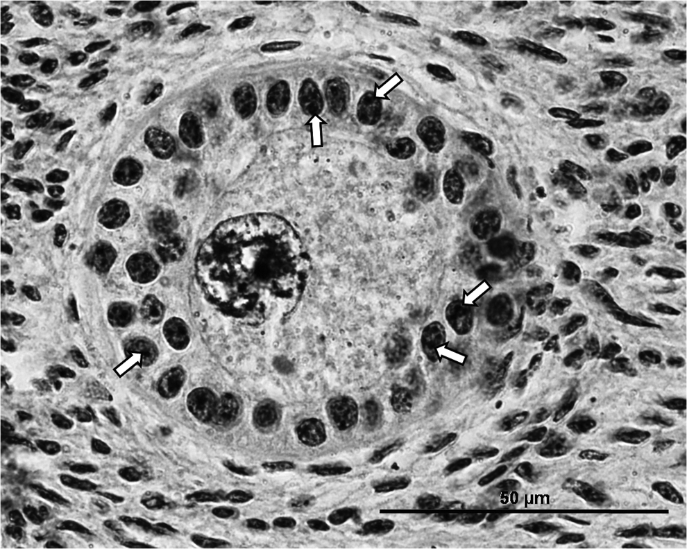
Bovine growing preantral follicles stained using the AgNOR technique, showing the nucleolar organizer regions (NOR) stained black by silver nitrate (white arrows).
Significant difference from the fresh control. †Significant difference from the frozen control. Capital letters represent significant difference between columns (culture time). Lower case represents significant difference between lines (experimental treatments) (p<0.05).
Preantral follicle viability analysis
The assessment of viability by membrane integrity as evidenced by trypan blue vital dye showed no difference in the percentage of viable follicles present between the fresh and frozen controls (Table 2). After 5 days of culture, there was a reduction in the percentage of viable follicles in fresh-cultured tissues, regardless of the media tested, compared to the fresh control (p<0.05). The analysis of follicles from the frozen-cultured group demonstrated that all media tested maintained follicle viability similar to the frozen control (p>0.05). Furthermore, McCoy medium was also able to maintain follicle viability similar to that of the fresh control. However, in both groups (fresh-cultured and frozen-cultured) no significant difference was observed among the media.
Significant difference from the fresh control. A,BWithin a column, means without a common letter differed. a,bWithin a row, means without a common letter differed (p<0.05). (−) absence (+) presence.
ROS and hormonal levels
There was no difference in ROS levels from the culture media obtained on days 1 and 5 of culture in the fresh-cultured tissues (Fig. 5), regardless of the medium used (p>0.05). As for the frozen-cultured tissues, irrespective of the media, the amount of ROS was lower at day 5 when compared to day 1 (p<0.05). Furthermore, M199 promoted an increase in ROS on day 1 in frozen-cultured tissues compared to fresh-cultured tissues.
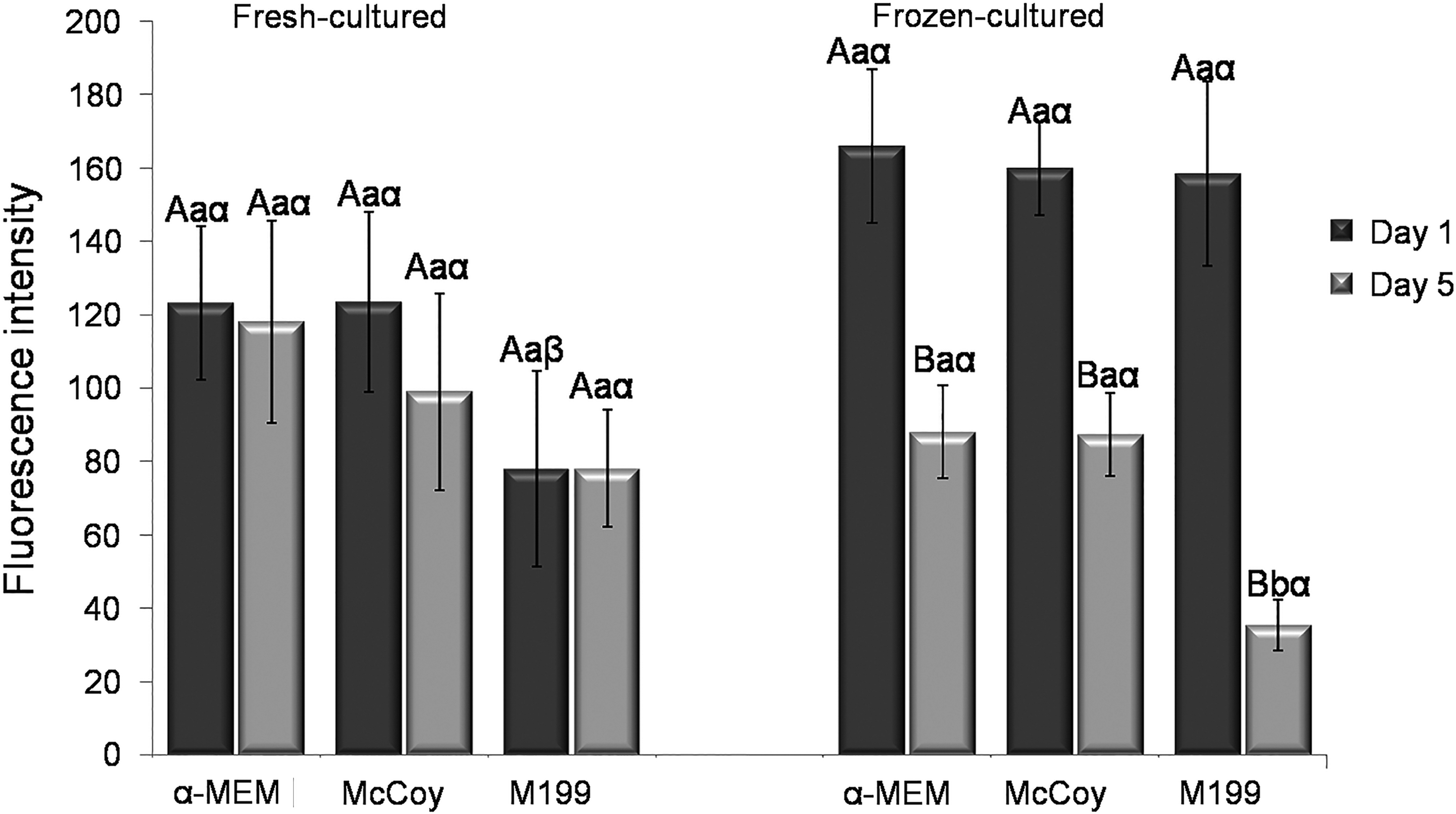
Quantification of reactive oxygen species from the media of the fresh-cultured and frozen-cultured tissues. Different uppercase letters (AB) indicate a significant difference between days of culture. Different lowercase letters (ab) indicate significant differences among media within the same day (1 or 5) and treatment (fresh or frozen). Different Greek symbols (αβ) indicate differences between treatments (fresh or frozen) within each medium and day of culture.
Hormonal production was 1.37–10.45 ng/mL for progesterone and 5–1299 pg/mL for estradiol. However, no significant difference in the levels of these hormones was observed between the fresh-cultured and frozen-cultured tissues, regardless of the medium used.
Discussion
In this study, the efficiency of three basic media (α-MEM, McCoy, and M199) was evaluated for the in vitro culture of bovine preantral follicles included in fresh or frozen ovarian tissue. The morphology and developmental competence of fresh-cultured or frozen-cultured preantral follicles was demonstrated to be influenced by the type of base medium used.
For fresh-cultured tissues, maintenance of normal morphology and follicular development was more efficient when the M199 medium was used. These results are in agreement with the findings of Rossetto et al., 20 who assessed the effect of different media (α-MEM, McCoy, M199) for the culture of isolated bovine preantral follicles that were not cryopreserved, and showed that M199 was more efficient than the other media with respect to the same parameters evaluated in the present study. On the other hand, our results showed that McCoy medium was more suitable for the culture of previously frozen tissues with respect to preserving the morphology and maintaining follicular viability, and promoted greater proliferation of granulosa cells, as demonstrated by the AgNOR. This can be attributed to the cryopreservation process, which can alter the natural conditions of the cellular environment, and thus modify their requirements for homeostasis and survival. In particular, cryopreservation can lead to retardation of follicular development and/or render cells more sensitive to degeneration occurring in vitro.9,21 These results provide strong evidence that fresh and cryopreserved preantral follicles have distinct requirements for proper in vitro development.
The histological analysis showed a lower percentage of normal follicles in the frozen-cultured group with αMEM and M199, but the viability analysis, based on the trypan blue technique, indicated a higher percentage of viable follicles in the frozen-cultured group compared to the fresh-cultured group. Although the morphology is not always correlated with viability, 22 we suggest that this difference between the histology and follicular viability test results is related to the follicular isolation procedure adopted in the present study. Cryopreserved follicular cells are considered more sensitive to laboratory processing techniques, but can withstand histological processing. If they show a high degree of degeneration, follicles may become lost and/or disintegrated during the mechanical processing of follicle isolation. Thus, the follicles derived from the frozen-cultured group had a higher degree of degeneration that was assessable using histological techniques, but possibly experienced a more pronounced loss during the isolation process, resulting in a higher proportion of viable follicles relative to identifiable follicles in the trypan blue viability analysis.
The nucleolar organizer regions (NOR) are structural-functional units of the nucleolus in which all the components necessary for the ribosomal RNA synthesis are located and the number of such regions is closely linked with cellular activity. Thus, cells with more intense proliferative activity exhibit a greater number of NOR. 23 This method of analysis has been widely used to evaluate the activity of cancer cell lines,24,25,26 and recently was also used to evaluate the activity of granulosa cells. 27 In the present study, besides the higher percentage of normal and growing follicles shown by histology on frozen-cultured tissue with McCoy medium, the AgNOR technique showed that the granulosa cells of these follicles had a higher mitotic activity when compared to follicles cultured in other media. Even with this increase in cellular activity as demonstrated by AgNOR, a reduction in the amount of ROS of D1 to D5 in the frozen group was observed. Possibly, this reduction is related to reduction in cell number, since the histological analysis showed a reduction in the number of normal follicles, consequently a smaller amount of ROS is produced.
Numerous studies have shown that various cell characteristics are altered by the cryopreservation process, including morphology, 6 ultrastructure, 28 and gene expression.29,30 Furthermore, most studies have used the same culture systems for fresh and cryopreserved follicles. Consequently, the results for the culture of previously cryopreserved cells have been unsatisfactory in general, which is partly because culture systems are developed based on the requirements of non-cryopreserved cells. Currently, several different types of culture media are commercially available for the in vitro culture of different cell types. However, these media show variation in their efficiency for the culture of preantral follicles, 20 which may be related to the composition of the medium with regard to the presence and concentration of contained substances. As demonstrated in this study, the procedure of freezing influences the efficiency of media, possibly by changes in cell metabolism that reflect changes in the requirement for cell survival and development.
The medium used for cell culture needs to meet the specific requirements of the cells to enable their normal growth and proliferation in vitro. In this context, efforts have been undertaken to characterize the metabolic activity of follicles in order to identify the requirements of these structures and facilitate development of a medium capable of promoting complete follicular development in vitro. Recently, it was shown that both the stroma and granulosa cells have glycolytic activity. 31 In another study, Harris et al. 32 found that primordial follicles have the ability to use glucose as the main carbohydrate source, suggesting that glycolysis is an important pathway to produce energy for this class of follicular cells.
Considering these observations, McCoy medium contains a higher glucose concentration than M199; thus, it is possible that cryopreserved follicles use glycolysis to obtain energy for the complete restoration of cellular activities. McCoy medium also has higher vitamin content than M199, especially those belonging to the B complex such as folic acid, pyridoxine, and cyanocobalamin. These vitamins are important for cell survival and development because they act as essential co-factors for the metabolism of carbon, which involves a complex metabolic network of interdependent biosynthetic pathways. 33 This network is involved in the synthesis of nucleotides and amino acids, 34 and also participates in other mechanisms such as the methylation of catecholamines and nuclear proteins such as histones.35,36
We formulated two hypotheses to explain the results of this study. The first hypothesis is that cells of the ovarian tissue require a higher energy intake after being subjected to stressful conditions of the freezing procedure. Thus, the increased efficiency demonstrated in McCoy medium may be related to its higher concentration of glucose compared with the other media tested. The second hypothesis is based on the importance of vitamins in various metabolic processes. 33 Thus, it is likely that follicles previously submitted to the freezing procedure have a greater requirement of these compounds, possibly due to the activation or increased activity of metabolic pathways required to repair sublethal damage induced by cryopreservation. 37
The results of this study allow us to conclude that the media, M199 and McCoy, are more suitable for the in vitro development of bovine preantral follicles contained in nonfrozen and cryopreserved by slow freezing ovarian tissues, respectively. As has already been shown that bovine and human follicles respond similarly, it is possible that in humans the use of specific culture systems for the cryopreserved tissue is also necessary. Thus, this experimental system is a valuable model for assessing the suitability of a culture system to promote the in vitro development of preantral follicles after cryopreservation.
Footnotes
Acknowledgments
This work was supported by CNPq and CAPES, Brazil. S.V. Castro and A.P.R Rodrigues are supported by a grant from CAPES.
Author Disclosure Statement
No competing financial interests exist.