
Abstract
Background:
RNA analysis of surgical specimens is one of the most useful methods for exploring biomarkers of advanced cancer. The most readily available source for RNA is formalin-fixed, paraffin-embedded (FFPE) specimens, but RNA isolated from FFPE tissue is of limited use. The PAXgene Tissue (PAX) system is a formalin-free system designed to improve the quality of molecular analysis without diminishing the quality of histopathological analysis. In this human colorectal cancer tissue study, we aimed to evaluate whether surgical specimens fixed with PAX can preserve high-quality RNA in comparison with FFPE and fresh-frozen tissue specimens.
Methods:
Ten consecutive advanced colorectal cancer patients undergoing colectomy were examined. Each specimen was processed in three ways: as frozen tissue, as PAX-fixed tissue, and as formalin-fixed tissue. RNA integrity numbers (RINs) were assessed using an Agilent Bioanalyzer. RNA transcript levels and stability were investigated by quantitative real-time PCR. We also evaluated the immunohistochemical intensity of Ki-67, CEA, and EGFR in the PAX samples.
Results:
The average RINs of RNA extracted from frozen and PAX samples were significantly higher than those from FFPE samples (p < 0.001). The cycle threshold (Ct) values were similar in PAX and frozen samples, but significantly increased in FFPE samples (p < 0.001). Most of the ΔCt values in the PAX samples did not differ significantly from those in the matched frozen samples. On the other hand, most of the ΔCt values in the FFPE samples differed significantly from those in the matched frozen samples. The immunohistochemical intensity in the PAX samples was well preserved.
Conclusions:
The quality of RNA extracted from PAX samples may be slightly inferior to that from frozen samples, but is greatly superior to that from FFPE samples.
Introduction
C
RNA analysis of surgical specimens is one of the most useful methods for exploring biomarkers of advanced cancer. RNA purity and integrity are critical elements for the overall success of RNA-based analyses, but RNA is susceptible to degradation. Although we have shown that the time from surgery to fixation can have an influence on RNA degradation, 4 many other factors including fixation and storage methods, tissue sample sizes, transport conditions, and sample storage conditions can also influence on RNA degradation.
Fixation and storage methods can have significant influences on RNA integrity. The most readily available source for RNA is usually formalin-fixed, paraffin-embedded (FFPE) specimens, but RNA isolated from FFPE tissue is of limited use for molecular analysis. 5 Formalin causes protein–nucleic acid cross-linking, which induces fragmentation of nucleic acids and lowers the quality of extracted RNA. 6 Recovery of high-quality RNA from FFPE samples is difficult,7,8 because RNA from these specimens may become degraded.9,10 The gold standard for RNA analysis remains fresh-frozen tissue, which is not the preferred state for accurate histological and morphological studies. Moreover, long-term storage of fresh-frozen tissue is expensive. Therefore, it is necessary to establish new, convenient fixation and storage methods to preserve RNA without affecting pathological examination.
The PAXgene tissue system (PAX; Qiagen, Hilden, Germany) is a commercially available tissue collection device. It is a formalin-free system designed to improve the quality of molecular analysis without diminishing the quality of histopathological analysis. In human melanoma and animal samples, PAX Tissue performed better than FFPE tissue in terms of RNA quality, quantity, and transcript stability. 11 Viertler et al. 12 evaluated the ability of PAX to preserve RNA in human malignant and nonmalignant tissues, including colon cancer. However, there have been no studies that include only colon cancer. Earlier studies evaluated PAX performance of RNA transcript stability not only by using cycle threshold (Ct) value, but also by the ΔCt and ΔΔCt methods.
In this human colorectal cancer tissue study, we aimed to evaluate whether surgical specimens fixed with PAX can preserve high-quality RNA in comparison with FFPE and fresh-frozen specimens and whether the results of RNA amplification analysis using PAX samples can produce the same results as using frozen samples. We evaluated detailed PAX performance of RNA transcript stability in colorectal cancer by using Ct values, ΔCt method and ΔΔCt method. By using the ΔΔCt method, we can clarify the differences between frozen samples and PAX or FFPE samples in a direct manner.
Materials and Methods
Patients and sample collection
We examined 10 consecutive advanced colorectal cancer patients undergoing colectomy from October 2012 to January 2013. Colorectal cancer specimens obtained from the primary site were collected immediately following the surgical extraction in the operating room. We extracted one piece (15 × 15 × 10 mm) and divided it into eight smaller pieces (one for frozen storage, one for PAXgene fixation, and six for formalin fixation). The study protocol was approved by the Gene Institutional Review Board of our institute, and written informed consent was obtained from all participants.
Tissue fixation and sample storage
Each sample was processed in three ways: as frozen samples (Frozen group); in PAXgene Tissue Containers (PAX group); or in non-buffered formalin (formalin) or neutral-buffered formalin (NBF) for 6 or 48 h (FO group). Frozen group samples were placed in RNAlater (Invitrogen, Paisley, UK), a fixative solution, and preserved at −80°C. PAX group samples were fixed using the PAXgene tissue system (Qiagen, Hilden, Germany) and embedded in paraffin (PAXgene-fixed, paraffin-embedded tissue/PFPE) according to the manufacturer's protocol.
Briefly, specimens were fixed in PAXgene Tissue FIX Solution for 2–4 h, followed by transfer to PAXgene Tissue STABILIZER and storage at −20°C. After 48 h, the specimens were dehydrated eight times in 99% ethanol for 85 min, immersed three times in chloroform for 2 h, and embedded in 56°C low-melting-temperature paraffin. FO group specimens were fixed using non-buffered 20% formalin or 10% or 20% NBF for 6 or 48 h. After fixation, the specimens were embedded in 60°C paraffin.
RNA extraction
Total RNA was isolated from each specimen using preservation-specific methods. In the Frozen group, RNA was extracted using an RNeasy Mini Kit (Qiagen) after 48–72 h of storage. In the PAX group, RNA was extracted using a PAXgene Tissue RNA Kit (Qiagen) at 48–72 h after fixation. In the FO groups, RNA was extracted using an RNeasy FFPE Kit (Qiagen) at 48–72 h after fixation. In the PAX and FO groups, RNA was extracted from five 7 μm thick slices cut directly before extraction.
Evaluation of RNA quality and yield
The concentration of extracted total RNA was measured using a NanoDrop 2000 spectrophotometer (Thermo Scientific, Waltham, MA). RNA purity was estimated by determining the 260 nm/280 nm absorbance ratio (A260/A280). Extracted RNA is considered to be of the highest quality when A260/A280 is within the range of 1.8–2.1.
RNA integrity was assessed using an Agilent Bioanalyzer (Agilent Technology, Palo Alto, CA) and RNA6000 Pico Kit (Agilent Technology). RNA integrity numbers (RINs) were calculated using Agilent 2100 Expert software. Extracted RNA is considered to be of the highest quality when the RIN is within the range of 7–10. Meanwhile, a RIN of 4–7 is good and a RIN of <4 is poor.13,14
Evaluation of RNA transcript stability
RNA transcript levels and stability were investigated by quantitative real-time PCR (qPCR). cDNA was synthesized using a High Capacity cDNA Reverse Transcription Kit (Applied Biosystems, Foster City, CA) according to the manufacturer's protocol. We used cDNA equivalent to 25 ng of RNA for real-time RT-PCR with SYBR Green in a 7500 real-time PCR system (Applied Biosystems). The total cycle number was set at 40 for all samples.
We evaluated five RNAs: GAPDH, GPX1, VDAC2, ABL1, and EGR1. GAPDH is an internal control housekeeping gene. GPX1 and VDAC2 are reference genes for OncotypeDX, which is the most up-to-date method to predict the risk of recurrence after curative resection in colorectal cancer patients. 2 ABL1 contains a nuclear localization signal and a DNA-binding domain through which it mediates DNA damage-repair functions. EGR1 inhibits apoptosis 15 and tumor cell invasion. 16 To evaluate RNA transcript stability and degradation, we used PCR primers designed for two amplicon lengths: long (about 500 bp) and short (about 100 bp). The primers used in this study are shown in Table 1.
Data analysis
We used Ct values as indicators of RNA transcript stability for each fixation method for the five genes. The target genes (GPX1, VDAC2, ABL1, EGR1) were normalized to the corresponding GAPDH internal control gene using the ΔCt method as follows: ΔCt = Ct (target gene) – Ct (GAPDH). The ΔΔCt method was performed to compare the RNA amplification analysis using frozen samples and PAX or FFPE samples as follows: ΔΔCt (gene) = ΔCt (PAX or FFPE) – ΔΔCt (Frozen). Using this analysis, if the expression levels of the target gene were not affected by RNA degradation, the values of ΔΔCt should be very close to 0.
Morphology
To determine morphology preservation, we evaluated FFPE and PFPE tissues using hematoxylin and eosin (H&E) staining. The histology of 3 μm sample sections from each fixation protocol was independently examined by two pathologists (TI and YM) who were blinded to the fixation method. Morphology assessment included overall morphology and nuclear, cytoplasmic, and membrane details. The pathologists scored PAX samples compared with the samples fixed in 20% formalin for 6 hours or those for 48 hours. We evaluated each end point (nucleus, cytoplasm, membrane) by using 5-point scale (PAX sample was considerably better: 2, better:1, equal:0, worse: −1, considerably worse: −2).
Immunohistochemistry
Paraffin-embedded sections (3 μm) were subjected to immunostaining using an automated slide staining system (Histostainer; Nichirei Japan). After deparaffinization, the tissue sections for Ki67 and CEA immunostaining were preheated in Heat Processor Solution pH 6 (Nichirei Japan) for 20 min at 100°C. The tissue sections were then incubated with an anti-Ki67 antibody (1: 200 dilution; Dako Japan), anti-CEA antibody (Histofine primary antibody; Nichirei Japan), or anti-epidermal growth factor receptor (EGFR) antibody (EGFR pharmDx kit; Dako Japan). Negative control tissue sections were prepared by omitting the primary antibody. Immunohistochemical review was performed by a pathologist (YM).
Statistical analysis
Statistical analyses were performed using SPSS 21.0 (IBM Japan, Tokyo, Japan). Repeated-measures ANOVA was used to evaluate RNA yield and RNA quality (A260/A280 and RIN). Repeated-measures ANOVA was also used to evaluate RNA transcript stability (Ct, ΔCt, and ΔΔCt) between the groups. Bonferroni analysis was used for post-hoc analysis. The Wilcoxon t-test was used to calculate mean Ct values between short amplicons and long amplicons for the same fixation method. Values of p < 0.05 were considered to indicate statistical significance for each analysis.
Results
RNA yields and quality
The clinical backgrounds of the 10 patients are shown in Table 2. The RNA yields from the frozen, PAX, and FO samples were similar, with no significant differences (Table 3; p = 0.22). In contrast, the average RINs of total RNA extracted from the frozen and PAX samples were significantly higher than those from the FO samples (p < 0.001; Table 3). Overall, 90% of the frozen samples showed high RINs (>7.0) and 80% of the PAX samples showed high RINs, while no FO samples showed high RINs.
tub1, Well differentiated type; tub2, Moderately differentiated type.
p < 0.01 ÷ significant difference vs. the Frozen group; **p < 0.01 ÷ significant difference vs. the PAX group.
NBF, neutral-buffered formalin; RIN, RNA integrity number.
RNA transcript stability and performance in qPCR
The Ct values were similar in the PAX and frozen samples, but significantly increased in the FO samples (Fig. 1A–E). Within the FO groups, there were no significant differences between formalin fixation methods (kind and fixation time). However, for all genes and all formalin fixation conditions (except the long amplicon of GAPDH in 10% NBF), the Ct values at 6 h were significantly lower than those at 48 h (p < 0.05; Table 4A, 4B).
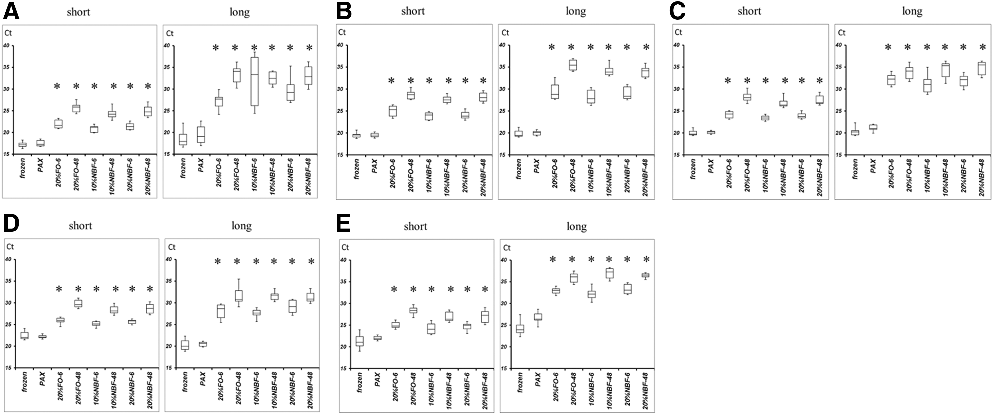
The differences of Ct values between frozen samples, PAX samples, and FO samples. Ct values of
NBF, neutral-buffered formalin.
For the short amplicons, none of the ΔCt values in the PAX samples (except for ABL1) showed significant differences compared with those in the matched frozen samples. Conversely, most of the ΔCt values in the FO samples (except for GPX1 in 20% NBF, VDAC2 in 10% NBF, and VDAC2 in 20% NBF) showed significant differences compared with those in the matched frozen samples (Fig. 2A–D). In many formalin samples, the long amplicon could not be amplified.
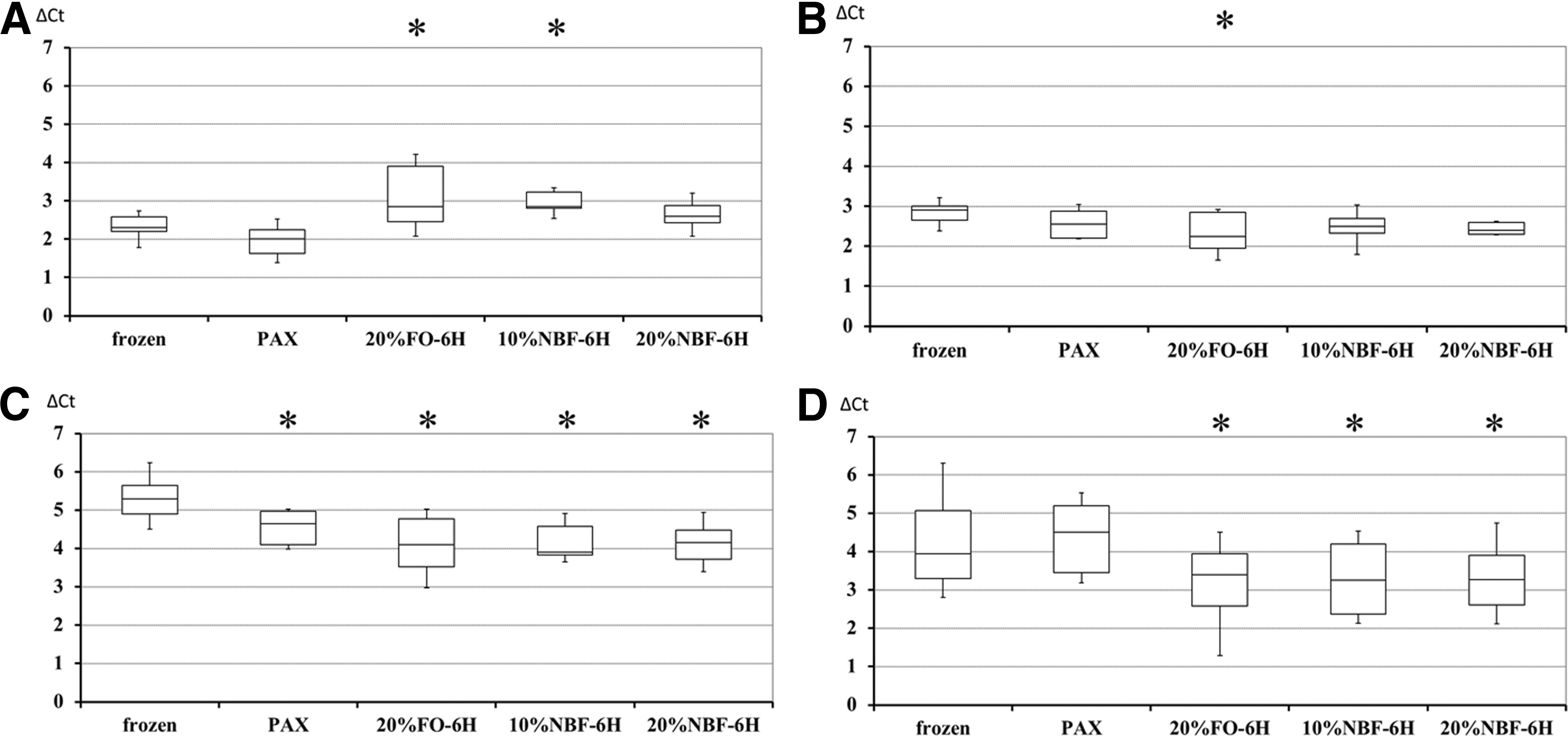
The differences of ΔCt values between frozen samples, PAX samples and FO samples. ΔCt value of
When the absolute value of ΔΔCt is <1 (expression level is lower than two-fold and higher than half quantity, since 21 = 2, and 2−1 = 0.5), the quality of the analysis is judged to be high. 17 In the PAX samples, the ratios of ΔΔCt < 1 values for GPX1, VDAC2, and ABL1 were over 70%. These results were better than those for the FO samples (Table 5A). Remarkably, none of the absolute ΔΔCt values exceeded 2 in the PAX samples, while some of the absolute ΔΔCt values for the FO samples did exceed 2 (Table 5B).
NBF, neutral-buffered formalin.
Morphology
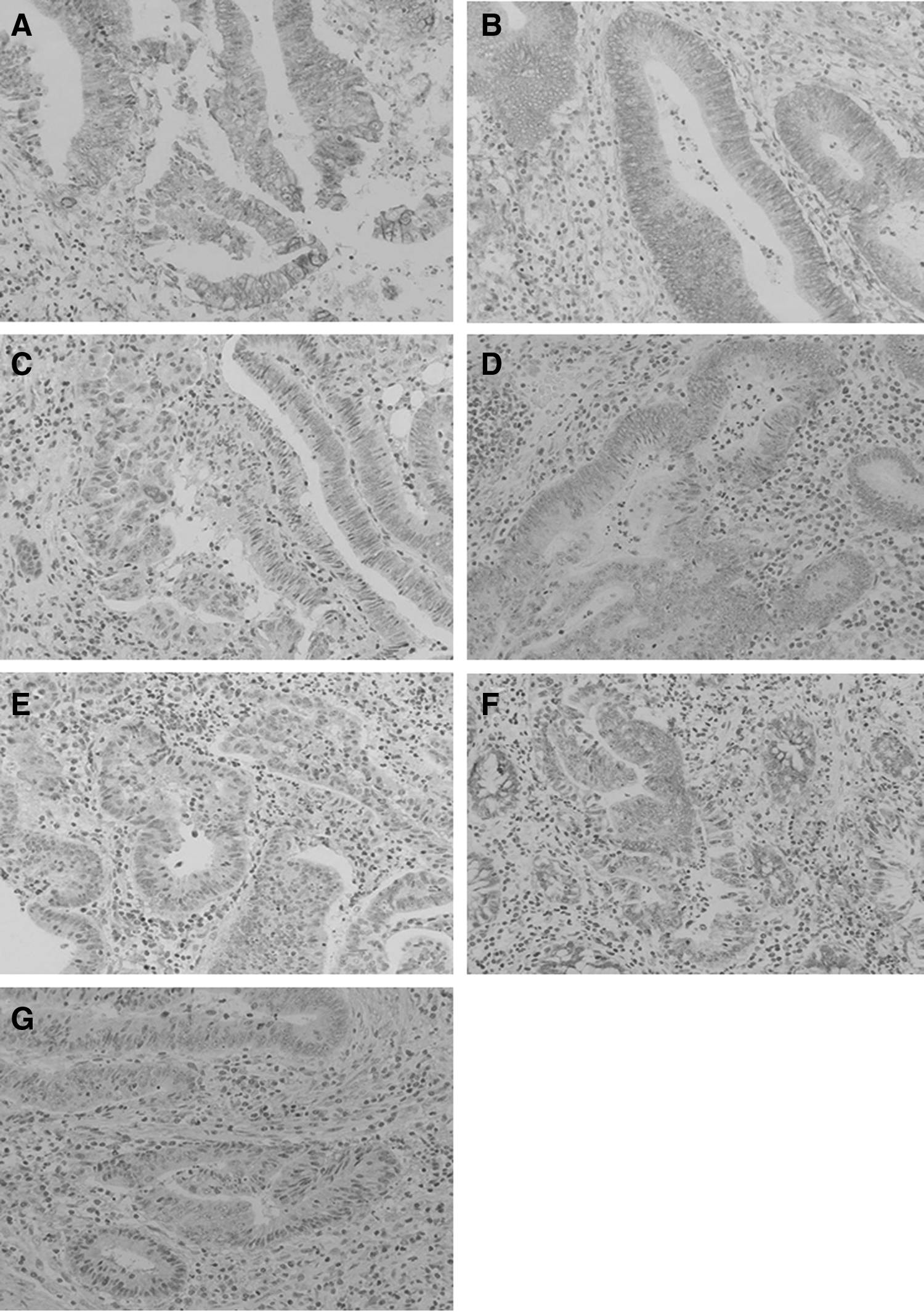
No samples were scored 2 or −2. In regard to nuclear and membranous, all samples were scored 1 or 0. In regard to cytoplasmic samples, Pathologist A scored −1 in one sample, and Pathologists B scored −1 in two samples. Other samples were scored as 0 by the two pathologists. The PAX samples also exhibited cellular contraction and the cells appeared smaller than those fixed by the other methods. However, the morphological changes of the cancer cells were limited, and did not influence the tumor diagnosis (Fig. 3A–H).
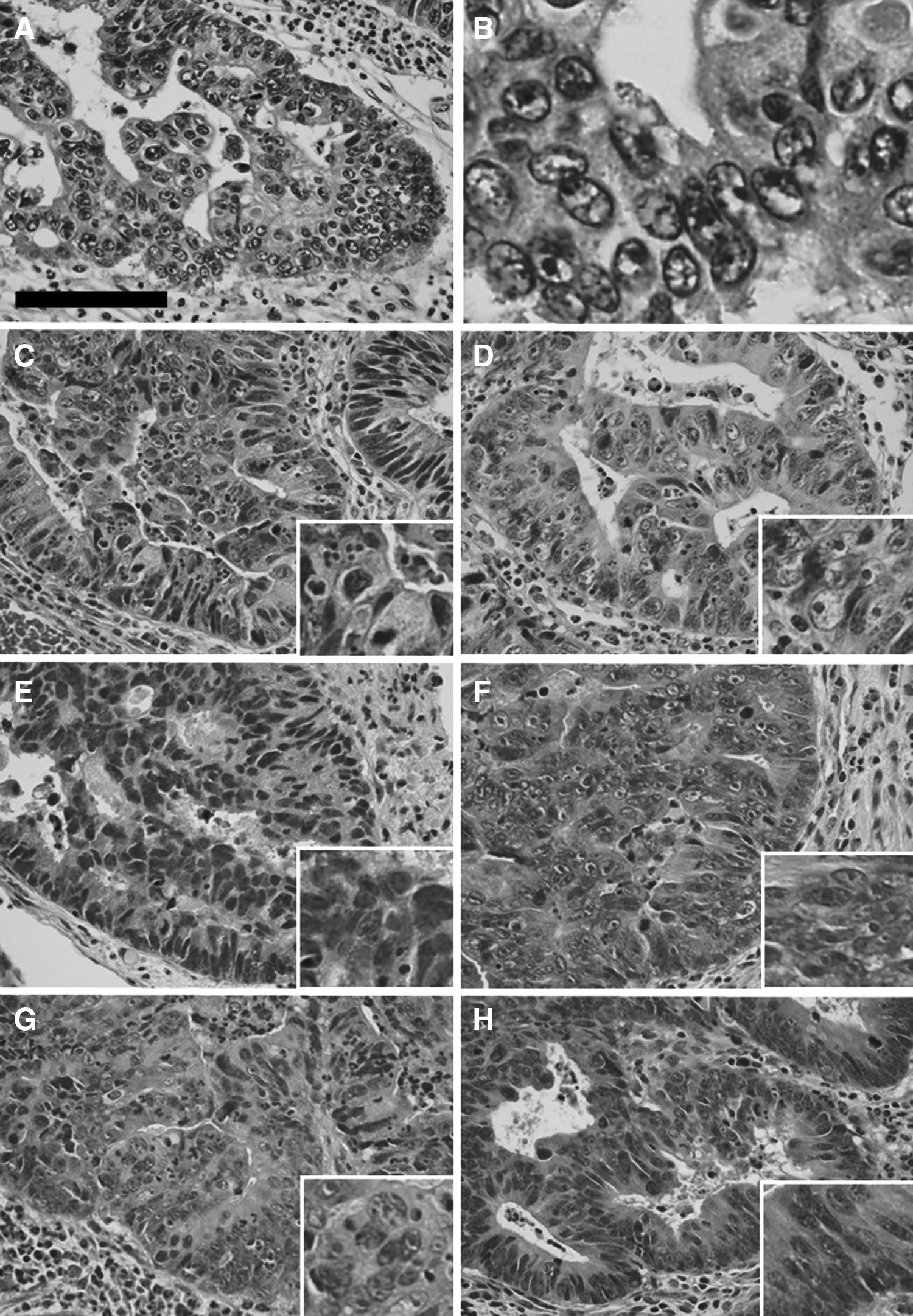
The morphology.
Immunohistochemistry
Immunohistochemical intensity of Ki-67, CEA, and EGFR of PAX fixed tissue was well preserved. Conversely, whereas immunohistochemical intensity of the tissues fixed in formalin for 6 h was preserved, that which was fixed for 48 h declined (Figs. 4, 5, 6).

The immunohistochemical study of Ki-67
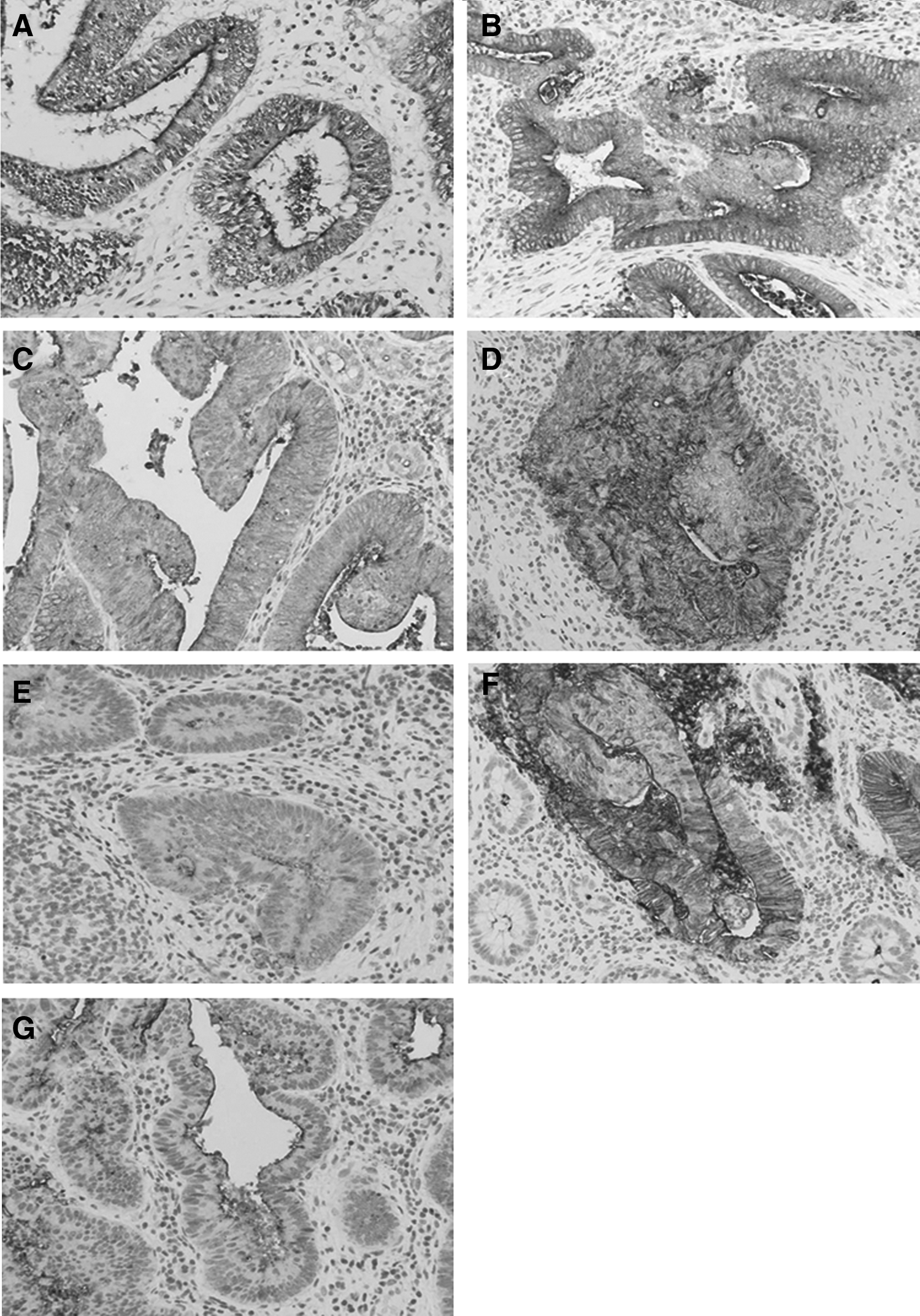
The immunohistochemical study of CEA.
The immunohistochemical study of EGFR.
Discussion
In this study, we have clearly demonstrated two important findings. First, the quality of mRNA extracted from PAX samples was similar to that from frozen samples, and greatly exceeded that from FFPE samples. The RINs and Ct values of the PAX samples did not differ significantly from those of the frozen samples, while the FO samples showed consistently lower RINs and higher Ct values (about 5 cycles higher for short amplicons and 10 cycles higher for long amplicons), compared with the Ct values for the corresponding frozen and PAX samples.
Second, the RNA amplification analysis using the PAX samples produced similar results to that using the frozen samples, while the RNA amplification analysis using the FFPE samples produced substantially different results from that using the frozen samples. The ΔCt and ΔΔCt values of the PAX samples were generally better than those of the FO samples.
Preservation method-specific differences were not shown in sample purity as measured by A260/A280 ratios, consistent with a previous report. 11 However, although the RINs of the frozen and PAX samples did not differ, the RINs of the FO samples were significantly lower than those of the frozen and PAX samples. It is important to evaluate RNA quality using a suitable method and not to rely on A260/A280 measurements. 17 A260/A280 ratios do not change even when the amount of contaminated protein changes greatly. 18 Therefore, in recent years, RNA integrity has been assessed with a bioanalyzer to measure the RIN.
A previous report showed that 80% of frozen colorectal samples had RINs of >7.0, 19 and 90% of frozen various samples had RINs of >6.5. 20 In this study, 90% of the frozen samples had RINs of >7.0 and all the frozen sample had RINs of >6.5, indicating that our frozen samples preserved high-quality RNA. Meanwhile, none of the FO samples had RINs of >7.0, and 80% of the PAX samples had RINs of >7.0. RINs above 5.5 are sufficient for most applications. 13 The success rate of high-quality array analysis was 100% (24/24) in samples with RINs of >5.0, but 30% in samples with RINs of <5.0. 21 In this study, all of the PAX samples had RINs above 5.5, compared with none of the FO samples, suggesting that PAX is a desirable fixation method for preservation of high-quality RNA.
None of the Ct values in the PAX samples showed significant differences compared with those in the frozen samples, and all of the Ct values in the FO samples were significantly higher than those in the frozen and PAX samples. Accordingly, the RNA transcript stability in the PAX samples was equivalent to that in the frozen samples, but higher than that in the FO samples. The differences in the Ct values between the PAX and FO samples were 5–10 (expression levels of 32–1024-fold), and these differences may greatly affect the reliability and reproducibility of downstream applications. Viertler et al. 12 also showed the Ct value differences (4–15) between PAX and FO samples.
As mentioned above, it has been reported by Viertler et al. 12 that Ct values of frozen sample have significant differences with that of formalin samples but PAX samples. However, it has not been reported how the differences can affect ΔCt. In this study, we demonstrated that only the ΔCt value of ABL1 showed a significant difference compared with the matched frozen samples in the PAX samples. We also demonstrated that most of the ΔCt values in the FO samples showed significant differences compared with the matched frozen samples in the 6 h FO samples, which showed better results than the 48 h FO samples. These findings suggest that low-quality RNA extracted from FO samples may be misleading for treatment selection. In addition, it is noteworthy that none of the absolute ΔΔCt values exceeded 2 in the PAX samples, suggesting the stability of PAX samples.
PAX has two additional merits. First, the PAX samples were not sensitive to overfixation or underfixation, because no adverse effects on mRNA quality were observed after either a prolonged fixation period (up to 120 h) or a short fixation period (3 h). 12 Conversely, the fixation time was more important than the kind (formalin or NBF) or concentration (10% or 20%) of formalin in terms of preserving high-quality RNA. The CT values after 6 h of fixation in formalin were significantly lower than those after 48 h of fixation in this study.
Second, PAX samples need no specialized equipment for storage or transport. For personalized medicine that requires RNA analysis, we often need to keep surgical specimens for long time periods. However, many general hospitals do not have deep freezers because they are expensive and require a large space. For multicenter studies using RNA analysis, PAX samples are easy to transport because they are stored at room temperature, whereas frozen samples need to be kept at −80°C during transportation.
PAX samples showed preserved morphology similar to the corresponding FFPE samples, consistent with two previous reports.12,22 These two reports also showed that PAX samples are useful for immunohistochemistry in the same way as our study. We can extract RNA from an adjacent section to that examined under a microscope, and this can prevent contamination of normal cells and maintain the quality of the study, whereas pathological examination using frozen samples requires great care.
Formalin is the most commonly used preservation material because it is a relatively cheap reagent compared with commercially available non-formalin fixatives, and also because morphology of tissues fixed with non-formalin fixatives is not exactly the same as that of tissues fixed with formalin. However, formalin is a carcinogen and a mutagen, and also an allergen. Moreover, the importance of RNA analysis is increasing with the progress of molecular biology. Therefore, the development of alternative agents to formalin is expected. The PAXgene Tissue System is one of the candidates because of its excellent biopreservation ability for RNA, morphology, and immunogenicity.
This study has several limitations. The first limitation is the storage period. In this study, we extracted RNA from all samples within 72 hours after acquisition. Although RNA quality decreases when extracted from FFPE samples after long-term storage, 23 we did not evaluate the quality of RNA extracted from PAX samples after long-term storage. Second, we only evaluated colorectal cancer samples and evaluated five genes. Thus, the usefulness of PAX for other kinds of malignancies remains unclear and the difference between OncoType DX scores using PAX samples and FFPE samples was not clarified. Third, it cannot be said that the RNA extracted from PAX samples is preserved in as high quality as that from frozen samples for analyses of all genes. Regarding ABL1, the Ct value of PAX samples was equal to that of frozen samples. However, the ΔCt of PAX samples was inferior to that of frozen samples. Nevertheless, our data demonstrate the impact of this new fixation and storage method on the quantification of gene expression and immunohistochemical intensity.
Conclusions
The quality of RNA extracted from PAX samples may be slightly inferior to that from frozen tissue, but is greatly superior to that from FFPE tissue. The immunohistochemical intensity in PAX samples is well preserved.
Footnotes
Author Disclosure Statement
No conflicting financial interests exist.