
Abstract
We established a standard breast cancer biobank at Harbin Medical University Cancer Hospital (HMUCH) in 2009. More than 100,000 biospecimens, including high-quality human breast cancer samples, matched blood samples, and adjacent normal tissues, were collected from patients and healthy donors in HMUCH and were then deposited in the repository. We reported the establishment of a biobank in our hospital and its crucial role in translational medicine research. We stored, processed, and distributed qualified biological specimens in accordance with international standard operating procedures. We also summarized the utilization of this biobank and its influence on research projects over the years since its establishment. Therefore, we can verify specific biomarkers that may aid in the development of targeted breast cancer therapies by using high-quality, well-annotated tissue samples provided by the biobank.
Introduction
A
With rapid growth of knowledge and innovations, biomedical research and healthcare are shifting toward the vision of personal medicine and translational medicine. 5 In this field, high-quality and well-annotated biospecimens are essential. Biobanking services must be improved rapidly to satisfy the needs of personal medicine and to enhance the quality and speed of basic and translational research. 6 Furthermore, the long-term storage of biological materials is an essential component of breast cancer research. The breast cancer biobank at Harbin Medical University Cancer Hospital (HMUCH) is the largest breast cancer research resource in Heilongjiang, China, which covers 38,312,224 inhabitants. The breast cancer biobank aims to establish a large repository of biological materials and information to gather, evaluate, store, and disseminate biospecimens in compliance with the International Society for Biological and Environmental Repositories Best Practices for Repositories 7 and to support future scientific investigation.
Using RNA yield and RNA integrity number (RIN), housekeeping gene expression, and morphological examination of frozen sections, we evaluated the quality of biological specimens stored under the following storage conditions, −80°C, −196°C, and RNAlater/−80°C, and different storage periods (from 2009 to 2014) to provide high-quality tissue samples to investigators. We summarized the major results of these investigations, focused on breast cancer research, and analyzed the influence of research projects that employed samples provided by our biobank over the years.
Materials and Methods
Tumor specimen collection and storage
Biological specimens were collected from women diagnosed with breast cancer and from healthy donors at HMUCH. Sample collection and use were in accordance with the ethical rules of Harbin Medical University. Fresh human pathologic and normal breast tissues were obtained directly after surgery was completed. Surgeons determined the specimens for sample collection on the basis of preoperative diagnoses. Diagnoses were achieved straightforwardly because of detailed clinical information, including mammography, ultrasonic examination, and biopsy; diagnoses were also verified through postoperative pathological analysis. After patient consent was obtained, tissues were collected by experienced residents under the supervision of practicing pathologists immediately after tissue specimens were removed from the body and before pathological diagnosis was performed. The samples were selected, registered, and frozen within a 10-minute window corresponding to cold ischemic time after the surgical specimen was removed from patients. The tissue was cut in cubes, and tissue fragments were stirred and divided into three grossly equal portions. One portion of the tissue fragments was immersed in 1.5 mL of RNAlater, RNA stabilization reagent (Ambion, Inc., Austin, TX). After 16 hours, the tissue fragments were transferred to a low-temperature freezer (RNAlater/−80°C). The samples requiring snap-freezing in the two remaining portions were frozen in liquid nitrogen (−196°C) immediately. After 16 hours, the two portions of tissues were stored in a low-temperature freezer (−80°C) and Thermo Scientific CryoPlus Sample Storage Systems (Thermo Scientific, Wilmington, DE) (−196°C), respectively.
Morphological characteristics and stress markers in frozen tissues
We selected 72 aliquots of breast cancer samples from 24 individuals. The three aliquots from one patient were stored under three different conditions (−80°C, −196°C, and RNAlater/−80°C). The aliquots were selected randomly each year from 2009 to 2014. Cryostat sections of tissues were generated with a cryostat equipped with the CryoJanes Tape-Transfer system (CryoJane; Instrumedics, Inc., Hackensack, NJ). The frozen tissue was cut into 4-μm-thick sections. These sections were then transferred on tape to an adhesive glass microscope slide (CryoJane). With a flash of ultraviolet light, the sections were permanently bonded to the glass. The sections were fixed with glutaraldehyde and stained with hematoxylin–eosin (H&E) in accordance with standard procedures. Histopathological assessment was quantified, as described by Mesker et al. 8 The carcinoma percentage (CP) was visually estimated by two experienced pathologists from the entire tumor area on the basis of morphological information (for clarity, we only provided CPs, but complementary to the CP are stromal percentages, for example, CP 70% implies a stromal percentage of 30%). Percentages were scored ranging from 20% to 90%. However, 10% and 100% were not found. Breast cancer is a heterogeneous disease with numerous clinical and histological types, 9 and the tumor–stroma ratio is significantly associated with several breast cancer subtypes.10,11 In this regard, CP was defined as CP-low if <50%, including 20%, 30%, and 40% tumors, and CP-high if ≥50%, including the values 50%, 60%, 70%, 80%, and 90%. Only tissues that satisfied the CP-high standard were considered fit for RNA quality evaluation. We also selected 24 aliquots of breast cancer samples stored at −80°C, which were selected randomly each year (from 2009 to 2014), to analyze the stress markers, ERK5 12 and Peroxiredoxin V, 13 by immunohistochemical detection. Cryostat sections were reacted with the corresponding purified rabbit monoclonal antibodies against ERK5 and Peroxiredoxin V (ABclonal Biotech Co., Ltd, Baltimore, MD, USA) overnight. For the second antibody, goat anti-rabbit horseradish peroxidase was used (dilution 1:400, from Dako). The specificity of the monoclonal antibodies to ERK5 and Peroxiredoxin V was confirmed by comparison with adjacent sections.
RNA isolation
We reselected 72 aliquots of breast cancer samples that satisfied the CP-high standard and were used to analyze RNA from 24 individuals. Three aliquots from one patient were stored under three different conditions (−80°C, −196°C, and RNAlater/−80°C), and 12 aliquots from the samples of four individuals were selected randomly each year (from 2009 to 2014). We then transferred and pulverized 45 mg of the frozen tissue with pestle and mortar containing liquid nitrogen. We then placed the powdered tissue into a tube containing 1 mL of TRIZOL Reagent (Invitrogen, Stockholm, Sweden). The homogenized samples were incubated for 5 minutes at room temperature to induce the complete dissociation of nucleoprotein complexes. The following additional steps were performed. Chloroform (0.2 mL) per 1 mL of TRIZOL reagent was then added, and the tubes were manually shaken for 15 seconds. The samples were centrifuged at 12,000 × g for 15 minutes at 4°C. The aqueous phase was transferred to a fresh tube, and 0.5 mL of isopropyl alcohol was added per 1 mL of TRIZOL reagent used for initial homogenization. Afterward, the samples were incubated at −20°C for 35 minutes and centrifuged at 12,000 × g for 10 minutes at 4°C. The supernatant was then removed, and RNA pellet was washed once with 80% ethanol. The RNA solution was centrifuged for 8 minutes at 12,000 × g, and the resulting supernatant was decanted. The RNA pellet was air-dried and subsequently dissolved in 50 μL of RNase-free water.
RNA yield and RIN measurement
The concentration of total RNA (ng/μL) was determined using a Nanodrop ND-1000 spectrophotometer (Thermo Scientific), which was also employed to calculate the total RNA yield (μg). RNA purity was assessed by measuring the A260/A280 ratio. The samples with the total RNA yield of at least 1 μg and an A260/A280 ratio of at least 1.8 were considered of sufficient quality for further analysis. RNA integrity can be reliably detected through microcapillary gel electrophoresis, from which RIN can be calculated. In literature, RNA with a RIN greater than six is generally considered of acceptable integrity for gene expression measurement.14,15 Thus, we defined that the tissue bank performance is adequate when the percentage of the samples with RIN ≥6 is higher than 80%. An Agilent 2100 Bioanalyzer and an RNA 6000 Pico Labchip kit (Agilent Biotechnologies, Palo Alto, CA) were utilized to evaluate the RIN of the tissue samples. The analysis was performed in accordance with the manufacturer's instructions.
cDNA synthesis and quantitative real-time polymerase chain reaction
cDNA was generated from 2 μg of total RNA by using the Transcriptor First-strand cDNA synthesis kit (Roche Diagnostics Corporation P.O., Mannheim, Germany). The process was conducted using the Gene Amp® PCR System 9700 (Applied Biosystems, Foster City, CA) in accordance with the manufacturer's instructions. Afterward, 2 μL of the cDNA solution was used for a 35-cycle SYBR Green PCR assay with the SYBR Green Universal PCR Master Mix (Applied Biosystems) with the primers described in Table 1. Real-time quantitative polymerase chain reaction (qPCR) was then performed with the ABI 7500 Fast Real-time PCR System (Applied Biosystems) under the following conditions: in stage 1, 95°C for 5 minutes; in stage 2, 95°C for 15 seconds, 60°C for 30 seconds, and 72°C for 30 seconds for 35 cycles. Expression levels were non-normalized by GADPH.
Statistical analysis
Differences in RNA yield, RIN, and relative expression of genes from the tissue samples under different storage conditions and for various durations were analyzed through two-way ANOVA and Kruskal–Wallis test. A p-value of >0.05 was considered significant.
Results
Composition of the breast cancer biobank
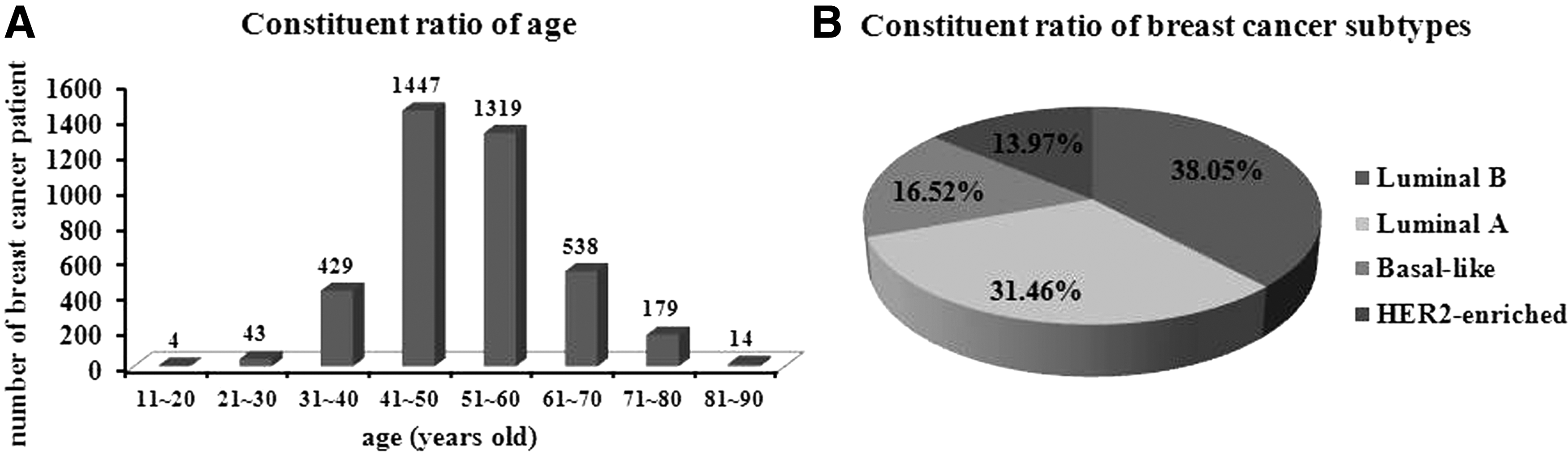
The total number of cancer cases reached 15,018 and the total sample copies reached 50,712 (9853 frozen tissues and 40,859 blood specimens) from 2009 to 2014. The mean age at diagnosis of breast cancer is consistent with that in previous studies. 16 Figure 1A shows the age distribution of the patients. Luminal B subtype accounted for 38.05% of all of the breast cancer tissues (Fig. 1B). Twice as many samples corresponded to the luminal A subtypes (34.16%) compared with basal-like subtypes (16.52%). The Her-2 enrichment subtype yielded the lowest percentage (13.97%). The databases are managed by an independent information management and security system (Avantech, Shanghai, China), which satisfies the regulatory requirements for storage and access.
Characterization of the breast cancer biobank sample.
Morphological characteristics and stress markers
The morphological examination of frozen sections revealed the intact breast cancer architecture in 72 aliquots of breast cancer samples preserved under different conditions (−80°C, −196°C, and RNAlater/−80°C), selected randomly each year (from 2009 to 2014), and stained with H&E. Signs of significant cellular decomposition could not be detected in all of the test samples, which were stored for four different durations (Fig. 2A). Routine H&E-stained slides from the most invasive part of the tumor were microscopically analyzed to determine CP by using 5× and 10× objectives. Of the 72 analyzed patients, 13 (18.06%) obtained low CPs and 59 (81.94%) achieved high CPs. Immunohistochemical detection showed that ERK5 and Peroxiredoxin V were overexpressed in all of the samples and did not significantly differ in various years (Fig. 2B).

Examination of frozen tissue morphology and stress marker examination.
RNA yield and RIN: effect of storage duration and condition
The RIN examination of the tissues in our bank (n = 72) indicated that 10.2% (n = 7) of the samples were unreliable for demanding downstream procedures (RIN <5). By contrast, 83.8% (n = 57) of the samples with RIN ≥6 are reliable for quantitative reverse transcription-PCR (RT-qPCR) and gene array analysis (Table 2). However, the storage conditions also significantly affected the RNA quality. Compared with the samples stored at −80°C, preservation in RNAlater/−80°C significantly improved the RNA yield and integrity (p < 0.05), as assessed by the concentration and RIN (Fig. 3). Compared with the samples stored at −196°C, the samples preserved at −80°C and in RNAlater exhibited no significant effect on the yield and RIN. The quality of the samples among different storage periods did not significantly change (from 2009 to 2014).

RNA yield and RIN of selected tissues from the breast cancer biobank in different storage conditions and different storage durations.
Samples were divided by RIN values. The samples with RIN < 5 are not reliable for demanding downstream genomic techniques; samples with 5 ≤ RIN <6 are only useful for RT-qPCR; and samples with RIN ≥6 are reliable for RT-qPCR and gene array analysis. N/D, not detected; RIN, RNA integrity number; RT-qPCR, quantitative reverse transcription-polymerase chain reaction.
Gene expression levels: effect of storage duration and condition
Six genes were analyzed in 54 aliquot samples on the basis of the A260/A280 ratio, total RNA yield, different storage conditions, and different durations to examine the effect of storage conditions and storage duration on gene expression. The genes were selected and used as markers of various cellular regulatory pathways: CFOs (immediate early response gene), TGFβ1 (growth factor), SMAD7 (signal transduction molecule), HIF1α (hypoxia response), Bcl2 (apoptosis regulator), and PCNA (cell cycle). 17 All of the gene expression levels remained stable when the tissue was preserved under different conditions and for different durations (Fig. 4). The results did not show any significant differences.

Gene expression levels under different storage conditions and durations. RNA isolated from selected tissues from the breast cancer biobank in different storage conditions and different storage durations. RNA was transcribed to cDNA and analyzed using primers for CFOs, TGFβ1, SMAD7, HIF1α, Bcl2, and PCNA. The results were non-normalized to GAPDH and presented in Ct values.
Utilization of biobank resources
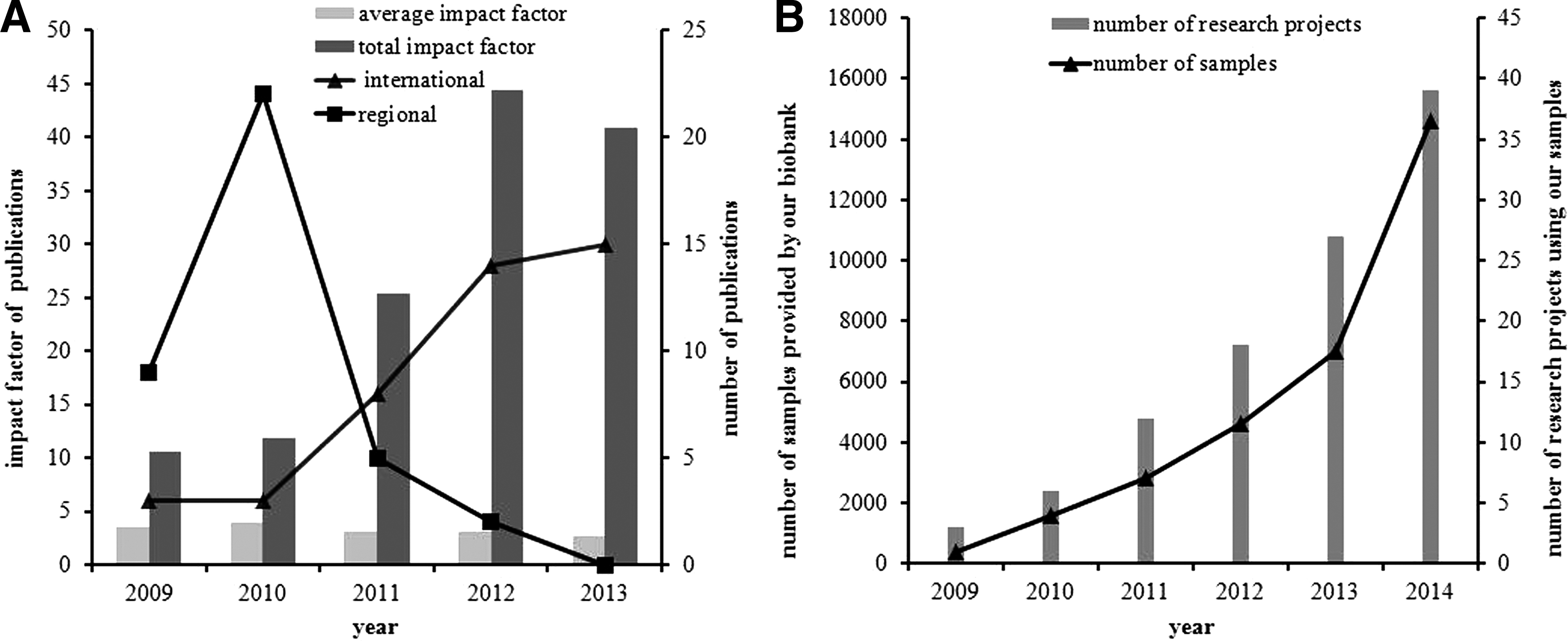
Since the breast cancer biobank was established, 43 articles that used human samples collected by our biobank have been published in international journals. Therefore, we determined the totality of publications by the researchers of our institute and collaborators in regional (Chinese) and international journals from 2009 to 2013. The average and total impact factors of publication were determined from 2009 to 2013. The number of research projects that requested access to samples from 2009 to 2014 and the number of biological specimens provided by our biobank were also retrieved.
The total impact factor of publications significantly increased from 10.5 in 2009 to 40.8 in 2013 (Fig. 5A) because of the increasing number of publications (from 12 in 2009 to 15 in 2013, Fig. 5A) and a significant transfer from Chinese regional journals to English international journals (25% of articles in English journals in 2009, 100% of articles in English journals in 2013, Fig. 5A). Most of our articles involved basic, clinical, and translational research on breast cancer and thus provided significant contributions to personalized and translational medicine. By the end of 2013, our students utilized human biological materials from our biobank to accomplish their academic dissertation. In 2014, our biobank provided 14,600 biosamples for 39 projects associated with academic dissertation. This value indicated a significant improvement with respect to that in 2009 (Fig. 5B).
Discussion
A biobank aims to establish storage of biological specimens with intact morphological characteristics, proteins, DNA, and RNA for research, diagnosis, treatment, and prevention of disease. The value of biological specimens for research is increased because of their prolonged storage periods; as such, investigators can design studies to identify biomarkers and responses to different drug treatments and prognosis. 18 Consequently, an optimal storage method is necessary to assure the quality of human biological samples stored in the biobank for their future use in a broader range of applications.
RNA is a thermodynamically stable molecule. However, the molecule is rapidly degraded in the presence of ubiquitous RNase enzymes because its ribonucleotides contain a free hydroxyl group in the pentose ring. 19 In live cells and tissues, the abundance of RNA is strictly regulated by various mechanisms related to their biological functional characteristics and sequence attributes.20–23 By contrast, RNA degradation in dying tissue and the decay of isolated RNA are not functions of normal cellular physiology; thus, these processes are less likely to be strictly regulated. Nevertheless, this phenomenon remains unclear and hence should be considered in studies that rely on RNA analyses because extracted RNA is often partly degraded and may not represent in vivo gene expression levels. 24 Therefore, circumventing RNA degradation is an important concern in the biobank. The Agilent 2100 Bioanalyzer has become the main tool used to analyze RNA, DNA, and protein samples since its introduction in 1999. 25 RIN 15 and other standardized RNA quality metrics provide specific experimental methods to assess sample quality.
Our results demonstrated that the storage conditions for the biospecimens adopted by our biobank are suitable for long-term (5-year biobank storage) sample preservation because RNA yield, RNA integrity, gene expression, and morphological examination of tissues did not significantly increase in breast cancer specimens. The samples preserved in RNAlater exhibited greater RNA integrity and RNA yield than the snap-frozen samples stored at −80°C. The higher RNA yield and RIN of the samples preserved in RNAlater may have been contributed by several factors. This phenomenon occurs possibly because RNA in small tissue fragments is preserved to a greater extent in RNAlater than at −80°C. This enhanced preservation may be due to the tissue penetration of stabilizers that prevent tissue desiccation or degradation by RNases. 26 The WHO-IARC document describes the minimum technical standards and protocols for biological resource centers and recommends that tissue samples should be stored at below −130°C. 27 Crawford 28 and Brockbank 29 demonstrated that storage temperatures below −135°C are required to preserve various biomarkers. The results between −80°C and −196°C storage conditions did not significantly differ in terms of RNA yield and RIN.
The storage period of biological specimens in our biobank is relatively short (presently up to 5 years). The results suggested that we should further verify the effect of different temperatures (−80°C and −196°C) over time. With regard to gene expression and morphological integrity, our biobank samples were relatively stable. These findings revealed that our biobank is a standardized breast cancer biobank that provides high-quality biological specimens to scholars, as verified from the feedback of researchers who adopted our biobank specimens.
Biological specimens collected by a biobank should be used in studies that aim to obtain knowledge through clinical research to appreciate the value of a biobank; thus, new treatments, diagnostic techniques, and prevention methods can be developed. With the recent progress in medical research, human biological specimens can be utilized to explore human disease mechanisms. Hence, the information obtained from these studies can be verified as biomarkers of diagnosis, disease progression, and therapeutic responses. These data can also provide a new basis for the development of novel treatments. This kind of research has also been advantageous to enhance a biobank's infrastructure. 30
For the past 5 years, our biobank has provided high-quality biological specimens to investigators involved in breast cancer research. With our biobank, the number of research projects and publications at our institution has significantly increased. Our findings and those described in other unpublished articles accounted for this significant increase, as indicated by the high impact factors of publications. Physicians, investigators, students, and collaborators at Harbin Medical University can utilize the resources for the advancement of breast cancer research. The number and quality of research projects performed by our institution's investigators and published in international journals have also significantly increased. Our biobank is a pioneer repository in Heilongjiang, China. It is the largest and earliest institution that governs the gathering, handling, storing, and tracking of human breast cancer biological specimens for personalized medicine and translational medicine research. Since the number of research projects and publications at our institution increased, our biobank has been remarkably enhanced in terms of infrastructure, quantity, quality, and variety of biological specimens and skills and knowledge of staff.
Footnotes
Acknowledgments
The authors express their utmost gratitude to the dedicated efforts of the physicians, faculty, and staff of the Department of Breast Surgery of Harbin Medical University Tumor Hospital in helping with the collection of tissue samples.
Author Disclosure Statement
No competing financial interests exist.